Related Research Articles

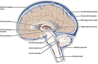
Cerebrospinal fluid (CSF) is a clear, colorless body fluid found within the tissue that surrounds the brain and spinal cord of all vertebrates.
In neuroanatomy, a plexus is a branching network of vessels or nerves. The vessels may be blood vessels or lymphatic vessels. The nerves are typically axons outside the central nervous system.

The ureters are tubes made of smooth muscle that propel urine from the kidneys to the urinary bladder. In a human adult, the ureters are usually 20–30 cm (8–12 in) long and around 3–4 mm (0.12–0.16 in) in diameter. The ureter is lined by urothelial cells, a type of transitional epithelium, and has an additional smooth muscle layer that assists with peristalsis in its lowest third.

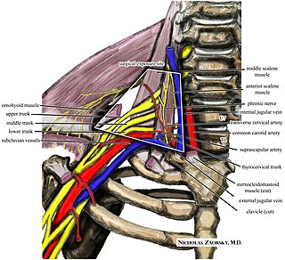
The brachial plexus is a network of nerves formed by the anterior rami of the lower four cervical nerves and first thoracic nerve. This plexus extends from the spinal cord, through the cervicoaxillary canal in the neck, over the first rib, and into the armpit, it supplies afferent and efferent nerve fibers the to chest, shoulder, arm, forearm, and hand.

A spinal nerve is a mixed nerve, which carries motor, sensory, and autonomic signals between the spinal cord and the body. In the human body there are 31 pairs of spinal nerves, one on each side of the vertebral column. These are grouped into the corresponding cervical, thoracic, lumbar, sacral and coccygeal regions of the spine. There are eight pairs of cervical nerves, twelve pairs of thoracic nerves, five pairs of lumbar nerves, five pairs of sacral nerves, and one pair of coccygeal nerves. The spinal nerves are part of the peripheral nervous system.

The myenteric plexus provides motor innervation to both layers of the muscular layer of the gut, having both parasympathetic and sympathetic input, whereas the submucous plexus provides secretomotor innervation to the mucosa nearest the lumen of the gut.

Thoracic outlet syndrome (TOS) is a condition in which there is compression of the nerves, arteries, or veins in the superior thoracic aperture the passageway from the lower neck to the armpit, also known as the thoracic outlet. There are three main types: neurogenic, venous, and arterial. The neurogenic type is the most common and presents with pain, weakness, paraesthesia, and occasionally loss of muscle at the base of the thumb. The venous type results in swelling, pain, and possibly a bluish coloration of the arm. The arterial type results in pain, coldness, and pallor of the arm.

Subconjunctival bleeding, also known as subconjunctival hemorrhage or subconjunctival haemorrhage, is bleeding from a small blood vessel over the whites of the eye. It results in a red spot in the white of the eye. There is generally little to no pain and vision is not affected. Generally only one eye is affected.

Choroid plexus cysts (CPCs) are cysts that occur within choroid plexus of the brain. They are the most common type of intraventricular cyst, occurring in 1% of all pregnancies.

The superior thoracic aperture, also known as the thoracic outlet, or thoracic inlet refers to the opening at the top of the thoracic cavity. It is also clinically referred to as the thoracic outlet, in the case of thoracic outlet syndrome. A lower thoracic opening is the inferior thoracic aperture.

The internal iliac artery is the main artery of the pelvis.

The obturator nerve in human anatomy arises from the ventral divisions of the second, third, and fourth lumbar nerves in the lumbar plexus; the branch from the third is the largest, while that from the second is often very small.

The vertebral vein is formed in the suboccipital triangle, from numerous small tributaries which spring from the internal vertebral venous plexuses and issue from the vertebral canal above the posterior arch of the atlas.

The renal plexus is a complex network of nerves formed by filaments from the celiac ganglia and plexus, aorticorenal ganglia, lower thoracic splanchnic nerves and first lumbar splanchnic nerve and aortic plexus.

The internal pudendal veins are a set of veins in the pelvis. They are the venae comitantes of the internal pudendal artery. Internal pudendal veins are enclosed by pudendal canal, with internal pudendal artery and pudendal nerve.

The superior hypogastric plexus is a plexus of nerves situated on the vertebral bodies anterior to the bifurcation of the abdominal aorta.

The vitelline veins are veins that drain blood from the yolk sac and the gut tube during gestation.

The inferior mesenteric vein begins in the rectum as the superior rectal vein, which has its origin in the hemorrhoidal plexus, and through this plexus communicates with the middle and inferior hemorrhoidal veins.

Sacral splanchnic nerves are splanchnic nerves that connect the inferior hypogastric plexus to the sympathetic trunk in the pelvis.
The Intrapulmonary nodes or Lymphatic Vessels of the Lungs originate in two plexuses, a superficial and a deep. The superficial plexus is placed beneath the pulmonary pleura. The deep accompanies the branches of the pulmonary vessels and the ramifications of the bronchi. In the case of the larger bronchi the deep plexus consists of two networks—one, submucous, beneath the mucous membrane, and another, peribronchial, outside the walls of the bronchi. In the smaller bronchi there is but a single plexus, which extends as far as the bronchioles, but fails to reach the alveoli, in the walls of which there are no traces of lymphatic vessels. The superficial efferents turn around the borders of the lungs and the margins of their fissures, and converge to end in some glands situated at the hilus; the deep efferents are conducted to the hilus along the pulmonary vessels and bronchi, and end in the tracheobronchial lymph nodes. Little or no anastomosis occurs between the superficial and deep lymphatics of the lungs, except in the region of the hilus. they are located in right fissure of lung near the heart
References
- ↑ "pericorneal plexus". Academic Dictionaries & Encyclopedias. Retrieved 2022-11-26.
- ↑ Chang, in-Hong; Garg, Nitin K.; Lunde, Elisa; Han, Kyu-Yeon; Jain, Sandeep; Azar, Dimitri T. (2012). "Corneal Neovascularization: An Anti-VEGF Therapy Review". Survey of Ophthalmology. 57 (5): 415–429. Retrieved 2022-11-26.
This article has not been added to any content categories . Please help out by adding categories to it so that it can be listed with similar articles, in addition to a stub category. (December 2022) |