Related Research Articles
LASIK or Lasik, commonly referred to as laser eye surgery or laser vision correction, is a type of refractive surgery for the correction of myopia, hyperopia, and an actual cure for astigmatism, since it is in cornea. LASIK surgery is performed by an ophthalmologist who uses a laser or microkeratome to reshape the eye's cornea in order to improve visual acuity. For most people, LASIK provides a long-lasting alternative to eyeglasses or contact lenses.

Photorefractive keratectomy (PRK) and laser-assisted sub-epithelial keratectomy (LASEK) are laser eye surgery procedures intended to correct a person's vision, reducing dependency on glasses or contact lenses. LASEK and PRK permanently change the shape of the anterior central cornea using an excimer laser to ablate a small amount of tissue from the corneal stroma at the front of the eye, just under the corneal epithelium. The outer layer of the cornea is removed prior to the ablation.

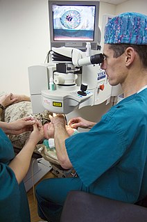
Eye surgery, also known as ocular surgery, is surgery performed on the eye or its adnexa, typically by an ophthalmologist. The eye is a very fragile organ, and requires extreme care before, during, and after a surgical procedure to minimise or prevent further damage. An expert eye surgeon is responsible for selecting the appropriate surgical procedure for the patient, and for taking the necessary safety precautions. Mentions of eye surgery can be found in several ancient texts dating back as early as 1800 BC, with cataract treatment starting in the fifth century BC. Today it continues to be a widely practiced type of surgery, with various techniques having been developed for treating eye problems.
Refractive eye surgery is optional eye surgery used to improve the refractive state of the eye and decrease or eliminate dependency on glasses or contact lenses. This can include various methods of surgical remodeling of the cornea (keratomileusis), lens implantation or lens replacement. The most common methods today use excimer lasers to reshape the curvature of the cornea. Refractive eye surgeries are used to treat common vision disorders such as myopia, hyperopia, presbyopia and astigmatism.

The Bowman's membrane is a smooth, acellular, nonregenerating layer, located between the superficial epithelium and the stroma in the cornea of the eye. It is composed of strong, randomly oriented collagen fibrils in which the smooth anterior surface faces the epithelial basement membrane and the posterior surface merges with the collagen lamellae of the corneal stroma proper.
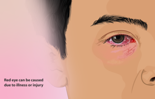
A red eye is an eye that appears red due to illness or injury. It is usually injection and prominence of the superficial blood vessels of the conjunctiva, which may be caused by disorders of these or adjacent structures. Conjunctivitis and subconjunctival hemorrhage are two of the less serious but more common causes.

Corneal transplantation, also known as corneal grafting, is a surgical procedure where a damaged or diseased cornea is replaced by donated corneal tissue. When the entire cornea is replaced it is known as penetrating keratoplasty and when only part of the cornea is replaced it is known as lamellar keratoplasty. Keratoplasty simply means surgery to the cornea. The graft is taken from a recently deceased individual with no known diseases or other factors that may affect the chance of survival of the donated tissue or the health of the recipient.
Recurrent corneal erosion is a disorder of the eyes characterized by the failure of the cornea's outermost layer of epithelial cells to attach to the underlying basement membrane. The condition is excruciatingly painful because the loss of these cells results in the exposure of sensitive corneal nerves. This condition can often leave patients with temporary blindness due to extreme light sensitivity (photophobia).

Corneal dystrophy is a group of rare hereditary disorders characterised by bilateral abnormal deposition of substances in the transparent front part of the eye called the cornea.
Corneal tattooing is the practice of tattooing the cornea of the human eye. Reasons for this practice include improvement of cosmetic appearance and the improvement of sight. Many different methods and procedures exist today, and there are varying opinions concerning the safety or success of this practice.

Band keratopathy is a corneal disease derived from the appearance of calcium on the central cornea. This is an example of metastatic calcification, which by definition, occurs in the presence of hypercalcemia.

Pellucid marginal degeneration (PMD), is a degenerative corneal condition, often confused with keratoconus. It typically presents with painless vision loss affecting both eyes. Rarely, it may cause acute vision loss with severe pain due to perforation of the cornea. It is typically characterized by a clear, bilateral thinning (ectasia) in the inferior and peripheral region of the cornea, although some cases affect only one eye. The cause of the disease remains unclear.

Meesmann corneal dystrophy (MECD) is a rare hereditary autosomal dominant disease that is characterized as a type of corneal dystrophy and a keratin disease. MECD is characterized by the formation of microcysts in the outermost layer of the cornea, known as the anterior corneal epithelium. The anterior corneal epithelium also becomes fragile. This usually affects both eyes rather than a single eye and worsens over time. There are two phenotypes, Meesmann corneal dystrophy 1 (MECD1) and Meesmann corneal dystrophy 2 (MECD2), which affect the genes KRT3 and KRT12, respectively. A heterozygous mutation in either of these genes will lead to a single phenotype. Many with Meesmann corneal dystrophy are asymptomatic or experience mild symptoms.

Ramón Castroviejo Briones (1904–1987) was a famous Spanish and American eye surgeon remembered for his achievements in corneal transplantation.

Reis-Bücklers corneal dystrophy, is a rare, corneal dystrophy of unknown cause, in which the Bowman's layer of the cornea undergoes disintegration. The disorder is inherited in an autosomal dominant fashion, and is associated with mutations in the gene TGFB1.
Epithelial basement membrane dystrophy (EBMD), is a disorder of the eye that can cause pain and dryness.

Lattice corneal dystrophy type, is a rare form of corneal dystrophy. It has no systemic manifestations, unlike the other type of the dystrophy, Lattice corneal dystrophy type II. Lattice corneal dystrophy was first described by Swiss ophthalmologist Hugo Biber in 1890.
Peter S. Hersh is an American ophthalmologist and specialist in LASIK eye surgery, keratoconus, and diseases of the cornea. He co-authored the article in the journal Ophthalmology that presented the results of the study that led to the first approval by the U.S. Food and Drug Administration (FDA) of the excimer laser for the correction of nearsightedness in the United States. Hersh was also medical monitor of the study that led to approval of corneal collagen crosslinking for the treatment of keratoconus.

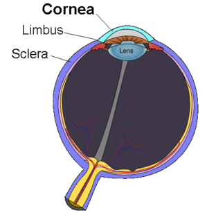
The human cornea is a transparent membrane which allows light to pass through it. The word corneal opacification literally means loss of normal transparency of cornea. The term corneal opacity is used particularly for the loss of transparency of cornea due to scarring. Transparency of the cornea is dependent on the uniform diameter and the regular spacing and arrangement of the collagen fibrils within the stroma. Alterations in the spacing of collagen fibrils in a variety of conditions including corneal edema, scars, and macular corneal dystrophy is clinically manifested as corneal opacity. The term "corneal blindness" is commonly used to describe blindness due to corneal opacity.
Exposure keratopathy is medical condition affecting the cornea of eyes. It can lead to corneal ulceration and permanent loss of vision due to corneal opacity.
References
- ↑ [PTK in the developing world]http://www.ophthalmologyweb.com/JournalUpdates.aspx?spid=23&jid=15009