Related Research Articles

Paranasal sinuses are a group of four paired air-filled spaces that surround the nasal cavity. The maxillary sinuses are located under the eyes; the frontal sinuses are above the eyes; the ethmoidal sinuses are between the eyes and the sphenoidal sinuses are behind the eyes. The sinuses are named for the facial bones in which they are located.
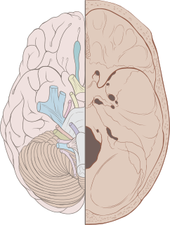
Cranial nerves are the nerves that emerge directly from the brain, of which there are conventionally considered twelve pairs. Cranial nerves relay information between the brain and parts of the body, primarily to and from regions of the head and neck, including the special senses of vision, taste, smell, and hearing.
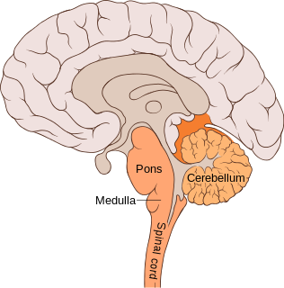
The pons is part of the brainstem, and in humans and other bipeds lies inferior to the midbrain, superior to the medulla oblongata and anterior to the cerebellum.
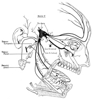
The trigeminal nerve (the fifth cranial nerve, or simply CN V) is a nerve responsible for sensation in the face and motor functions such as biting and chewing; it is the most complex of the cranial nerves. Its name ("trigeminal" = tri-, or three, and - geminus, or twin: thrice-twinned) derives from the fact that each of the two nerves (one on each side of the pons) has three major branches: the ophthalmic nerve (V1), the maxillary nerve (V2), and the mandibular nerve (V3). The ophthalmic and maxillary nerves are purely sensory, whereas the mandibular nerve supplies motor as well as sensory (or "cutaneous") functions. Adding to the complexity of this nerve is the fact that autonomic nerve fibers as well as special sensory fibers (taste) are contained within it.

The mandibular nerve (V3) is the largest of the three divisions of the trigeminal nerve, the fifth cranial nerve (CN V).

Trigeminal neuralgia is a long-term pain disorder that affects the trigeminal nerve. It is a form of neuropathic pain. There are two main types: typical and atypical trigeminal neuralgia. The typical form results in episodes of severe, sudden, shock-like pain in one side of the face that lasts for seconds to a few minutes. Groups of these episodes can occur over a few hours. The atypical form results in a constant burning pain that is less severe. Episodes may be triggered by any touch to the face. Both forms may occur in the same person. It is regarded to be one of the most painful disorders known to medicine, and often results in depression..

The scalp is the anatomical area bordered by the human face at the front, and by the neck at the sides and back.
A lemniscus is a bundle of secondary sensory fibers in the brainstem. The medial lemniscus and lateral lemniscus terminate in specific relay nuclei of the diencephalon. The trigeminal lemniscus is sometimes considered as the cephalic part of the medial lemniscus.

The ophthalmic nerve (V1) is one of three divisions of the trigeminal nerve (CN V). It has three branches that provide sensory innervation to the eye, skin of the upper face and anterior scalp.
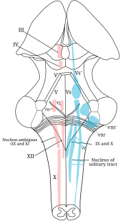
A cranial nerve nucleus is a collection of neurons in the brain stem that is associated with one or more cranial nerves. Axons carrying information to and from the cranial nerves form a synapse first at these nuclei. Lesions occurring at these nuclei can lead to effects resembling those seen by the severing of nerve(s) they are associated with. All the nuclei except that of the trochlear nerve supply nerves of the same side of the body.

The spinal trigeminal nucleus is a nucleus in the medulla that receives information about deep/crude touch, pain, and temperature from the ipsilateral face. In addition to the trigeminal nerve, the facial, glossopharyngeal, and vagus nerves also convey pain information from their areas to the spinal trigeminal nucleus. Thus the spinal trigeminal nucleus receives input from cranial nerves V, VII, IX, and X.

The trigeminal motor nucleus contains motor neurons that innervate muscles of the first branchial arch, namely the muscles of mastication, the tensor tympani, tensor veli palatini, mylohyoid, and anterior belly of the digastric. This nucleus is located in the mid-pons.
The trigeminal ganglion is a sensory ganglion of the trigeminal nerve that occupies a cavity in the dura mater, covering the trigeminal impression near the apex of the petrous part of the temporal bone.
Hypoesthesia or numbness is a common side effect of various medical conditions which manifests as a reduced sense of touch or sensation, or a partial loss of sensitivity to sensory stimuli. In everyday speech this is generally referred to as numbness.
The principal sensory nucleus of trigeminal nerve is a group of second-order neurons which have cell bodies in the caudal pons.
The mesencephalic nucleus of trigeminal nerve is involved with reflex proprioception of the periodontium and of the muscles of mastication in the jaw that functions to prevent biting down hard enough to lose a tooth. To subserve this reflex protective function, mechanoreceptive nerves in the periodontal ligament sense tooth movement and project to the mesencephalic nucleus. Likewise, afferent fibers from muscle spindles, the sensory organs of skeletal muscle, are stimulated by the stretch of hard contraction of jaw muscles. The temporomandibular joints and the Golgi tendon organs of the jaw muscles do not project to the mesencephalic nucleus. The mesencephalic nucleus is one of four trigeminal nerve nuclei, three sensory and one motor. The other two sensory nuclei are the chief sensory nucleus mediating conscious facial touch and the spinal trigeminal nucleus, mediating pain and temperature in the head, and is of importance in headache. The trigeminal motor nucleus innervates the muscles of mastication, mylohyoid, anterior belly of digastric, tensor veli palatini, and tensor tympani.
The ventral trigeminal tract, ventral trigeminothalamic tract, anterior trigeminal tract, or anterior trigeminothalamic tract, is a tract composed of second order neuronal axons. These fibers carry sensory information about discriminative and crude touch, conscious proprioception, pain, and temperature from the head, face, and oral cavity. The ventral trigeminal tract connects the two major components of the brainstem trigeminal complex – the principal, or main sensory nucleus and the spinal trigeminal nucleus, to the ventral posteromedial nucleus of the thalamus.
The trigeminal lemniscus, also called the trigeminothalamic tract, is composed of the ventral trigeminal tract, and the dorsal trigeminal tract – nerve tracts that convey tactile, pain, and temperature impulses from the skin of the face, the mucous membranes of the nasal and oral cavities, and the eye, as well as proprioceptive information from the facial and masticatory muscles.
The dorsal trigeminal tract, dorsal trigeminothalamic tract, or posterior trigeminothalamic tract, is composed of second-order neuronal axons. These fibers carry sensory information about discriminative touch and conscious proprioception in the oral cavity from the principal nucleus of the trigeminal nerve to the ventral posteromedial (VPM) nucleus of the thalamus.

Trigeminal trophic syndrome is a rare disease caused by the interruption of peripheral or central sensory pathways of the trigeminal nerve. A slowly enlarging, uninflammed ulcer can occur in the area that has suffered the trigeminal nerve damage; including but not limited to the cheek beside the ala nasi. These sores affect the skin supplied by the sensory component of the trigeminal nerve. Similar lesions may also occur in the corners of the eyes, inside the ear canal, on the scalp or inside the mouth.
References
- ↑ James, William D.; Berger, Timothy G.; et al. (2006). Andrews' Diseases of the Skin: clinical Dermatology. Saunders Elsevier. ISBN 978-0-7216-2921-6.
| This cutaneous condition article is a stub. You can help Wikipedia by expanding it. |