
In aphasia, a person may be unable to comprehend or unable to formulate language because of damage to specific brain regions. The major causes are stroke and head trauma; prevalence is hard to determine, but aphasia due to stroke is estimated to be 0.1–0.4% in the Global North. Aphasia can also be the result of brain tumors, epilepsy, autoimmune neurological diseases, brain infections, or neurodegenerative diseases.

Expressive aphasia is a type of aphasia characterized by partial loss of the ability to produce language, although comprehension generally remains intact. A person with expressive aphasia will exhibit effortful speech. Speech generally includes important content words but leaves out function words that have more grammatical significance than physical meaning, such as prepositions and articles. This is known as "telegraphic speech". The person's intended message may still be understood, but their sentence will not be grammatically correct. In very severe forms of expressive aphasia, a person may only speak using single word utterances. Typically, comprehension is mildly to moderately impaired in expressive aphasia due to difficulty understanding complex grammar.

In neuroscience and psychology, the term language center refers collectively to the areas of the brain which serve a particular function for speech processing and production. Language is a core system that gives humans the capacity to solve difficult problems and provides them with a unique type of social interaction. Language allows individuals to attribute symbols to specific concepts, and utilize them through sentences and phrases that follow proper grammatical rules. Finally, speech is the mechanism by which language is orally expressed.
Wernicke's aphasia, also known as receptive aphasia, sensory aphasia, fluent aphasia, or posterior aphasia, is a type of aphasia in which individuals have difficulty understanding written and spoken language. Patients with Wernicke's aphasia demonstrate fluent speech, which is characterized by typical speech rate, intact syntactic abilities and effortless speech output. Writing often reflects speech in that it tends to lack content or meaning. In most cases, motor deficits do not occur in individuals with Wernicke's aphasia. Therefore, they may produce a large amount of speech without much meaning. Individuals with Wernicke's aphasia often suffer of anosognosia – they are unaware of their errors in speech and do not realize their speech may lack meaning. They typically remain unaware of even their most profound language deficits.

A head injury is any injury that results in trauma to the skull or brain. The terms traumatic brain injury and head injury are often used interchangeably in the medical literature. Because head injuries cover such a broad scope of injuries, there are many causes—including accidents, falls, physical assault, or traffic accidents—that can cause head injuries.

Broca's area, or the Broca area, is a region in the frontal lobe of the dominant hemisphere, usually the left, of the brain with functions linked to speech production.
Aphasiology is the study of language impairment usually resulting from brain damage, due to neurovascular accident—hemorrhage, stroke—or associated with a variety of neurodegenerative diseases, including different types of dementia. These specific language deficits, termed aphasias, may be defined as impairments of language production or comprehension that cannot be attributed to trivial causes such as deafness or oral paralysis. A number of aphasias have been described, but two are best known: expressive aphasia and receptive aphasia.

Neuropsychology is a branch of psychology concerned with how a person's cognition and behavior are related to the brain and the rest of the nervous system. Professionals in this branch of psychology focus on how injuries or illnesses of the brain affect cognitive and behavioral functions.

Brain injury (BI) is the destruction or degeneration of brain cells. Brain injuries occur due to a wide range of internal and external factors. In general, brain damage refers to significant, undiscriminating trauma-induced damage.
Anomic aphasia is a mild, fluent type of aphasia where individuals have word retrieval failures and cannot express the words they want to say. By contrast, anomia is a deficit of expressive language, and a symptom of all forms of aphasia, but patients whose primary deficit is word retrieval are diagnosed with anomic aphasia. Individuals with aphasia who display anomia can often describe an object in detail and maybe even use hand gestures to demonstrate how the object is used, but cannot find the appropriate word to name the object. Patients with anomic aphasia have relatively preserved speech fluency, repetition, comprehension, and grammatical speech.
Wernicke's area, also called Wernicke's speech area, is one of the two parts of the cerebral cortex that are linked to speech, the other being Broca's area. It is involved in the comprehension of written and spoken language, in contrast to Broca's area, which is primarily involved in the production of language. It is traditionally thought to reside in Brodmann area 22, which is located in the superior temporal gyrus in the dominant cerebral hemisphere, which is the left hemisphere in about 95% of right-handed individuals and 70% of left-handed individuals.
In neurology, conduction aphasia, also called associative aphasia, is an uncommon form of difficulty in speaking (aphasia). It is caused by damage to the parietal lobe of the brain. An acquired language disorder, it is characterised by intact auditory comprehension, coherent speech production, but poor speech repetition. Affected people are fully capable of understanding what they are hearing, but fail to encode phonological information for production. This deficit is load-sensitive as the person shows significant difficulty repeating phrases, particularly as the phrases increase in length and complexity and as they stumble over words they are attempting to pronounce. People have frequent errors during spontaneous speech, such as substituting or transposing sounds. They are also aware of their errors and will show significant difficulty correcting them.
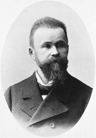
CarlWernicke was a German physician, anatomist, psychiatrist and neuropathologist. He is known for his influential research into the pathological effects of specific forms of encephalopathy and also the study of receptive aphasia, both of which are commonly associated with Wernicke's name and referred to as Wernicke encephalopathy and Wernicke's aphasia, respectively. His research, along with that of Paul Broca, led to groundbreaking realizations of the localization of brain function, specifically in speech. As such, Wernicke's area has been named after the scientist.

Global aphasia is a severe form of nonfluent aphasia, caused by damage to the left side of the brain, that affects receptive and expressive language skills as well as auditory and visual comprehension. Acquired impairments of communicative abilities are present across all language modalities, impacting language production, comprehension, and repetition. Patients with global aphasia may be able to verbalize a few short utterances and use non-word neologisms, but their overall production ability is limited. Their ability to repeat words, utterances, or phrases is also affected. Due to the preservation of the right hemisphere, an individual with global aphasia may still be able to express themselves through facial expressions, gestures, and intonation. This type of aphasia often results from a large lesion of the left perisylvian cortex. The lesion is caused by an occlusion of the left middle cerebral artery and is associated with damage to Broca's area, Wernicke's area, and insular regions which are associated with aspects of language.

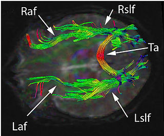
In neuroanatomy, the arcuate fasciculus is a bundle of axons that generally connects Broca's area and Wernicke's area in the brain. It is an association fiber tract connecting caudal temporal lobe and inferior frontal lobe.
Transcortical sensory aphasia (TSA) is a kind of aphasia that involves damage to specific areas of the temporal lobe of the brain, resulting in symptoms such as poor auditory comprehension, relatively intact repetition, and fluent speech with semantic paraphasias present. TSA is a fluent aphasia similar to Wernicke's aphasia, with the exception of a strong ability to repeat words and phrases. The person may repeat questions rather than answer them ("echolalia").

Brodmann area 22 is a Brodmann's area that is cytoarchitecturally located in the posterior superior temporal gyrus of the brain. In the left cerebral hemisphere, it is one portion of Wernicke's area. The left hemisphere BA22 helps with generation and understanding of individual words. On the right side of the brain, BA22 helps to discriminate pitch and sound intensity, both of which are necessary to perceive melody and prosody. Wernicke's area is active in processing language and consists of the left Brodmann area 22 and Brodmann area 40, the supramarginal gyrus.

Mixed transcortical aphasia is the least common of the three transcortical aphasias. This type of aphasia can also be referred to as "Isolation Aphasia". This type of aphasia is a result of damage that isolates the language areas from other brain regions. Broca's, Wernicke's, and the arcuate fasiculus are left intact; however, they are isolated from other brain regions.

The lateralization of brain function is the tendency for some neural functions or cognitive processes to be specialized to one side of the brain or the other. The median longitudinal fissure separates the human brain into two distinct cerebral hemispheres, connected by the corpus callosum. Although the macrostructure of the two hemispheres appears to be almost identical, different composition of neuronal networks allows for specialized function that is different in each hemisphere.
Paraphasia is a type of language output error commonly associated with aphasia, and characterized by the production of unintended syllables, words, or phrases during the effort to speak. Paraphasic errors are most common in patients with fluent forms of aphasia, and come in three forms: phonemic or literal, neologistic, and verbal. Paraphasias can affect metrical information, segmental information, number of syllables, or both. Some paraphasias preserve the meter without segmentation, and some do the opposite. However, most paraphasias affect both partially.
