In neuroanatomy, dura mater is a thick membrane made of dense irregular connective tissue that surrounds the brain and spinal cord. It is the outermost of the three layers of membrane called the meninges that protect the central nervous system. The other two meningeal layers are the arachnoid mater and the pia mater. It envelops the arachnoid mater, which is responsible for keeping in the cerebrospinal fluid. It is derived primarily from the neural crest cell population, with postnatal contributions of the paraxial mesoderm.

In human anatomy, the spinal canal, vertebral canal or spinal cavity is an elongated body cavity enclosed within the dorsal bony arches of the vertebral column, which contains the spinal cord, spinal roots and dorsal root ganglia. It is a process of the dorsal body cavity formed by alignment of the vertebral foramina. Under the vertebral arches, the spinal canal is also covered anteriorly by the posterior longitudinal ligament and posteriorly by the ligamentum flavum. The potential space between these ligaments and the dura mater covering the spinal cord is known as the epidural space. Spinal nerves exit the spinal canal via the intervertebral foramina under the corresponding vertebral pedicles.

In anatomy, the epidural space is the potential space between the dura mater and vertebrae (spine).

In human anatomy, the artery of Adamkiewicz is the largest anterior segmental medullary artery. It typically arises from a 9th to 12th left posterior intercostal artery, enters through the L2-L3 intervertebral foramen to join the anterior spinal artery and supply much of the inferior half of the spinal cord.

The arachnoid mater is one of the three meninges, the protective membranes that cover the brain and spinal cord. It is so named because of its resemblance to a spider web. The arachnoid mater is a derivative of the neural crest mesoectoderm in the embryo.

The conus medullaris or conus terminalis is the tapered, lower end of the spinal cord. It occurs near lumbar vertebral levels 1 (L1) and 2 (L2), occasionally lower. The upper end of the conus medullaris is usually not well defined, however, its corresponding spinal cord segments are usually S1–S5.

The rectal venous plexus is the venous plexus surrounding the rectum. It consists of an internal and an external rectal plexus. It is drained by the superior, middle, and inferior rectal veins. It forms a portosystemic (portocaval) anastomosis. This allows rectally administered medications to bypass first pass metabolism.

The femoral canal is the medial compartment of the three compartments of the femoral sheath. It is conical in shape. The femoral canal contains lymphatic vessels, and adipose and loose connective tissue, as well as - sometimes - a deep inguinal lymph node. The function of the femoral canal is to accommodate the distension of the femoral vein when venous return from the leg is increased or temporarily restricted.

The intervertebral foramen is an opening between two pedicles of adjacent vertebra in the articulated spine. Each intervertebral foramen gives passage to a spinal nerve and spinal blood vessels, and lodges a posterior (dorsal) root ganglion. Cervical, thoracic, and lumbar vertebrae all have intervertebral foramina.
A nerve root is the initial segment of a nerve leaving the central nervous system. Nerve roots can be classified as:

The internal vertebral venous plexuses lie within the vertebral canal in the epidural space, embedded within epidural fat. They receive tributaries from bones, red bone marrow, and spinal cord. They are arranged into four interconnected, vertically oriented vessels - two situated anteriorly, and two posteriorly:

The intervertebral veins accompany the spinal nerves through the intervertebral foramina to drain the internal vertebral venous plexuses into the external vertebral venous plexuses. They drain into vertebral vein, intercostal veins, lumbar veins, and lateral sacral veins. Upper posterior intercostal veins may additionally drain via brachiocephalic vens. They may drain to ascending lumbar veins. They may drain into the inferior vena cava directly, reaching it by winding around the surface of the vertebral body.

Anterior spinal veins are veins that receive blood from the anterior spinal cord.

Segmental medullary arteries are arteries of varying size in the thoracolumbar region that arise from segmental arteries of this region and pass through intervertebral foramina to supply the spinal cord. They may join the anterior spinal artery.
The anterior and posterior radicular arteries are 32 pairs of small/ variously sized arteries that enter an intervertebral foramen, bifurcating within it to form an anterior and a posterior radicular artery which accompany the anterior root and posterior root of a spinal nerve, respectively. They supply the corresponding spinal cord segment as well as the anterior and posterior root of the spinal nerve and its sensory ganglion.

The iliolumbar vein is the vena comitans of the iliolumbar artery.
Vascular myelopathy refers to an abnormality of the spinal cord in regard to its blood supply. The blood supply is complicated and supplied by two major vessel groups: the posterior spinal arteries and the anterior spinal arteries—of which the Artery of Adamkiewicz is the largest. Both the posterior and anterior spinal arteries run the entire length of the spinal cord and receive anastomotic (conjoined) vessels in many places. The anterior spinal artery has a less efficient supply of blood and is therefore more susceptible to vascular disease. Whilst atherosclerosis of spinal arteries is rare, necrosis in the anterior artery can be caused by disease in vessels originating from the segmental arteries such as atheroma or aortic dissection.

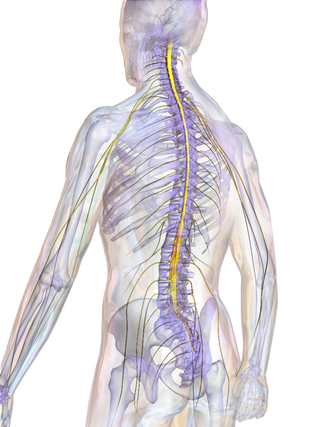
The spinal cord is a long, thin, tubular structure made up of nervous tissue that extends from the medulla oblongata in the brainstem to the lumbar region of the vertebral column (backbone) of vertebrate animals. The center of the spinal cord is hollow and contains a structure called the central canal, which contains cerebrospinal fluid. The spinal cord is also covered by meninges and enclosed by the neural arches. Together, the brain and spinal cord make up the central nervous system.
The epidural venous plexus is a venous plexus embedded within the epidural fat of the vertebral canal. It is situated within the anterior epidural space. The plexus extends from the skull base to the sacrum. It is surrounded by sparse fat. It drains into the cavernous sinus of the cranial cavity; it also communicates with the radicular veins.
The cerebrospinal venous system (CSVS) consists of the interconnected venous systems of the brain and the spine.


