Radionuclide cisternogram

Last updated
Radionuclide cisternogram
Medical diagnostics
Purpose determine if there is abnormal CSF flow within the brain

A radionuclide cisternogram is a medical imaging study which involves injecting a radionuclide by lumbar puncture (spinal tap) into a patient's cerebral spinal fluid (CSF) to determine if there is abnormal CSF flow within the brain and spinal canal which can be altered by hydrocephalus, Arnold–Chiari malformation, syringomyelia, or an arachnoid cyst. [1] It may also evaluate a suspected leak (also known as a CSF fistula) from the CSF cavity into the nasal cavity. A leak can also be confirmed by the presence of beta-2 transferrin in fluid collected from the nose before this more invasive procedure is performed.

Medical imaging technique and process of creating visual representations of the interior of a body

Medical imaging is the technique and process of creating visual representations of the interior of a body for clinical analysis and medical intervention, as well as visual representation of the function of some organs or tissues (physiology). Medical imaging seeks to reveal internal structures hidden by the skin and bones, as well as to diagnose and treat disease. Medical imaging also establishes a database of normal anatomy and physiology to make it possible to identify abnormalities. Although imaging of removed organs and tissues can be performed for medical reasons, such procedures are usually considered part of pathology instead of medical imaging.

A radionuclide is an atom that has excess nuclear energy, making it unstable. This excess energy can be used in one of three ways: emitted from the nucleus as gamma radiation; transferred to one of its electrons to release it as a conversion electron; or used to create and emit a new particle from the nucleus. During those processes, the radionuclide is said to undergo radioactive decay. These emissions are considered ionizing radiation because they are powerful enough to liberate an electron from another atom. The radioactive decay can produce a stable nuclide or will sometimes produce a new unstable radionuclide which may undergo further decay. Radioactive decay is a random process at the level of single atoms: it is impossible to predict when one particular atom will decay. However, for a collection of atoms of a single element the decay rate, and thus the half-life (t1/2) for that collection can be calculated from their measured decay constants. The range of the half-lives of radioactive atoms have no known limits and span a time range of over 55 orders of magnitude.

Lumbar puncture Procedure to collect cerebrospinal fluid

Lumbar puncture (LP), also known as a spinal tap, is a medical procedure in which a needle is inserted into the spinal canal, most commonly to collect cerebrospinal fluid (CSF) for diagnostic testing. The main reason for a lumbar puncture is to help diagnose diseases of the central nervous system, including the brain and spine. Examples of these conditions include meningitis and subarachnoid hemorrhage. It may also be used therapeutically in some conditions. Increased intracranial pressure is a contraindication, due to risk of brain matter being compressed and pushed toward the spine. Sometimes, lumbar puncture cannot be performed safely. It is regarded as a safe procedure, but post-dural-puncture headache is a common side effect.

The patient may be instructed to not eat or drink, or take medications such as aspirin or other blood thinners before the procedure. Pledgets can be inserted into the nasal cavity before the procedure when a CSF leak is suspected.

Aspirin medication

Aspirin, also known as acetylsalicylic acid (ASA), is a medication used to treat pain, fever, or inflammation. Specific inflammatory conditions which aspirin is used to treat include Kawasaki disease, pericarditis, and rheumatic fever. Aspirin given shortly after a heart attack decreases the risk of death. Aspirin is also used long-term to help prevent further heart attacks, ischaemic strokes, and blood clots in people at high risk. It may also decrease the risk of certain types of cancer, particularly colorectal cancer. For pain or fever, effects typically begin within 30 minutes. Aspirin is a nonsteroidal anti-inflammatory drug (NSAID) and works similarly to other NSAIDs but also suppresses the normal functioning of platelets.

The patient's spinal fluid is injected with a radiopharmaceutical tracer, such as DTPA tagged with indium 111, through a lumbar puncture (spinal tap). The tracer will diffuse up the spinal column and into the intracranial ventricles and the subarachnoid spaces around the brain. The progress of the tracer's diffusion through the CSF will be recorded by a nuclear medicine gamma camera. Images are usually taken immediately, at 6 hours, and at 24 hours. The patient may be asked to return for 48- and 72-hour follow-up scans.

Indium (49In) consists of two primordial nuclides, with the most common (~ 95.7%) nuclide (115In) being measurably though weakly radioactive. Its spin-forbidden decay has a half life of 4.41×10¹⁴ years.

Diffusion Statistical movement of molecules or atoms from a region of high concentration (or high chemical potential) to a region of low concentration (or low chemical potential)

Diffusion is the net movement of molecules or atoms from a region of higher concentration to a region of lower concentration. Diffusion is driven by a gradient in chemical potential of the diffusing species.

Gamma camera

A gamma camera (γ-camera), also called a scintillation camera or Anger camera, is a device used to image gamma radiation emitting radioisotopes, a technique known as scintigraphy. The applications of scintigraphy include early drug development and nuclear medical imaging to view and analyse images of the human body or the distribution of medically injected, inhaled, or ingested radionuclides emitting gamma rays.

The pledgets will be removed and either imaged with a gamma camera or counted using a gamma counter. If the tracer has leaked onto the pledget through the skull, it will appear on the gamma camera image or register abnormal counts allowing the diagnostician to determine the location of the leak within the sinus cavity. The site of the CSF leak can be plugged with fat or muscle by endoscopic surgery.

Gamma counter

A gamma counter is a machine to measure gamma radiation emitted by a radionuclide. Unlike survey meters, gamma counters are designed to measure small samples of radioactive material, typically with automated measurement and movement of multiple samples.

Headaches following the procedure are common, but should fade in 3–5 days. Drinking caffeinated liquids, as well as bed rest, is often recommended, though at least one scientific paper disputes the practice.

Bed rest

Bed rest, also referred to as the rest-cure, is a medical treatment in which a person lies in bed for most of the time to try to cure an illness. Bed rest refers to voluntarily lying in bed as a treatment and not being confined to bed because of a health impairment which physically prevents leaving bed. The practice is still used although a 1999 systematic review found no benefits for any of the 17 conditions studied and no proven benefit for any conditions at all, beyond that imposed by symptoms.
Related Research Articles

Cerebrospinal fluid clear colorless bodily fluid found in the brain and spine

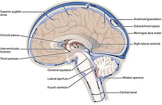
Cerebrospinal fluid (CSF) is a clear, colorless body fluid found in the brain and spinal cord. It is produced by the specialised ependymal cells in the choroid plexuses of the ventricles of the brain, and absorbed in the arachnoid granulations. There is about 125mL of CSF at any one time, and about 500 mL is generated every day. CSF acts as a cushion or buffer for the brain, providing basic mechanical and immunological protection to the brain inside the skull. CSF also serves a vital function in cerebral autoregulation of cerebral blood flow.

Syringomyelia congenital disorder of nervous system

Syringomyelia is a generic term referring to a disorder in which a cyst or cavity forms within the spinal cord. This cyst, called a syrinx, can expand and elongate over time, destroying the spinal cord. The damage may result in loss of feeling, paralysis, weakness, and stiffness in the back, shoulders, and extremities. Syringomyelia may also cause a loss of the ability to feel extremes of hot or cold, especially in the hands. It may also lead to a cape-like bilateral loss of pain and temperature sensation along the upper chest and arms. Each patient experiences a different combination of symptoms. These symptoms typically vary depending on the extent and, often more critically, on the location of the syrinx within the spinal cord.

Ventricular system set of structures containing cerebrospinal fluid in the brain

The ventricular system is a set of four interconnected cavities (ventricles) in the brain, where the cerebrospinal fluid (CSF) is produced. Within each ventricle is a region of choroid plexus, a network of ependymal cells involved in the production of CSF. The ventricular system is continuous with the central canal of the spinal cord, allowing for the flow of CSF to circulate. All of the ventricular system and the central canal of the spinal cord are lined with ependyma, a specialised form of epithelium.

Pneumoencephalography

Pneumoencephalography was a common medical procedure in which most of the cerebrospinal fluid (CSF) was drained from around the brain by means of a lumbar puncture and replaced with air, oxygen, or helium to allow the structure of the brain to show up more clearly on an X-ray image. It was derived from ventriculography, an earlier and more primitive method where the air is injected through holes drilled in the skull.

Spinal anaesthesia

Spinal anaesthesia, also called spinal block, subarachnoid block, intradural block and intrathecal block, is a form of neuraxial regional anaesthesia involving the injection of a local anaesthetic or opioid into the subarachnoid space, generally through a fine needle, usually 9 cm (3.5 in) long. It is a safe and effective form of anesthesia performed by nurse anesthetists and anesthesiologists which can be used as an alternative to general anesthesia commonly in surgeries involving the lower extremities and surgeries below the umbilicus. The local anesthetic or opioid injected into the cerebrospinal fluid provides anesthesia, analgesia, and motor and sensory blockade. The tip of the spinal needle has a point or small bevel. Recently, pencil point needles have been made available.

Myelography radiographic examination of spinal cord

Myelography is a type of radiographic examination that uses a contrast medium to detect pathology of the spinal cord, including the location of a spinal cord injury, cysts, and tumors. Historically the procedure involved the injection of a radiocontrast agent into the cervical or lumbar spine, followed by several X-ray projections. Today, myelography has largely been replaced by the use of MRI scans, although the technique is still sometimes used under certain circumstances – though now usually in conjunction with CT rather than X-ray projections.

Neoplastic or malignant meningitis, also called carcinomatous meningitis, leptomeningeal carcinoma, leptomeningeal carcinomatosis, leptomeningeal metastasis, meningeal carcinomatosis, meningeal metastasis, and meningitis carcinomatosa, is the development of meningitis due to infiltration of the subarachnoid space by cancerous cells. Malignant cells come from primary cancer such as breast cancer or from a primary brain tumor like medulloblastoma. Neoplastic Meningitis (NM) was first reported in the 1870s with the most common cause being breast cancer, lung cancer, and malignant melanoma.

A suboccipital puncture or cisternal puncture is a diagnostic procedure that can be performed in order to collect a sample of cerebrospinal fluid (CSF) for biochemical, microbiological, and cytological analysis, or rarely to relieve increased intracranial pressure. It is done by inserting a needle through the skin below the external occipital protuberance into the cisterna magna and is an alternative to lumbar puncture. Indications for its use are limited. Subarachnoid hemorrhage and direct puncture of brain tissue are the most common major complications. Fluoroscopic guidance decreases the risk for complications. The use of this procedure in humans was first described by Ayer in 1920.

Post-dural-puncture headache (PDPH) is a complication of puncture of the dura mater. The headache is severe and described as "searing and spreading like hot metal," involving the back and front of the head, and spreading to the neck and shoulders, sometimes involving neck stiffness. It is exacerbated by movement, and sitting or standing, and relieved to some degree by lying down. Nausea, vomiting, pain in arms and legs, hearing loss, tinnitus, vertigo, dizziness and paraesthesia of the scalp are common.

Epidural blood patch

An epidural blood patch is a surgical procedure that uses autologous blood in order to close one or many holes in the dura mater of the spinal cord, usually as a result of a previous lumbar puncture. The procedure can be used to relieve post dural puncture headaches caused by lumbar puncture. A small amount of the patient's blood is injected into the epidural space near the site of the original puncture; the resulting blood clot then "patches" the meningeal leak. The procedure carries the typical risks of any epidural puncture. However, even though it is often effective, further intervention is sometimes necessary.

Mollarets meningitis Human disease

Mollaret's meningitis is a recurrent or chronic inflammation of the protective membranes covering the brain and spinal cord, known collectively as the meninges. Since Mollaret's meningitis is a recurrent, benign (non-cancerous), aseptic meningitis, it is also referred to as benign recurrent lymphocytic meningitis. It was named for Pierre Mollaret, the French neurologist who first described it in 1944.

Spontaneous cerebrospinal fluid leak

A spontaneous cerebrospinal fluid leak (SCSFL) is a medical condition in which the cerebrospinal fluid (CSF) surrounding the human brain and spinal cord leaks out of the surrounding protective dural sac for no apparent reason. The dura, a tough, inflexible tissue, is the outermost of the three layers of the meninges, the system of membranes surrounding the brain and spinal cord.

Metrizamide chemical compound

Metrizamide is a non-ionic radiopaque contrast agent. A density gradient medium for the centrifugation of biological particles.

Orthostatic headache

Orthostatic headache is a medical condition in which a person develops a headache while vertical and the headache is relieved when horizontal. Previously it was often misdiagnosed as different primary headache disorders such as migraine or tension headaches. Increasing awareness of the symptom and its causes has prevented delayed or missed diagnosis.

A cerebrospinal fluid leak is a medical condition where the cerebrospinal fluid (CSF) in the brain or spinal cord leaks out of one or more holes or tears in the dura mater.

DMSA scan a radionuclide scan that assesses renal morphology, structure and function

A DMSA scan is a radionuclide scan that uses dimercaptosuccinic acid (DMSA) in assessing renal morphology, structure and function. Radioactive technetium-99m is combined with DMSA and injected into a patient, followed by imaging with a gamma camera after 2-3 hours. A DMSA scan is usually static imaging, other radiotracers like DTPA & MAG3 are usually used for dynamic imaging to assess renal excretion.

A lumbar–peritoneal shunt is a technique to channelise the cerebrospinal fluid (CSF) from the lumbar thecal sac into the peritoneal cavity.

Thecal sac Posterior disc

The thecal sac or dural sac is the membranous sheath (theca) or tube of dura mater that surrounds the spinal cord and the cauda equina. The thecal sac contains the cerebrospinal fluid which provides nutrients and buoyancy to the spinal cord. From the skull the tube adheres to bone at the foramen magnum and extends down to the second sacral vertebra where it tapers to cover over the filum terminale. Along most of the spinal canal it is separated from the inner surface by the epidural space. The sac has projections that follow the spinal nerves along their paths out of the vertebral canal which become the dural root sheaths.

Lymphocytic pleocytosis

Lymphocytic pleocytosis is an abnormal increase in the amount of lymphocytes in the cerebrospinal fluid (CSF). It is usually considered to be a sign of infection or inflammation within the nervous system, and is encountered in a number of neurological diseases, such as pseudomigraine, Susac's syndrome, and encephalitis. While lymphocytes make up roughly a quarter of all white blood cells (WBC) in the body, they are generally rare in the CSF. Under normal conditions, there are usually less than 5 white blood cells per µL of CSF. In a pleocytic setting, the number of lymphocytes can jump to more than 1,000 cells per µL. Increases in lymphocyte count are often accompanied by an increase in cerebrospinal protein concentrations in addition to pleocytosis of other types of white blood cells.

References

  1. Battal, B; Kocaoglu, M; Bulakbasi, N; Husmen, G; Tuba Sana, H; Tayfun, C (2011). "Cerebrospinal fluid flow imaging by using phase-contrast MR technique". The British Journal of Radiology. 84: 758–765. doi:10.1259/bjr/66206791. PMC   3473435 Lock-green.svg. PMID   21586507.