Related Research Articles

Diffraction is the interference or bending of waves around the corners of an obstacle or through an aperture into the region of geometrical shadow of the obstacle/aperture. The diffracting object or aperture effectively becomes a secondary source of the propagating wave. Italian scientist Francesco Maria Grimaldi coined the word diffraction and was the first to record accurate observations of the phenomenon in 1660.

Microscopy is the technical field of using microscopes to view objects and areas of objects that cannot be seen with the naked eye. There are three well-known branches of microscopy: optical, electron, and scanning probe microscopy, along with the emerging field of X-ray microscopy.

Holography is a technique that enables a wavefront to be recorded and later reconstructed. It is best known as a method of generating three-dimensional images, and has a wide range of other uses, including data storage, microscopy, and interferometry. In principle, it is possible to make a hologram for any type of wave.

Angular resolution describes the ability of any image-forming device such as an optical or radio telescope, a microscope, a camera, or an eye, to distinguish small details of an object, thereby making it a major determinant of image resolution. It is used in optics applied to light waves, in antenna theory applied to radio waves, and in acoustics applied to sound waves. The colloquial use of the term "resolution" sometimes causes confusion; when an optical system is said to have a high resolution or high angular resolution, it means that the perceived distance, or actual angular distance, between resolved neighboring objects is small. The value that quantifies this property, θ, which is given by the Rayleigh criterion, is low for a system with a high resolution. The closely related term spatial resolution refers to the precision of a measurement with respect to space, which is directly connected to angular resolution in imaging instruments. The Rayleigh criterion shows that the minimum angular spread that can be resolved by an image-forming system is limited by diffraction to the ratio of the wavelength of the waves to the aperture width. For this reason, high-resolution imaging systems such as astronomical telescopes, long distance telephoto camera lenses and radio telescopes have large apertures.

In optics, any optical instrument or system – a microscope, telescope, or camera – has a principal limit to its resolution due to the physics of diffraction. An optical instrument is said to be diffraction-limited if it has reached this limit of resolution performance. Other factors may affect an optical system's performance, such as lens imperfections or aberrations, but these are caused by errors in the manufacture or calculation of a lens, whereas the diffraction limit is the maximum resolution possible for a theoretically perfect, or ideal, optical system.
In optics, the Fraunhofer diffraction equation is used to model the diffraction of waves when plane waves are incident on a diffracting object, and the diffraction pattern is viewed at a sufficiently long distance from the object, and also when it is viewed at the focal plane of an imaging lens. In contrast, the diffraction pattern created near the diffracting object and is given by the Fresnel diffraction equation.

The point spread function (PSF) describes the response of a focused optical imaging system to a point source or point object. A more general term for the PSF is the system's impulse response; the PSF is the impulse response or impulse response function (IRF) of a focused optical imaging system. The PSF in many contexts can be thought of as the shapeless blob in an image that should represent a single point object. We can consider this as a spatial impulse response function. In functional terms, it is the spatial domain version of the optical transfer function (OTF) of an imaging system. It is a useful concept in Fourier optics, astronomical imaging, medical imaging, electron microscopy and other imaging techniques such as 3D microscopy and fluorescence microscopy.

Gopalasamudram Narayanan Ramachandran, or G.N. Ramachandran, FSR was an Indian physicist who was known for his work that led to his creation of the Ramachandran plot for understanding peptide structure. He was the first to propose a triple-helical model for the structure of collagen. He subsequently went on to make other major contributions in biology and physics.

Confocal microscopy, most frequently confocal laser scanning microscopy (CLSM) or laser scanning confocal microscopy (LSCM), is an optical imaging technique for increasing optical resolution and contrast of a micrograph by means of using a spatial pinhole to block out-of-focus light in image formation. Capturing multiple two-dimensional images at different depths in a sample enables the reconstruction of three-dimensional structures within an object. This technique is used extensively in the scientific and industrial communities and typical applications are in life sciences, semiconductor inspection and materials science.

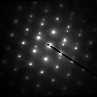
Selected area (electron) diffraction is a crystallographic experimental technique typically performed using a transmission electron microscope (TEM). It is a specific case of electron diffraction used primarily in material science and solid state physics as one of the most common experimental techniques. Especially with appropriate analytical software, SAD patterns (SADP) can be used to determine crystal orientation, measure lattice constants or examine its defects.

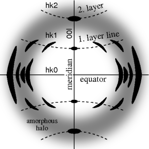
Fiber diffraction is a subarea of scattering, an area in which molecular structure is determined from scattering data. In fiber diffraction, the scattering pattern does not change, as the sample is rotated about a unique axis. Such uniaxial symmetry is frequent with filaments or fibers consisting of biological or man-made macromolecules. In crystallography, fiber symmetry is an aggravation regarding the determination of crystal structure, because reflections are smeared and may overlap in the fiber diffraction pattern. Materials science considers fiber symmetry a simplification, because almost the complete obtainable structure information is in a single two-dimensional (2D) diffraction pattern exposed on photographic film or on a 2D detector. 2 instead of 3 co-ordinate directions suffice to describe fiber diffraction.
The equivalent spherical diameter of an irregularly shaped object is the diameter of a sphere of equivalent geometric, optical, electrical, aerodynamic or hydrodynamic behavior to that of the particle under investigation.
Diffraction topography is a imaging technique based on Bragg diffraction. Diffraction topographic images ("topographies") record the intensity profile of a beam of X-rays diffracted by a crystal. A topography thus represents a two-dimensional spatial intensity mapping (image) of the X-rays diffracted in a specific direction, so regions which diffract substantially will appear brighter than those which do not. This is equivalent to the spatial fine structure of a Laue reflection. Topographs often reveal the irregularities in a non-ideal crystal lattice. X-ray diffraction topography is one variant of X-ray imaging, making use of diffraction contrast rather than absorption contrast which is usually used in radiography and computed tomography (CT). Topography is exploited to a lesser extent with neutrons, and is the same concept as dark field imaging in an electron microscope.
Arthur William Pryor was an Australian physicist known for his contributions to neutron diffraction and infrared laser isotope separation. Pryor authored and co-authored a number of papers in the field of crystallography and he also co-authored, with B. T. M. Willis, the book Thermal Vibrations in Crystallography.
In crystallography, mosaicity is a measure of the spread of crystal plane orientations. A mosaic crystal is an idealized model of an imperfect crystal, imagined to consist of numerous small perfect crystals (crystallites) that are to some extent randomly misoriented. Empirically, mosaicities can be determined by measuring rocking curves. Diffraction by mosaics is described by the Darwin–Hamilton equations.

Paul Anthony Midgley FRS is a Professor of Materials Science in the Department of Materials Science and Metallurgy at the University of Cambridge and a fellow of Peterhouse, Cambridge.

John Charles Howorth Spence ForMemRS HonFRMS was Richard Snell Professor of Physics at Arizona State University and Director of Science at the National Science Foundation BioXFEL Science and Technology Center.
Richard Hugh Sibson is a New Zealand structural geologist and emeritus professor at the University of Otago, who has received numerous honors and awards for his work in the field of earthquake research. He has caused a 'fundamental shift' in the interpretation of the relationship between earthquakes and fault zone geology and on the origin of fault-hosted mineral deposits.

Serial femtosecond crystallography (SFX) is a form of X-ray crystallography developed for use at X-ray free-electron lasers (XFELs). Single pulses at free-electron lasers are bright enough to generate resolvable Bragg diffraction from sub-micron crystals. However, these pulses also destroy the crystals, meaning that a full data set involves collecting diffraction from many crystals. This method of data collection is referred to as serial, referencing a row of crystals streaming across the X-ray beam, one at a time.

Janos Hajdu is a Swedish/Hungarian scientist, who has made contributions to biochemistry, biophysics, and the science of X-ray free-electron lasers. He is a professor of molecular biophysics at Uppsala University and a leading scientist at the European Extreme Light Infrastructure ERIC in Prague.
References
- ↑ "Rick Millane Elected as Fellow of the Royal Society" . Retrieved 27 November 2017.
- ↑ "T. K. Sidley Medal". iucr.org. Retrieved 27 November 2017.
- ↑ "Rick Millane". cmdt.org.nz. Retrieved 27 November 2017.
- ↑ "Rick Millane". canterbury.ac.nz. Retrieved 27 November 2017.