
Magnetic resonance imaging (MRI) is a medical imaging technique used in radiology to form pictures of the anatomy and the physiological processes of the body. MRI scanners use strong magnetic fields, magnetic field gradients, and radio waves to generate images of the organs in the body. MRI does not involve X-rays or the use of ionizing radiation, which distinguishes it from computed tomography (CT) and positron emission tomography (PET) scans. MRI is a medical application of nuclear magnetic resonance (NMR) which can also be used for imaging in other NMR applications, such as NMR spectroscopy.

Medical imaging is the technique and process of imaging the interior of a body for clinical analysis and medical intervention, as well as visual representation of the function of some organs or tissues (physiology). Medical imaging seeks to reveal internal structures hidden by the skin and bones, as well as to diagnose and treat disease. Medical imaging also establishes a database of normal anatomy and physiology to make it possible to identify abnormalities. Although imaging of removed organs and tissues can be performed for medical reasons, such procedures are usually considered part of pathology instead of medical imaging.
Neuroimaging is a medical technique that allows doctors and researchers to take pictures of the inner workings of the body or brain of a patient. It can show areas with heightened activity, areas with high or low blood flow, the structure of the patients brain/body, as well as certain abnormalities. Neuroimaging is most often used to find the specific location of certain diseases or birth defects such as tumors, cancers, or clogged arteries. Neuroimaging first came about as a medical technique in the 1880's with the invention of the human circulation balance and has since lead to other inventions such as the x-ray, air ventriculography, cerebral angiography, PET/SPECT scans, magnetoencephalography, and xenon CT scanning.

Neuroimaging is the use of quantitative (computational) techniques to study the structure and function of the central nervous system, developed as an objective way of scientifically studying the healthy human brain in a non-invasive manner. Increasingly it is also being used for quantitative research studies of brain disease and psychiatric illness. Neuroimaging is highly multidisciplinary involving neuroscience, computer science, psychology and statistics, and is not a medical specialty. Neuroimaging is sometimes confused with neuroradiology.
Kenneth Kin Man Kwong is a Hong Kong-born American nuclear physicist. He is a pioneer in human brain imaging. He received his bachelor's degree in Political Science in 1972 from the University of California, Berkeley. He went on to receive his Ph.D. in physics from the University of California, Riverside studying photon-photon collision interactions.
MRI contrast agents are contrast agents used to improve the visibility of internal body structures in magnetic resonance imaging (MRI). The most commonly used compounds for contrast enhancement are gadolinium-based contrast agents (GBCAs). Such MRI contrast agents shorten the relaxation times of nuclei within body tissues following oral or intravenous administration.
Perfusion is the passage of fluid through the lymphatic system or blood vessels to an organ or a tissue. The practice of perfusion scanning is the process by which this perfusion can be observed, recorded and quantified. The term perfusion scanning encompasses a wide range of medical imaging modalities.
Burton Drayer, MD, FACR, FANN, is an American radiologist and nationally recognized authority on the use of computed tomography and magnetic resonance imaging for diagnosing neurological disorders. From 2003 to 2008, he served as president, The Mount Sinai Hospital. As of 2020, he is the Charles M. and Marilyn Newman Professor and System Chair, Radiology, for The Mount Sinai Health System and Icahn School of Medicine at Mount Sinai Hospital in New York City.

Positron emission tomography–magnetic resonance imaging (PET–MRI) is a hybrid imaging technology that incorporates magnetic resonance imaging (MRI) soft tissue morphological imaging and positron emission tomography (PET) functional imaging.

Magnetic resonance imaging of the brain uses magnetic resonance imaging (MRI) to produce high quality two-dimensional or three-dimensional images of the brain and brainstem as well as the cerebellum without the use of ionizing radiation (X-rays) or radioactive tracers.

Computed tomography of the head uses a series of X-rays in a CT scan of the head taken from many different directions; the resulting data is transformed into a series of cross sections of the brain using a computer program. CT images of the head are used to investigate and diagnose brain injuries and other neurological conditions, as well as other conditions involving the skull or sinuses; it used to guide some brain surgery procedures as well. CT scans expose the person getting them to ionizing radiation which has a risk of eventually causing cancer; some people have allergic reactions to contrast agents that are used in some CT procedures.
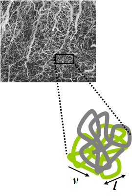
Intravoxel incoherent motion (IVIM) imaging is a concept and a method initially introduced and developed by Le Bihan et al. to quantitatively assess all the microscopic translational motions that could contribute to the signal acquired with diffusion MRI. In this model, biological tissue contains two distinct environments: molecular diffusion of water in the tissue, and microcirculation of blood in the capillary network (perfusion). The concept introduced by D. Le Bihan is that water flowing in capillaries mimics a random walk (Fig.1), as long as the assumption that all directions are represented in the capillaries is satisfied.

Jürgen Klaus Hennig is a German chemist and medical physicist. Internationally he is considered to be one of the pioneers of Magnetic Resonance Imaging for clinical diagnostics. He is the Scientific Director of the Department of Diagnostic Radiology and Chairman of the Magnetic Resonance Development and Application Center (MRDAC) at the University Medical Center Freiburg. In the year 2003 he was awarded the Max Planck Research Award in the category of Biosciences and Medicine.

Val Murray Runge is an American and Swiss professor of radiology and the editor-in-chief of Investigative Radiology. Runge was one of the early researchers to investigate the use of gadolinium-based contrast agents for magnetic resonance imaging (MRI), giving the first presentation in this field, followed two years later by the first presentation of efficacy. His research also pioneered many early innovations in MRI, including the use of tilted planes and respiratory gating. His publication on multiple sclerosis in 1984 represented the third and largest clinical series investigating the role of MRI in this disease, and the first to show characteristic abnormalities on MRI in patients whose CT was negative.
Bruce Rosen is an American physicist and radiologist and a leading expert in the area of functional neuroimaging. His research for the past 30 years has focused on the development and application of physiological and functional nuclear magnetic resonance techniques, as well as new approaches to combine functional magnetic resonance imaging (fMRI) data with information from other modalities such as positron emission tomography (PET), magnetoencephalography (MEG) and noninvasive optical imaging. The techniques his group has developed to measure physiological and metabolic changes associated with brain activation and cerebrovascular insult are used by research centers and hospitals throughout the world.
Ferenc Andras Jolesz was a Hungarian-American physician and scientist best known for his research on image guided therapy, the process by which information derived from diagnostic imaging is used to improve the localization and targeting of diseased tissue to monitor and control treatment during surgical and interventional procedures. He pioneered the field of Magnetic Resonance Imaging-guided interventions and introduced of a variety of new medical procedures based on novel combinations of imaging and therapy delivery.
The history of magnetic resonance imaging (MRI) includes the work of many researchers who contributed to the discovery of nuclear magnetic resonance (NMR) and described the underlying physics of magnetic resonance imaging, starting early in the twentieth century. MR imaging was invented by Paul C. Lauterbur who developed a mechanism to encode spatial information into an NMR signal using magnetic field gradients in September 1971; he published the theory behind it in March 1973.
Alan Edward Zimmer, M.D. was an American neuroradiologist, specializing in duplex neurovascular and magnetic resonance imaging (MRI). In the 1960s, Zimmer helped bring early neuroradiology methological advancements developed in Sweden to radiologists in the United States. He also conducted early research related to the emerging technologies of computer axial tomography and MRI as these procedures began to revolutionize radiology in the 1970s and '80s. As New Jersey’s senior neuroradiologist, Zimmer was consulted frequently by physicians, hospitals, and the courts to help diagnosis injuries and disease related to the head, neck, and spine. Zimmer was chief of neuroradiology at the University of Medicine and Dentistry of New Jersey (UMDNJ) from 1983 until his death.
Denis Le Bihan is a medical doctor, physicist, member of the Institut de France, member of the French Academy of Technologies and director since 2007 of NeuroSpin, an institution of the Atomic Energy and Alternative Energy Commission (CEA) in Saclay, dedicated to the study of the brain by magnetic resonance imaging (MRI) with a very high magnetic field. Denis Le Bihan has received international recognition for his outstanding work, introducing new imaging methods, particularly for the study of the human brain, as evidenced by the many international awards he has received, such as the Gold Medal of the International Society of Magnetic Resonance in Medicine (2001), the coveted Lounsbery Prize, the Louis D. Prize from the Institut de France, the prestigious Honda Prize (2012), the Louis-Jeantet Prize (2014), the Rhein Foundation Award (2021). His work has focused on the introduction, development and application of highly innovative methods, notably diffusion MRI.
Brian Worthington was the first radiologist to be elected a Fellow of the Royal Society and is acknowledged as a pioneer in clinical magnetic resonance imaging. He was born in Oldham, England and was educated at Hulme Grammar School, training at Guy's Hospital after graduating in physiology and medicine. After graduation his career developed rapidly, particularly in the field of MRI research and he was subsequently admitted as a Fellow of the Royal College of Radiologists.
