Related Research Articles

The blood vessels are the components of the circulatory system that transport blood throughout the human body. These vessels transport blood cells, nutrients, and oxygen to the tissues of the body. They also take waste and carbon dioxide away from the tissues. Blood vessels are needed to sustain life, because all of the body's tissues rely on their functionality.
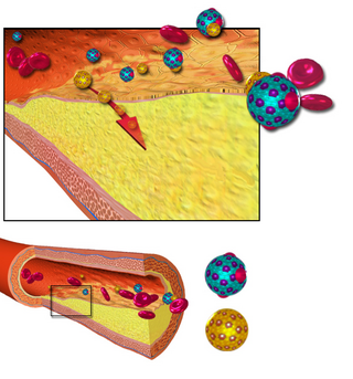
Arteriosclerosis is the thickening, hardening, and loss of elasticity of the walls of arteries. This process gradually restricts the blood flow to one's organs and tissues and can lead to severe health risks brought on by atherosclerosis, which is a specific form of arteriosclerosis caused by the buildup of fatty plaques, cholesterol, and some other substances in and on the artery walls. It can be brought on by smoking, a bad diet, or many genetic factors.

The portal vein or hepatic portal vein (HPV) is a blood vessel that carries blood from the gastrointestinal tract, gallbladder, pancreas and spleen to the liver. This blood contains nutrients and toxins extracted from digested contents. Approximately 75% of total liver blood flow is through the portal vein, with the remainder coming from the hepatic artery proper. The blood leaves the liver to the heart in the hepatic veins.

Hypertensive retinopathy is damage to the retina and retinal circulation due to high blood pressure.
Microangiopathy is a disease of the microvessels, small blood vessels in the microcirculation. It can be contrasted to coronary heart disease, an angiopathy that affects the larger vessels.

The microcirculation is the circulation of the blood in the smallest blood vessels, the microvessels of the microvasculature present within organ tissues. The microvessels include terminal arterioles, metarterioles, capillaries, and venules. Arterioles carry oxygenated blood to the capillaries, and blood flows out of the capillaries through venules into veins.

The juxtaglomerular apparatus is a structure in the kidney that regulates the function of each nephron, the functional units of the kidney. The juxtaglomerular apparatus is named because it is next to (juxta-) the glomerulus.

An arteriole is a small-diameter blood vessel in the microcirculation that extends and branches out from an artery and leads to capillaries.

Amlodipine, sold under the brand name Norvasc among others, is a calcium channel blocker medication used to treat high blood pressure and coronary artery disease. It is taken by mouth.

The glomerulus is a network of small blood vessels (capillaries) known as a tuft, located at the beginning of a nephron in the kidney. Each of the two kidneys contains about one million nephrons. The tuft is structurally supported by the mesangium, composed of intraglomerular mesangial cells. The blood is filtered across the capillary walls of this tuft through the glomerular filtration barrier, which yields its filtrate of water and soluble substances to a cup-like sac known as Bowman's capsule. The filtrate then enters the renal tubule of the nephron.
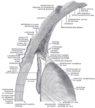
The conjunctiva is a thin mucous membrane that lines the inside of the eyelids and covers the sclera. It is composed of non-keratinized, stratified squamous epithelium with goblet cells, stratified columnar epithelium and stratified cuboidal epithelium. The conjunctiva is highly vascularised, with many microvessels easily accessible for imaging studies.

Hypertensive kidney disease is a medical condition referring to damage to the kidney due to chronic high blood pressure. It manifests as hypertensive nephrosclerosis. It should be distinguished from renovascular hypertension, which is a form of secondary hypertension, and thus has opposite direction of causation.

In human anatomy, the splenic vein is a blood vessel that drains blood from the spleen, the stomach fundus and part of the pancreas. It is part of the hepatic portal system.

Arteriolosclerosis is a form of cardiovascular disease involving hardening and loss of elasticity of arterioles or small arteries and is most often associated with hypertension and diabetes mellitus. Types include hyaline arteriolosclerosis and hyperplastic arteriolosclerosis, both involved with vessel wall thickening and luminal narrowing that may cause downstream ischemic injury. The following two terms whilst similar, are distinct in both spelling and meaning and may easily be confused with arteriolosclerosis.

Congestive hepatopathy, is liver dysfunction due to venous congestion, usually due to congestive heart failure. The gross pathological appearance of a liver affected by chronic passive congestion is "speckled" like a grated nutmeg kernel; the dark spots represent the dilated and congested hepatic venules and small hepatic veins. The paler areas are unaffected surrounding liver tissue. When severe and longstanding, hepatic congestion can lead to fibrosis; if congestion is due to right heart failure, it is called cardiac cirrhosis.
Charcot–Bouchard aneurysms are aneurysms of the brain vasculature which occur in small blood vessels. Charcot–Bouchard aneurysms are most often located in the lenticulostriate vessels of the basal ganglia and are associated with chronic hypertension. Charcot–Bouchard aneurysms are a common cause of cerebral hemorrhage.
Arteriovenous nicking, also known as AV nicking, is the phenomenon where, on examination of the eye, a small artery (arteriole) is seen crossing a small vein (venule), which results in the compression of the vein with bulging on either side of the crossing. This is most commonly seen in eye disease caused by high blood pressure.
Lipohyalinosis is a cerebral small vessel disease affecting the small arteries, arterioles or capillaries in the brain. Originally defined by C. Miller Fisher as 'segmental arteriolar wall disorganisation', it is characterized by vessel wall thickening and a resultant reduction in luminal diameter. Fisher considered this small vessel disease to be the result of hypertension, induced in the acute stage by fibrinoid necrosis that would lead to occlusion and hence lacunar stroke. However, recent evidence suggests that endothelial dysfunction as a result of inflammation is a more likely cause for it. This may occur subsequent to blood–brain barrier failure, and lead to extravasation of serum components into the brain that are potentially toxic. Lacunar infarction could thus occur in this way, and the narrowing – the hallmark feature of lipohyalinosis – may merely be a feature of the swelling occurring around it that squeezes on the structure.
Robert Salus was an Austrian ophthalmologist known for describing Salus's sign. He studied at the German University in Prague, gaining his M.D. in 1902. He was habilitated in ophthalmology in 1909 and became professor of ophthalmology in Prague in 1916. He described rubeosis iridis in 1928, and vascular changes in hypertension in 1939.
A resistance artery is small diameter blood vessel in the microcirculation that contributes significantly to the creation of the resistance to flow and regulation of blood flow. Resistance arteries are usually small arteries or arterioles and include precapillary sphincters. Having thick muscular walls and narrow lumen they contribute the most to the resistance to blood flow. Degree of the contraction of vascular smooth muscle in the wall of a resistance artery is directly connected to the size of the lumen.
References
- ↑ Hypertension at Medscape
- ↑ Sebastian Wolf, Berndt Kirchof, Martin Reim. The ocular fundus, page 131. Thieme, 2005. ISBN 978-1-58890-338-9. Google books
- ↑ Salus, Robert; Aldstein, Ernst (1939). "The fundus oculi in generalized hypertension and arteriosclerosis". Arch Ophthalmol. 21 (3): 505–508. doi:10.1001/archopht.1939.00860030113011 . Retrieved 2009-04-12.
| | This medical sign article is a stub. You can help Wikipedia by expanding it. |