Sebaceous adenitis

Last updated
Sebaceous adenitis and hair loss in a dog Sebadenitis.jpg
Sebaceous adenitis and hair loss in a dog

Sebaceous adenitis in an uncommon skin disease found in some breeds of dog, and more rarely in cats, rabbits and horses. [1] characterised by an inflammatory response against the dog's sebaceous glands (glands found in the hair follicles in the skin dermis), which can lead to the destruction of the gland. It was first described in veterinary literature in the 1980s. [2]

Contents

Signs

There are two expressions of this condition, one for long or double coated breeds and one for short coated breeds, both with differing presentations. [3]

Cause

Anatomy of human skin, which shows the location of human and canine sebaceous glands Skin.png
Anatomy of human skin, which shows the location of human and canine sebaceous glands

The signs of sebaceous adenitis are caused by an inflammatory disease process which affects the sebaceous glands of the skin. [5] The cause of the inflammatory disease is unknown. [6] Different breeds of dogs may have different underlying causes of the disease. [6]

Research is currently underway to find if there is a genetic predisposition for sebaceous adenitis; the exact mode of inheritance remains unknown. [7]

In Standard Poodles, sebaceous adenitis is most likely an autosomal recessive inherited disease, with variable expression. [5]

Diagnosis

In general, sebaceous adenitis is underdiagnosed in dogs. [1] Diagnosis confirmation requires multiple punch biopsies analysed by a dermopathologist who will comment on the condition of the sebaceous glands, revealing granulomatous or pyogranulomatous inflammation surrounding the sebaceous glands or even complete destruction of sebaceous glands. [8]

Other conditions with similar presentations include: bacterial folliculitis and demodicosis, dermatophytosis, endocrinopathy, pemphigus foliaceus, zinc responsive dermatosis, vitamin A-responsive dermatosis, ichthyosis, and nutritional deficiencies. [9] As well as, superficial pyoderma, primary idiopathic seborrhea and other endocrine diseases. [8]

Treatment

There is no cure for this condition. [10] Treatment is generally lifelong [3] and takes the form of bathing and soaking in mineral oils and washing with antibiotic shampoos to try to alleviate symptoms and slow the condition's progression. [11] Antiseptic and antibiotic shampoos (chlorhexidine or benzoyl peroxide) are used to manage further secondary bacterial infection. [9] For some breeds, cyclosporine or corticosteroids and immunosuppressant drugs may be effective, [3] and it is postulated, through some studies, that large doses of vitamin A given orally may result in some improvement. [3]

It has been suggested that the more aggressively one applies the topical methods of treatment, the less aggressively one needs to employ the immunosuppressant therapy. The suggestion is that this phenomenon may be due to a cyclic feedback whereby secondary infection, when not aggressively treated with topical therapy, increases and contributes to further sebaceous gland inflammation. [9]

Topical therapy

This forms a major and critical part in the disease treatment and the shampoo treatment can need to be applied as often as 3 to 4 times per week. [9] An antiseborrheic shampoo removes the scale blocking the follicles. The mineral oil soak, whereby the oil remains on the affected animal for at least 2 hours, is needed to replace epidermal lipids as well as to restore normal epidermal barrier function. The oil is then removed through the process of many baths. This oil treatment needs to be repeated at least once a week for 4 to 7 weeks until new hair growth is observed. [9] Once new hair growth is observed, topical treatment can be decreased to every 2 to 4 weeks.

Immunosuppressant therapy

Immunosuppressant and anti-inflammatory therapy serves to stop on-going destruction of the sebaceous glands. Like other inflammatory diseases, most animals receive an initial course to stop the inflammation and treatment is tapered off to the lowest dose that keeps the disease in remission. [9] Oral cyclosporine may be used. [9] Corticosteroids (e.g. prednisone) are used only if pruritus is a major clinical feature. [9]

Dietary supplementation

Commonly used dietary supplements include:

Epidemiology

While the condition has been seen in over 60 breeds of dog (including cross breeds), [9] [12] certain breeds have been found to be more susceptible than others to sebaceous adenitis:

Breeds also mentioned in scientific literature as having some susceptibility include:

Sebaceous adenitis has no sex-predisposition. [1] Sebaceous adenitis also occurs in cats, [8] rabbits, [3] [17] and horses. [18]

Etymology

Sebaceous refers to the gland which is affected by the disease. Adenitis is a general term referring to the inflammation of a gland.

See also

Related Research Articles

<span class="mw-page-title-main">Akita (dog)</span> Dog breed

The Akita is a Japanese dog breed of large size. Originating from the mountains of northern Japan, the Akita has a short double coat similar to that of many other northern spitz breeds. Historically, they were used by matagi for guarding and the hunting of bears.

<span class="mw-page-title-main">Dandruff</span> Skin condition of the scalp

Dandruff is a skin condition that mainly affects the scalp. Symptoms include flaking and sometimes mild itchiness. It can result in social or self-esteem problems. A more severe form of the condition, which includes inflammation of the skin, is known as seborrhoeic dermatitis.

<span class="mw-page-title-main">Sebaceous gland</span> Gland to lubricate the hair and skin

A sebaceous gland, or oil gland, is a microscopic exocrine gland in the skin that opens into a hair follicle to secrete an oily or waxy matter, called sebum, which lubricates the hair and skin of mammals. In humans, sebaceous glands occur in the greatest number on the face and scalp, but also on all parts of the skin except the palms of the hands and soles of the feet. In the eyelids, meibomian glands, also called tarsal glands, are a type of sebaceous gland that secrete a special type of sebum into tears. Surrounding the female nipple, areolar glands are specialized sebaceous glands for lubricating the nipple. Fordyce spots are benign, visible, sebaceous glands found usually on the lips, gums and inner cheeks, and genitals.

<span class="mw-page-title-main">Poodle</span> Dog breed

The Poodle, called the Pudel in German and the Caniche in French, is a breed of water dog. The breed is divided into four varieties based on size, the Standard Poodle, Medium Poodle, Miniature Poodle and Toy Poodle, although the Medium Poodle is not universally recognised. They have a distinctive thick, curly coat that comes in many colors and patterns, with only solid colors recognized by breed registries. Poodles are active and intelligent, and are particularly able to learn from humans. Poodles tend to live 10–18 years, with smaller varieties tending to live longer than larger ones.

Adenitis is a general term for an inflammation of a gland. Often it is used to refer to lymphadenitis which is the inflammation of a lymph node.

<span class="mw-page-title-main">Demodicosis</span> Medical condition

Demodicosis, also called Demodex folliculitis in humans and demodectic mange or red mange in animals, is caused by a sensitivity to and overpopulation of Demodex spp. as the host's immune system is unable to keep the mites under control.

<span class="mw-page-title-main">Dog coat</span> Hair that covers its body

The coat of the domestic dog refers to the hair that covers its body. Dogs demonstrate a wide range of coat colors, patterns, textures, and lengths.

<span class="mw-page-title-main">Cradle cap</span> Human medical condition

Cradle cap causes crusty or oily scaly patches on a baby's scalp. The condition is not painful or itchy, but it can cause thick white or yellow scales that are not easy to remove. Cradle cap most commonly begins sometime in the first three months but can occur in later years. Similar symptoms in older children are more likely to be dandruff than cradle cap. The rash is often prominent around the ear, the eyebrows or the eyelids. It may appear in other locations as well, where it is called infantile seborrhoeic dermatitis. Cradle cap is just a special—and more benign—case of this condition. The exact cause of cradle cap is not known. Cradle cap is not spread from person to person (contagious). It is also not caused by poor hygiene. It is not an allergy, and it is not dangerous. Cradle cap often lasts a few months. In some children, the condition can last until age 2 or 3.

<span class="mw-page-title-main">Comedo</span> Medical condition

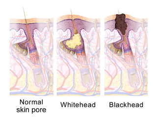
A comedo is a clogged hair follicle (pore) in the skin. Keratin combines with oil to block the follicle. A comedo can be open (blackhead) or closed by skin (whitehead) and occur with or without acne. The word "comedo" comes from the Latin comedere, meaning "to eat up", and was historically used to describe parasitic worms; in modern medical terminology, it is used to suggest the worm-like appearance of the expressed material.

<span class="mw-page-title-main">Dog health</span> Health of dogs

The health of dogs is a well studied area in veterinary medicine.

Follicular dysplasia is a genetic disease of dogs causing alopecia, also called hair loss. It is caused by hair follicles that are misfunctioning due to structural abnormality. There are several types, some affecting only certain breeds. Diagnosis is achieved through a biopsy, and treatment is rarely successful. Certain breeds, such as the Mexican Hairless Dog and Chinese Crested Dog, are bred specifically for alopecia.

Dogs, as with all mammals, have natural odors. Natural dog odor can be unpleasant to dog owners, especially when dogs are kept inside the home, as some people are not used to being exposed to the natural odor of a non-human species living in proximity to them. Dogs may also develop unnatural odors as a result of skin disease or other disorders or may become contaminated with odors from other sources in their environment.

<span class="mw-page-title-main">Gingival enlargement</span> Human disease

Gingival enlargement is an increase in the size of the gingiva (gums). It is a common feature of gingival disease. Gingival enlargement can be caused by a number of factors, including inflammatory conditions and the side effects of certain medications. The treatment is based on the cause. A closely related term is epulis, denoting a localized tumor on the gingiva.

<span class="mw-page-title-main">Feline acne</span> Medical condition

Feline acne is a problem seen in cats primarily involving the formation of blackheads accompanied by inflammation on the cat's chin and surrounding areas that can cause lesions, alopecia, and crusty sores. In many cases symptoms are mild and the disease does not require treatment. Mild cases will resemble dirt on the cat's chin, but the "dirt" will not brush off. More severe cases, however, may respond slowly to treatment and seriously detract from the health and appearance of the cat. Feline acne can affect cats of any age, sex or breed, although Persian cats are also likely to develop acne on the face and in the skin folds. This problem can happen once, be reoccurring, or even persistent throughout the cat's life.

<span class="mw-page-title-main">Pyotraumatic dermatitis</span> Skin disease of dogs and cats

Pyotraumatic dermatitis, also known as a hot spot or acute moist dermatitis, is a common infection of the skin surface of dogs, particularly those with thick or long coats. It occurs following self-inflicted trauma of the skin. Pyotraumatic dermatitis rarely affects cats.

<span class="mw-page-title-main">Dog grooming</span> Care and cleaning of a dog

Dog grooming refers to both the hygienic care and cleaning of a dog, as well as a process by which a dog's physical appearance is enhanced for showing or other types of competition. A dog groomer is a person who earns their living grooming dogs.

Hypoadrenocorticism in dogs, or, as it is known in people, Addison's disease, is an endocrine system disorder that occurs when the adrenal glands fail to produce enough hormones for normal function. The adrenal glands secrete glucocorticoids such as cortisol and mineralocorticoids such as aldosterone; when proper amounts of these are not produced, the metabolic and electrolyte balance is upset. Mineralocorticoids control the amount of potassium, sodium, and water in the body. Hypoadrenocorticism is fatal if left untreated.

Scarring hair loss, also known as cicatricial alopecia, is the loss of hair which is accompanied with scarring. This is in contrast to non scarring hair loss.

Infantile acne is a form of acneiform eruption that occurs in infants from 6 weeks to 1 year of age. Typical symptoms include inflammatory and noninflammatory lesions, papules and pustules most commonly present on the face. No cause of infantile acne has been established but it may be caused by increased sebaceous gland secretions due to elevated androgens, genetics and the fetal adrenal gland causing increased sebum production. Infantile acne can resolve by itself by age 1 or 2. However, treatment options include topical benzyl peroxide, topical retinoids and topical antibiotics in most cases.

References

  1. 1 2 3 4 5 Craig, Mark (2006). "Clinical refresher: Canine sebaceous adenitis". Companion Animal. 11 (5): 62–8. doi:10.1111/j.2044-3862.2006.tb00066.x.
  2. Spaterna, A.; Antognoni, M.T.; Cappuccini, S.; Tesei, B. (2003). "Sebaceous Adenitis in the Dog: Three Cases". Veterinary Research Communications. 27: 441–443. doi:10.1023/B:VERC.0000014199.39879.bb. PMID   14535449. S2CID   30352408.
  3. 1 2 3 4 5 6 7 8 Linek, Monika (2008). "Sebaceous adenitis in the dog" (PDF). Veterinary Focus. 18 (1): 12–16. doi:10.1055/s-0034-1381781. Archived from the original (PDF) on 22 March 2012. Retrieved 10 April 2011.
  4. "Sebaceous Adenitis". Canine Inherited Disorders Database. 1998. Archived from the original on June 5, 2011. Retrieved 10 April 2011.
  5. 1 2 Rosser, EJ Jr (2014). "Therapy for sebaceous adenitis". In Bonagura, JD; Twedt, DC (eds.). Kirk's current veterinary therapy (15th ed.). St. Louis, Mo.: Elsevier/Saunders. ISBN   9780323227629.
  6. 1 2 Paterson, Sue (2009). "Chapter 17: Keratinisation defects". Manual of Skin Diseases of the Dog and Cat (2nd ed.). Chichester: John Wiley & Sons. pp. 277–291. ISBN   9781444309324.
  7. Koch, Sandra N. (June 1, 2009 – November 30, 2010). "01346-A: Genetic Basis of Sebaceous Adenitis in Dogs". University of Minnesota. Archived from the original on 2011-10-02. Retrieved 7 April 2011.
  8. 1 2 3 4 5 6 7 8 Hall, Jan A. (2005). "Congenital and Hereditary Defects in Skin Disease". Ontario Veterinary College, University of Guelph, Canada: Omnibooks Online. Retrieved 2 June 2011.[ permanent dead link ]
  9. 1 2 3 4 5 6 7 8 9 10 11 Angus, DVM, DACVD, John C. (2009). "How I Treat Sebaceous Adenitis". Animal Dermatology Clinic, Pasadena, CA, USA: Omnibooks Online. p. 1. Retrieved 2 June 2011.{{cite web}}: CS1 maint: multiple names: authors list (link)
  10. "Sebaceous Adenitis". Orthopedic Foundation for Animals. Retrieved 2019-09-27.
  11. "Inflammatory Skin Disease in Dogs" . Retrieved 10 April 2011.
  12. Mr Charlie Walker BVetMed CertVD MRCVS (2010). "Skin: idiopathic/granulomatous sebaceous adenitis". Cambridge CB21 4EN, UK: VetStream. Archived from the original on 22 March 2012. Retrieved 19 July 2011.{{cite web}}: CS1 maint: location (link)
  13. 1 2 3 Pfeiffer, Ina (April 1 – June 30, 2006). "577-AT: Sebaceous Adenitis in the Akita". Archived from the original on 2011-10-02. Retrieved 7 April 2011.
  14. Hernblad Tevell, E; Bergvall, K; Egenvall, A (2008). "Sebaceous adenitis in Swedish dogs, a retrospective study of 104 cases". Acta Veterinaria Scandinavica. 50 (1): 11. doi:10.1186/1751-0147-50-11. PMC   2412885 . PMID   18501018.
  15. 1 2 3 4 5 6 7 Gross, Thelma Lee; Peter J. Ihrke; Emily J. Walder; Verena K. Affolter (2005). Skin diseases of the dog and cat: clinical and histopathologic diagnosis (second ed.). Wiley-Blackwell. pp. 186–8. ISBN   978-0-632-06452-6.
  16. Frazer, Megan; Anthea E. Schick; Thomas P. Lewis; Edward Jazic (June 2011). "Sebaceous adenitis in Havanese dogs: a retrospective study of the clinical presentation and incidence". Veterinary Dermatology. 22 (3): 267–274. doi:10.1111/j.1365-3164.2010.00942.x. PMID   21210878.
  17. Anna Meredith; Stephen White (2010). "Sebaceous adenitis". Cambridge CB21 4EN, UK: VetStream. Archived from the original on 11 October 2013. Retrieved 19 July 2011.{{cite web}}: CS1 maint: location (link)
  18. Osborne, Christina (2006). "Sebaceous adenitis in a 7-year-old Arabian gelding". The Canadian Veterinary Journal. 47 (6): 583–6. PMC   1461412 . PMID   16808233.

Further reading