Related Research Articles

The lungs are the primary organs of the respiratory system in humans and many other animals including a few fish and some snails. In mammals and most other vertebrates, two lungs are located near the backbone on either side of the heart. Their function in the respiratory system is to extract oxygen from the atmosphere and transfer it into the bloodstream, and to release carbon dioxide from the bloodstream into the atmosphere, in a process of gas exchange. Respiration is driven by different muscular systems in different species. Mammals, reptiles and birds use their different muscles to support and foster breathing. In early tetrapods, air was driven into the lungs by the pharyngeal muscles via buccal pumping, a mechanism still seen in amphibians. In humans, the main muscle of respiration that drives breathing is the diaphragm. The lungs also provide airflow that makes vocal sounds including human speech possible.

A capillary is a small blood vessel from 5 to 10 micrometres (μm) in diameter, and having a wall one endothelial cell thick. They are the smallest blood vessels in the body: they convey blood between the arterioles and venules. These microvessels are the site of exchange of many substances with the interstitial fluid surrounding them. Substances which exit include water, oxygen, and glucose; substances which enter include water, carbon dioxide, uric acid, lactic acid, urea and creatinine. Lymph capillaries connect with larger lymph vessels to drain lymphatic fluid collected in the microcirculation.

The respiratory system is a biological system consisting of specific organs and structures used for gas exchange in animals and plants. The anatomy and physiology that make this happen varies greatly, depending on the size of the organism, the environment in which it lives and its evolutionary history. In land animals the respiratory surface is internalized as linings of the lungs. Gas exchange in the lungs occurs in millions of small air sacs called alveoli in mammals and reptiles, but atria in birds. These microscopic air sacs have a very rich blood supply, thus bringing the air into close contact with the blood. These air sacs communicate with the external environment via a system of airways, or hollow tubes, of which the largest is the trachea, which branches in the middle of the chest into the two main bronchi. These enter the lungs where they branch into progressively narrower secondary and tertiary bronchi that branch into numerous smaller tubes, the bronchioles. In birds the bronchioles are termed parabronchi. It is the bronchioles, or parabronchi that generally open into the microscopic alveoli in mammals and atria in birds. Air has to be pumped from the environment into the alveoli or atria by the process of breathing which involves the muscles of respiration.

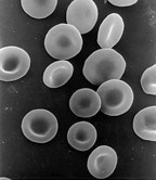
Red blood cells (RBCs), also referred to as red cells, red blood corpuscles (in humans or other animals not having nucleus in red blood cells), haematids, erythroid cells or erythrocytes (from Greek erythros for "red" and kytos for "hollow vessel", with -cyte translated as "cell" in modern usage), are the most common type of blood cell and the vertebrate's principal means of delivering oxygen (O2) to the body tissues—via blood flow through the circulatory system. RBCs take up oxygen in the lungs, or in fish the gills, and release it into tissues while squeezing through the body's capillaries.

A pulmonary alveolus is a hollow cup-shaped cavity found in the lung parenchyma where gas exchange takes place. Lung alveoli are found in the acini at the beginning of the respiratory zone. They are located sparsely in the respiratory bronchioles, line the walls of the alveolar ducts, and are more numerous in the blind-ended alveolar sacs. The acini are the basic units of respiration, with gas exchange taking place in all the alveoli present. The alveolar membrane is the gas exchange surface, surrounded by a network of capillaries. Across the membrane oxygen is diffused into the capillaries and carbon dioxide released from the capillaries into the alveoli to be breathed out.
Diffusing capacity of the lung (DL) measures the transfer of gas from air in the lung, to the red blood cells in lung blood vessels. It is part of a comprehensive series of pulmonary function tests to determine the overall ability of the lung to transport gas into and out of the blood. DL, especially DLCO, is reduced in certain diseases of the lung and heart. DLCO measurement has been standardized according to a position paper by a task force of the European Respiratory and American Thoracic Societies.

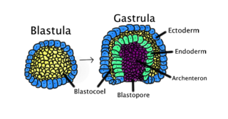
In developmental biology, gastrulation is a phase early in the embryonic development of most animals, during which the blastula is reorganized into a multilayered structure known as the gastrula. Before gastrulation, the embryo is a continuous epithelial sheet of cells; by the end of gastrulation, the embryo has begun differentiation to establish distinct cell lineages, set up the basic axes of the body, and internalized one or more cell types including the prospective gut.

The chorion is the outermost fetal membrane around the embryo in mammals, birds and reptiles (amniotes). It develops from an outer fold on the surface of the yolk sac, which lies outside the zona pellucida, known as the vitelline membrane in other animals. In insects it is developed by the follicle cells while the egg is in the ovary.
Gas exchange is the physical process by which gases move passively by diffusion across a surface. For example, this surface might be the air/water interface of a water body, the surface of a gas bubble in a liquid, a gas-permeable membrane, or a biological membrane that forms the boundary between an organism and its extracellular environment.

Among animals which produce eggs, the yolk is the nutrient-bearing portion of the egg whose primary function is to supply food for the development of the embryo. Some types of egg contain no yolk, for example because they are laid in situations where the food supply is sufficient or because the embryo develops in the parent's body, which supplies the food, usually through a placenta. Reproductive systems in which the mother's body supplies the embryo directly are said to be matrotrophic; those in which the embryo is supplied by yolk are said to be lecithotrophic. In many species, such as all birds, and most reptiles and insects, the yolk takes the form of a special storage organ constructed in the reproductive tract of the mother. In many other animals, especially very small species such as some fish and invertebrates, the yolk material is not in a special organ, but inside the egg cell (ovum).
A syncytium or symplasm is a multinucleate cell which can result from multiple cell fusions of uninuclear cells, in contrast to a coenocyte, which can result from multiple nuclear divisions without accompanying cytokinesis. The term may also refer to cells interconnected by specialized membranes with gap junctions, as seen in the heart muscle cells and certain smooth muscle cells, which are synchronized electrically in an action potential.
Organogenesis is the phase of embryonic development that starts at the end of gastrulation and continues until birth. During organogenesis, the three germ layers formed from gastrulation form the internal organs of the organism.

Human fertilization is the union of a human egg and sperm, usually occurring in the ampulla of the fallopian tube. The result of this union is the production of a zygote cell, or fertilized egg, initiating prenatal development. Scientists discovered the dynamics of human fertilization in the nineteenth century.
Intussusceptive angiogenesis also known as splitting angiogenesis, is a type of angiogenesis, the process whereby a new blood vessel is created. By intussusception a new blood vessel is created by splitting of an existing blood vessel in two. Intussusception occurs in normal development as well as in pathologic conditions involving wound healing, tissue regeneration, inflammation as colitis or myocarditis, lung fibrosis, and tumors amongst others.

In biology, placentation refers to the formation, type and structure, or arrangement of the placenta. The function of placentation is to transfer nutrients, respiratory gases, and water from maternal tissue to a growing embryo, and in some instances to remove waste from the embryo. Placentation is best known in live-bearing mammals (theria), but also occurs in some fish, reptiles, amphibians, a diversity of invertebrates, and flowering plants. In vertebrates, placentas have evolved more than 100 times independently, with the majority of these instances occurring in squamate reptiles.

Bird anatomy, or the physiological structure of birds' bodies, shows many unique adaptations, mostly aiding flight. Birds have a light skeletal system and light but powerful musculature which, along with circulatory and respiratory systems capable of very high metabolic rates and oxygen supply, permit the bird to fly. The development of a beak has led to evolution of a specially adapted digestive system. These anatomical specializations have earned birds their own class in the vertebrate phylum.

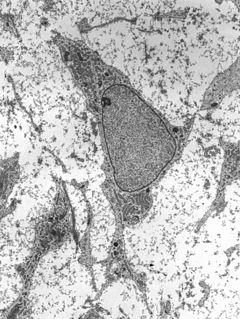
Mesenchyme is a type of connective tissue found mostly during the embryonic development of bilateral triploblast animals.

Human embryonic development, or human embryogenesis, refers to the development and formation of the human embryo. It is characterised by the processes of cell division and cellular differentiation of the embryo that occurs during the early stages of development. In biological terms, the development of the human body entails growth from a one-celled zygote to an adult human being. Fertilisation occurs when the sperm cell successfully enters and fuses with an egg cell (ovum). The genetic material of the sperm and egg then combine to form a single cell called a zygote and the germinal stage of development commences. Embryonic development in the human, covers the first eight weeks of development; at the beginning of the ninth week the embryo is termed a fetus. Human embryology is the study of this development during the first eight weeks after fertilisation. The normal period of gestation (pregnancy) is about nine months or 40 weeks.

In avian gastrulation, Koller's sickle is a local thickening of cells at the posterior edge of the upper layer of the area pellucida called the epiblast. Koller's sickle is crucial for avian development, due to its critical role in inducing the differentiation of various avian body parts. Koller's sickle induces primitive streak and Hensen's node, which are major components of avian gastrulation. Avian gastrulation is a process by which developing cells in an avian embryo move relative to one another in order to form the three germ layers.

The Chorioallantoic Membrane (CAM), also known as the chorioallantois, is a highly vascularized membrane found in the eggs of certain amniotes like birds and reptiles. It is formed by the fusion of the mesodermal layers of two extra-embryonic membranes – the chorion and the allantois. It is the avian homologue of the mammalian placenta. It is the outermost extra-embryonic membrane which lines the non-vascular egg shell membrane.
References
- ↑ Makanya AN, Hlushchuk R, Duncker HR, Djonov V (2006). "Epithelial transformations in the establishment of the blood-gas barrier in the developing chick embryo lung". Developmental Dynamics. 235: 68–81. doi:10.1002/dvdy.20627. PMID 16258963. S2CID 28152754.[ dead link ]
- ↑ Makanya, AN; Hlushchuk, R; Djonov, V (31 August 2011). "The pulmonary blood-gas barrier in the avian embryo: inauguration, development and refinement". Respiratory Physiology & Neurobiology. 178 (1): 30–8. doi:10.1016/j.resp.2011.03.028. PMID 21477666. S2CID 207506433.
| This cell biology article is a stub. You can help Wikipedia by expanding it. |