Septate junction

Last updated
Sepate junction in developing trachea in Drosophila Septatejunction.jpg
Sepate junction in developing trachea in Drosophila

Septate junctions are intercellular junctions found in invertebrate epithelial cells, appearing as ladder-like structures under electron microscopy. They are thought to provide structural strength and a barrier to solute diffusion through the intercellular space. They are considered somewhat analogous to the (vertebrate) tight junctions; however, tight and septate junctions are different in many ways. Known insect homologues of tight junction components are components of conserved signalling pathways that localize to either adherens junctions, the subapical complex, or the marginal zone. [1] Recent studies show that septate junctions are also identified in the myelinated nerve fibers of the vertebrates. [2] [3]

Contents

Structure

The main trait of septate junctions structure is that cross-bridges or septa are in the ladder-like shape and cover the 15–20 nm intermembrane space of cell–cell contacts. Septate junctions are in a tight arrangement which is parallel to each other. [4]

For the septate junctions, several components are related to the function or the morphology of septate junctions, like Band 4.1-Coracle, Discs-large, fasciclin III, Neurexin IV (NRX) and so on. [5] [6] Band 4.1-Coracle is necessary for the interaction of the cell. [6] Discs-large, a key component of septate junctions, is needed for the growth control. [7] [8] Fasciclin III acts as an adhesion protein. [6] Neurexin IV (NRX) is a required transmembrane protein for the formation of septate junctions. For example, the glial–glial septate junctions that lack NRX will cause the blood barriers to break down. [5] Gliotactin (Gli), is also a necessary a transmembrane protein for the formation of pleated septate junctions. [4] [9] Tsp2A and Undicht are newly identified components that are needed for the formation of smooth septate junctions and septate junctions. [10] [11]

There are three known claudins contained in the septate junctions, Megatrachea (Mega), Sinuous (Sinu) and Kune-kune (Kune). Among these three claudins, Kune-kune (Kune) plays a more central role in septate junctions organization and function. [12]

Function

There are several functions of septate junctions.

For the septate junctions in the vertebrates, they play some roles of tight junctions. [14]

Na+/K+ ATPase works for the function of septate junctions. [16]

Classification

In Drosophila melanogaster , there are two types of septate junctions, smooth SJs (sSJs) and pleated SJs(pSJs). sSJs and pSJs are distributed in different tissues. sSJs are in gut endoderm and Malpighian tubules, while pSJs are in the ectodermally derived epithelia. [5] sSJs and pSJs vary in shape but have the same function. [4]

See also

Related Research Articles

Cell junctions are a class of cellular structures consisting of multiprotein complexes that provide contact or adhesion between neighboring cells or between a cell and the extracellular matrix in animals. They also maintain the paracellular barrier of epithelia and control paracellular transport. Cell junctions are especially abundant in epithelial tissues. Combined with cell adhesion molecules and extracellular matrix, cell junctions help hold animal cells together.

Tight junction

Tight junctions, also known as occluding junctions or zonulae occludentes are multiprotein junctional complexes whose general function is to prevent leakage of transported solutes and water and seals the paracellular pathway. Tight junctions may also serve as leaky pathways by forming selective channels for small cations, anions, or water. Tight junctions are present mostly in vertebrates. The corresponding junctions that occur in invertebrates are septate junctions.

Claudin

Claudins are a family of proteins which, along with occludin, are the most important components of the tight junctions. Tight junctions establish the paracellular barrier that controls the flow of molecules in the intercellular space between the cells of an epithelium. They have four transmembrane domains, with the N-terminus and the C-terminus in the cytoplasm.

Occludin Mammalian protein found in Homo sapiens

Occludin is an enzyme that oxidizes NADH. It was first identified in epithelial cells as a 65 kDa integral plasma-membrane protein localized at the tight junctions. Together with Claudins, and zonula occludens-1 (ZO-1), occludin has been considered a staple of tight junctions, and although it was shown to regulate the formation, maintenance, and function of tight junctions, its precise mechanism of action remained elusive and most of its actions were initially attributed to conformational changes following selective phosphorylation, and its redox-sensitive dimerization. However, mounting evidence demonstrated that occludin is not only present in epithelial/endothelial cells, but is also expressed in large quantities in cells that do not have tight junctions but have very active metabolism: pericytes, neurons and astrocytes, oligodendrocytes, dendritic cells, monocytes/macrophages lymphocytes, and myocardium. Recent work, using molecular modeling, supported by biochemical and live-cell experiments in human cells demonstrated that occludin is a NADH oxidase that influences critical aspects of cell metabolism like glucose uptake, ATP production and gene expression. Furthermore, manipulation of occludin content in human cells is capable of influencing the expression of glucose transporters, and the activation of transcription factors like NFkB, and histone deacetylases like sirtuins, which proved capable of diminishing HIV replication rates in infected human macrophages under laboratory conditions.

CLDN1

Claudin-1 is a protein that in humans is encoded by the CLDN1 gene. It belongs to the group of claudins.

CLDN4

Claudin 4, also known as CLDN4, is a protein which in humans is encoded by the CLDN4 gene. It belongs to the group of claudins.

CLDN5

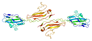
Claudin-5 is a protein that in humans is encoded by the CLDN5 gene. It belongs to the group of claudins.

CLDN3

Claudin 3, also known as CLDN3, is a protein which in humans is encoded by the CLDN3 gene. It is a member of the claudin protein family.

CLDN7 Protein-coding gene in the species Homo sapiens

Claudin-7 is a protein that in humans is encoded by the CLDN7 gene. It belongs to the group of claudins.

CLDN6

Claudin-6 is a protein that in humans is encoded by the CLDN6 gene. It belongs to the group of claudins. The knockout mice of mouse homolog exhibit no phenotype, indicating that claudin-6 is dispensable for normal development and homeostasis.

CLDN2

Claudin-2 is a protein that in humans is encoded by the CLDN2 gene. It belongs to the group of claudins.

CLDN16

Claudin-16 is a protein that in humans is encoded by the CLDN16 gene. It belongs to the group of claudins.

CLDN14

Claudin-14 is a protein that in humans is encoded by the CLDN14 gene. It belongs to a related family of proteins called claudins.

CLDN9

Claudin-9 is a protein that in humans is encoded by the CLDN9 gene. It belongs to the group of claudins.

CLDN10

Claudin-10 is a protein that in humans is encoded by the CLDN10 gene. It belongs to the group of claudins.

CLDN15

Claudin-15 is a protein that in humans is encoded by the CLDN15 gene. It belongs to the group of claudins. Among its related pathways are Blood-Brain Barrier and Immune Cell Transmigration: VCAM-1/CD106 Signaling Pathways and Tight junction. GO annotations related to this gene include identical protein binding and structural molecule activity. An important paralog of this gene is CLDN10.

CLDN19

Claudin-19 is a protein that in humans is encoded by the CLDN19 gene. It belongs to the group of claudins. Claudin-19 has been implicated in magnesium transport.

CLDN18

Claudin-18 is a protein that in humans is encoded by the CLDN18 gene. It belongs to the group of claudins.

Intestinal epithelium

The intestinal epithelium is the single cell layer that form the luminal surface (lining) of both the small and large intestine (colon) of the gastrointestinal tract. Composed of simple columnar epithelial cells, it serves two main functions: absorbing useful substances into the body and restricting the entry of harmful substances. As part of its protective role, the intestinal epithelium forms an important component of the intestinal mucosal barrier. Certain diseases and conditions are caused by functional defects in the intestinal epithelium. On the other hand, various diseases and conditions can lead to its dysfunction which, in turn, can lead to further complications.

Tight junction proteins are molecules situated at the tight junctions of epithelial, endothelial and myelinated cells. This mutliprotein junctional complex has a regulatory function in passage of ions, water and solutes through the paracellular pathway. It can also coordinate the motion of lipids and proteins between the apical and basolateral surfaces of the plasma membrane. Thereby tight junction conducts signaling molecules, that influence the differentiation, proliferation and polarity of cells. So tight junction plays a key role in maintenance of osmotic balance and trans-cellular transport of tissue specific molecules. Nowadays is known more than 40 different proteins, that are involved in these selective TJ channels.

References

  1. Matter K, Balda MS (March 2003). "Signalling to and from tight junctions". Nature Reviews. Molecular Cell Biology. 4 (3): 225–36. doi:10.1038/nrm1055. PMID   12612641.
  2. 1 2 3 Faivre-Sarrailh C, Banerjee S, Li J, Hortsch M, Laval M, Bhat MA (October 2004). "Drosophila contactin, a homolog of vertebrate contactin, is required for septate junction organization and paracellular barrier function". Development. 131 (20): 4931–42. doi: 10.1242/dev.01372 . PMID   15459097.
  3. Genova JL, Fehon RG (June 2003). "Neuroglian, Gliotactin, and the Na+/K+ ATPase are essential for septate junction function in Drosophila". The Journal of Cell Biology. 161 (5): 979–89. doi:10.1083/jcb.200212054. PMC   2172966 . PMID   12782686.
  4. 1 2 3 Schulte J, Tepass U, Auld VJ (June 2003). "Gliotactin, a novel marker of tricellular junctions, is necessary for septate junction development in Drosophila". The Journal of Cell Biology. 161 (5): 991–1000. doi:10.1083/jcb.200303192. PMC   2172969 . PMID   12782681.
  5. 1 2 3 4 5 Baumgartner S, Littleton JT, Broadie K, Bhat MA, Harbecke R, Lengyel JA, Chiquet-Ehrismann R, Prokop A, Bellen HJ (December 1996). "A Drosophila neurexin is required for septate junction and blood-nerve barrier formation and function". Cell. 87 (6): 1059–68. CiteSeerX   10.1.1.580.1295 . doi:10.1016/s0092-8674(00)81800-0. PMID   8978610.
  6. 1 2 3 4 Lamb RS, Ward RE, Schweizer L, Fehon RG (December 1998). "Drosophila coracle, a member of the protein 4.1 superfamily, has essential structural functions in the septate junctions and developmental functions in embryonic and adult epithelial cells". Molecular Biology of the Cell. 9 (12): 3505–19. doi:10.1091/mbc.9.12.3505. PMC   25665 . PMID   9843584.
  7. Woods DF, Bryant PJ (August 1991). "The discs-large tumor suppressor gene of Drosophila encodes a guanylate kinase homolog localized at septate junctions". Cell. 66 (3): 451–64. doi:10.1016/0092-8674(81)90009-x. PMID   1651169.
  8. Ward, Robert E.; Lamb, Rebecca S.; Fehon, Richard G. (1998-03-23). "A Conserved Functional Domain ofDrosophilaCoracle Is Required for Localization at the Septate Junction and Has Membrane-organizing Activity". The Journal of Cell Biology. 140 (6): 1463–1473. doi:10.1083/jcb.140.6.1463. ISSN   0021-9525. PMC   2132682 . PMID   9508778.
  9. 1 2 Kolosov, Dennis; Jonusaite, Sima; Donini, Andrew; Kelly, Scott P.; O'Donnell, Michael J. (2019-05-07). "Septate junction in the distal ileac plexus of larval lepidopteran Trichoplusia ni: alterations in paracellular permeability during ion transport reversal". The Journal of Experimental Biology. 222 (11): jeb204750. doi: 10.1242/jeb.204750 . ISSN   0022-0949. PMID   31064858.
  10. Izumi, Yasushi; Motoishi, Minako; Furuse, Kyoko; Furuse, Mikio (2016-04-01). "A tetraspanin regulates septate junction formation inDrosophilamidgut". Development. 143 (7): e1.1. doi:10.1242/dev.137646. ISSN   0950-1991.
  11. Petri, Johanna; Syed, Mubarak Hussain; Rey, Simone; Klämbt, Christian (February 2019). "Non-Cell-Autonomous Function of the GPI-Anchored Protein Undicht during Septate Junction Assembly". Cell Reports. 26 (6): 1641–1653.e4. doi: 10.1016/j.celrep.2019.01.046 . ISSN   2211-1247. PMID   30726744.
  12. Nelson, Kevin S.; Furuse, Mikio; Beitel, Greg J. (2010-04-20). "The Drosophila Claudin Kune-kune Is Required for Septate Junction Organization and Tracheal Tube Size Control". Genetics. 185 (3): 831–839. doi:10.1534/genetics.110.114959. ISSN   0016-6731. PMC   2907205 . PMID   20407131.
  13. Wu VM. Sinuous and varicose function to organize the septate junction complex and regulate tracheal tube size control (Ph. D. thesis). Northwestern University. ISBN   978-0-542-74446-4. OCLC   157010952.
  14. 1 2 3 Llimargas, M. (2004-01-01). "Lachesin is a component of a septate junction-based mechanism that controls tube size and epithelial integrity in the Drosophila tracheal system". Development. 131 (1): 181–190. doi: 10.1242/dev.00917 . ISSN   0950-1991. PMID   14681183.
  15. Dubey, Pankaj; Kapoor, Tushna; Gupta, Samir; Shirolikar, Seema; Ray, Krishanu (2018-07-01). "Atypical septate junctions maintain the somatic enclosure around maturing spermatids and prevent premature sperm release in Drosophila testis". doi: 10.1101/360115 .Cite journal requires |journal= (help)
  16. Paul SM, Ternet M, Salvaterra PM, Beitel GJ (October 2003). "The Na+/K+ ATPase is required for septate junction function and epithelial tube-size control in the Drosophila tracheal system". Development. 130 (20): 4963–74. doi: 10.1242/dev.00691 . PMID   12930776.