Panniculitis is a group of diseases whose hallmark is inflammation of subcutaneous adipose tissue. Symptoms include tender skin nodules, and systemic signs such as weight loss and fatigue.

Cutaneous larva migrans is a skin disease in humans, caused by the larvae of various nematode parasites of the hookworm family (Ancylostomatidae). The parasites live in the intestines of dogs, cats, and wild animals; they should not be confused with other members of the hookworm family for which humans are definitive hosts, namely Ancylostoma duodenale and Necator americanus.

Urticaria pigmentosa (also known as generalized eruption of cutaneous mastocytosis (childhood type) ) is the most common form of cutaneous mastocytosis. It is a rare disease caused by excessive numbers of mast cells in the skin that produce hives or lesions on the skin when irritated.

Livedo reticularis is a common skin finding consisting of a mottled reticulated vascular pattern that appears as a lace-like purplish discoloration of the skin. The discoloration is caused by reduction in blood flow through the arterioles that supply the cutaneous capillaries, resulting in deoxygenated blood showing as blue discoloration. This can be a secondary effect of a condition that increases a person's risk of forming blood clots, including a wide array of pathological and nonpathological conditions. Examples include hyperlipidemia, microvascular hematological or anemia states, nutritional deficiencies, hyper- and autoimmune diseases, and drugs/toxins.

In medicine, nodules are small firm lumps, usually greater than 1 cm in diameter. If filled with fluid they are referred to as cysts. Smaller raised soft tissue bumps may be termed papules.
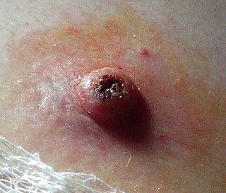
Keratoacanthoma (KA) is a common low-grade rapidly-growing skin tumour that is believed to originate from the hair follicle and can resemble squamous cell carcinoma.

Erythema toxicum neonatorum is a common, non-threatening rash in newborns. It appears in 4-70% of newborns within the first week of life, and it typically improves within 1–2 weeks. It only occurs during the newborn period, but may appear slightly later in premature babies. The rash has a variable appearance. It typically includes blotchy red spots, often with overlying firm, yellow-white bumps or pus-filled boils. There may be only a few or many lesions. The lesions can appear almost anywhere on the body, and individual lesions may appear and disappear within hours. There are no other symptoms associated with erythema toxicum neonatorum, and the rash does not have any long-term effects on the skin. Erythema toxicum neonatorum is not harmful and does not require any treatment.
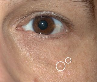
Syringomas are benign eccrine sweat duct tumors, typically found clustered on eyelids, although they may also be found in the armpits, abdomen, chest, neck, scalp, or groin area, including genitals, in a symmetric pattern. They are skin-colored or yellowish firm, rounded bumps, 1–3 mm in diameter, and may be confused with xanthoma, milia, hidrocystoma, trichoepithelioma, and xanthelasma. They are more common in women and are most commonly found in middle-aged Asian women. While they can present at any time in life, they typically present during adolescence. They are usually not associated with any other symptoms, although can sometimes cause itchiness or irritation.

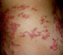
Erythema migrans or erythema chronicum migrans is an expanding rash often seen in the early stage of Lyme disease, and can also be caused by southern tick-associated rash illness (STARI). It can appear anywhere from one day to one month after a tick bite. This rash does not represent an allergic reaction to the bite, but rather an actual skin infection of one of the Lyme bacteria species from the genus Borrelia. The rash's name comes from New Latin for "migrating redness".

Granuloma annulare (GA) is a common, sometimes chronic skin condition which presents as reddish bumps on the skin arranged in a circle or ring. It can initially occur at any age, though two-thirds of patients are under 30 years old, and it is seen most often in children and young adults. Females are two times as likely to have it than males.

Erythema annulare centrifugum (EAC), is a descriptive term for a class of skin lesion presenting redness (erythema) in a ring form (anulare) that spreads from a center (centrifugum). It was first described by Darier in 1916. Many different terms have been used to classify these types of lesions and it is still controversial on what exactly defines EAC. Some of the types include annular erythema, erythema perstans, erythema gyratum perstans, erythema gyratum repens, darier erythema and erythema figuratum perstans.
Gianotti–Crosti syndrome, also known as infantile papular acrodermatitis, papular acrodermatitis of childhood, and papulovesicular acrolocated syndrome, is a reaction of the skin to a viral infection. Hepatitis B virus and Epstein–Barr virus are the most frequently reported pathogens. Other viruses implicated are hepatitis A virus, hepatitis C virus, cytomegalovirus, coxsackievirus, adenovirus, enterovirus, rotavirus, rubella virus, HIV, and parainfluenza virus.

Dissecting cellulitis of the scalp, also known as dissecting folliculitis, perifolliculitis capitis abscedens et suffodiens of Hoffman, perifolliculitis abscedens et suffodiens, or folliculitis abscedens et suffodiens, is an inflammatory condition of the scalp that can lead to scarring alopecia, which begins with deep inflammatory nodules, primarily over occiput, that progresses to coalescing regions of boggy scalp. Boggy tissue has a high fluid level that results in a spongy feeling. Isotretinoin proves to be the medicine of choice for the treatment of the disease.
Reactive perforating collagenosis is a rare, familial, nonpuritic skin disorder characterized by papules that grow in a diameter of 4 to 6mm and develop a central area of umbilication to which keratinous material is lodged. The cause of reactive perforating collagenosis is unknown.

Transient neonatal pustular melanosis (TNPM), also known as pustular melanosis, is a transient rash common in newborns. It is vesiculopustular and made up of 1–3 mm fluid-filled lesions that rupture, leaving behind a collarette of scale and a brown macule.[3] This rash occurs only in the newborn stage, usually appearing a few days after birth[2], but is sometimes already present at birth[3]. The rash usually fades over three to four weeks but may linger for up to three months after birth.[3] It can occur anywhere on the body, including the palms and soles.[1][2][3]
Keratoderma climactericum is a skin condition characterized by hyperkeratosis of the palms and soles beginning at about the time of menopause.

Livedoid vasculopathy is a chronic cutaneous disease seen predominantly in young to middle-aged women. One acronym used to describe its features is "Painful purpuric ulcers with reticular pattern of the lower extremities" (PURPLE).
Symmetrical drug-related intertriginous and flexural exanthema (SDRIFE), more popularly known as baboon syndrome because of its resemblance to the distinctive red buttocks displayed by female baboons, is a systemic dermatitis characterized by well-demarcated patches of erythema distributed symmetrically on the buttocks. The cause of the syndrome may be drug-related, i.e. induced by systemic administration of hydroxyzine penicillin, iodinated radio contrast media and others.

Topical steroid withdrawal, also known as red burning skin and steroid dermatitis, has been reported in people who apply topical steroids for 2 weeks or longer and then discontinue use. Symptoms affect the skin and include redness, a burning sensation, and itchiness, which may then be followed by peeling.
Histopathology of dermatitis can be performed in uncertain cases of inflammatory skin condition that remain uncertain after history and physical examination.