Related Research Articles

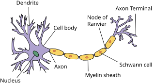
Myelin is a lipid-rich material that surrounds nerve cell axons to insulate them and increase the rate at which electrical impulses are passed along the axon. The myelinated axon can be likened to an electrical wire with insulating material (myelin) around it. However, unlike the plastic covering on an electrical wire, myelin does not form a single long sheath over the entire length of the axon. Rather, myelin sheaths the nerve in segments: in general, each axon is encased with multiple long myelinated sections with short gaps in between called nodes of Ranvier.

Pelizaeus–Merzbacher disease is an X-linked neurological disorder that damages oligodendrocytes in the central nervous system. It is caused by mutations in proteolipid protein 1 (PLP1), a major myelin protein. It is characterized by a decrease in the amount of insulating myelin surrounding the nerves (hypomyelination) and belongs to a group of genetic diseases referred to as leukodystrophies.

A demyelinating disease is any disease of the nervous system in which the myelin sheath of neurons is damaged. This damage impairs the conduction of signals in the affected nerves. In turn, the reduction in conduction ability causes deficiency in sensation, movement, cognition, or other functions depending on which nerves are involved.

Krabbe disease (KD) is a rare and often fatal lysosomal storage disease that results in progressive damage to the nervous system. KD involves dysfunctional metabolism of sphingolipids and is inherited in an autosomal recessive pattern. The disease is named after the Danish neurologist Knud Krabbe (1885–1961).

Leukodystrophies are a group of, usually, inherited disorders, characterized by degeneration of the white matter in the brain. The word leukodystrophy comes from the Greek roots leuko, "white", dys, "abnormal" and troph, "growth". The leukodystrophies are caused by imperfect growth or development of the glial cells which produce the myelin sheath, the fatty insulating covering around nerve fibers. Leukodystrophies may be classified as hypomyelinating or demyelinating diseases, respectively, depending on whether the damage is present before birth or occurs after. Other demyelinating diseases are usually not congenital and have a toxic or autoimmune cause.
Metachromatic leukodystrophy (MLD) is a lysosomal storage disease which is commonly listed in the family of leukodystrophies as well as among the sphingolipidoses as it affects the metabolism of sphingolipids. Leukodystrophies affect the growth and/or development of myelin, the fatty covering which acts as an insulator around nerve fibers throughout the central and peripheral nervous systems. MLD involves cerebroside sulfate accumulation. Metachromatic leukodystrophy, like most enzyme deficiencies, has an autosomal recessive inheritance pattern.

Myelin basic protein (MBP) is a protein believed to be important in the process of myelination of nerves in the nervous system. The myelin sheath is a multi-layered membrane, unique to the nervous system, that functions as an insulator to greatly increase the velocity of axonal impulse conduction. MBP maintains the correct structure of myelin, interacting with the lipids in the myelin membrane.
Sulfatide, also known as 3-O-sulfogalactosylceramide, SM4, or sulfated galactocerebroside, is a class of sulfolipids, specifically a class of sulfoglycolipids, which are glycolipids that contain a sulfate group. Sulfatide is synthesized primarily starting in the endoplasmic reticulum and ending in the Golgi apparatus where ceramide is converted to galactocerebroside and later sulfated to make sulfatide. Of all of the galactolipids that are found in the myelin sheath, one fifth of them are sulfatide. Sulfatide is primarily found on the extracellular leaflet of the myelin plasma membrane produced by the oligodendrocytes in the central nervous system and in the Schwann cells in the peripheral nervous system. However, sulfatide is also present on the extracellular leaflet of the plasma membrane of many cells in eukaryotic organisms.
Galactosylceramidase is an enzyme that in humans is encoded by the GALC gene. Galactosylceramidase is an enzyme which removes galactose from ceramide derivatives (galactosylceramides).

Arylsulfatase A is an enzyme that breaks down sulfatides, namely cerebroside 3-sulfate into cerebroside and sulfate. In humans, arylsulfatase A is encoded by the ARSA gene.

Proteolipid protein 1 (PLP1) is a form of myelin proteolipid protein (PLP). Mutations in PLP1 are associated with Pelizaeus–Merzbacher disease. It is a 4 transmembrane domain protein which is proposed to bind other copies of itself on the extracellular side of the membrane. In a myelin sheath, as the layers of myelin wraps come together, PLP will bind itself and tightly hold the cellular membranes together.

Nuclear receptor TLX also known as NR2E1 is a protein that in humans is encoded by the NR2E1 gene. TLX is a member of the nuclear receptor family of intracellular transcription factors.
In molecular cloning and biology, a gene knock-in refers to a genetic engineering method that involves the one-for-one substitution of DNA sequence information in a genetic locus or the insertion of sequence information not found within the locus. Typically, this is done in mice since the technology for this process is more refined and there is a high degree of shared sequence complexity between mice and humans. The difference between knock-in technology and traditional transgenic techniques is that a knock-in involves a gene inserted into a specific locus, and is thus a "targeted" insertion. It is the opposite of gene knockout.
A humanized mouse is a mouse carrying functioning human genes, cells, tissues, and/or organs. Humanized mice are commonly used as small animal models in biological and medical research for human therapeutics.
Protective autoimmunity is a condition in which cells of the adaptive immune system contribute to maintenance of the functional integrity of a tissue, or facilitate its repair following an insult. The term ‘protective autoimmunity’ was coined by Prof. Michal Schwartz of the Weizmann Institute of Science (Israel), whose pioneering studies were the first to demonstrate that autoimmune T lymphocytes can have a beneficial role in repair, following an injury to the central nervous system (CNS). Most of the studies on the phenomenon of protective autoimmunity were conducted in experimental settings of various CNS pathologies and thus reside within the scientific discipline of neuroimmunology.
The NSG mouse is a brand of immunodeficient laboratory mice, developed and marketed by Jackson Laboratory, which carries the strain NOD.Cg-Prkdcscid Il2rgtm1Wjl/SzJ. NSG branded mice are among the most immunodeficient described to date. NSG branded mice lack mature T cells, B cells, and natural killer (NK) cells. NSG branded mice are also deficient in multiple cytokine signaling pathways, and they have many defects in innate immunity. The compound immunodeficiencies in NSG branded mice permit the engraftment of a wide range of primary human cells, and enable sophisticated modeling of many areas of human biology and disease. NSG branded mice were developed in the laboratory of Dr. Leonard Shultz at Jackson Laboratory, which owns the NSG trade mark.
A knockout mouse, or knock-out mouse, is a genetically modified mouse in which researchers have inactivated, or "knocked out", an existing gene by replacing it or disrupting it with an artificial piece of DNA. They are important animal models for studying the role of genes which have been sequenced but whose functions have not been determined. By causing a specific gene to be inactive in the mouse, and observing any differences from normal behaviour or physiology, researchers can infer its probable function.

Myelin regulatory factor, also known as myelin gene regulatory factor (MRF), is a protein that in humans is encoded by the MYRF gene.
Mice with severe combined immunodeficiency (SCIDs) are often used in the research of human disease. Human immune cells are used to develop human lymphoid organs within these immunodeficient mice, and many different types of SCID mouse models have been developed. These mice allow researchers to study the human immune system and human disease in a small animal model.
Myelinoids or myelin organoids are three dimensional in vitro cultured model derived from human pluripotent stem cells (hPSCs) that represent various brain regions, spinal cord or the peripheral nervous system in early fetal human development. They have the capacity to recapitulate aspects of brain developmental processes, microenvironments, cell to cell interaction, structural organization and cellular composition. The differentiating aspect dictating whether an organoid is deemed a cerebral organoid/brain organoid or myelinoid is the presence of myelination and compact myelin formation that is a defining feature of myelinoids. Due to the complex nature of the human brain, there is a need for model systems which can closely mimic complicated biological processes. Myelinoids provide a unique in vitro model through which myelin pathology, neurodegenerative diseases, developmental processes and therapeutic screening can be accomplished.
References
- ↑ Molineaux SM, Engh H, de Ferra F, Hudson L, Lazzarini RA (1986). "Recombination within the myelin basic protein gene created the dysmyelinating shiverer mouse mutation". Proc Natl Acad Sci U S A. 83 (19): 7542–7546. Bibcode:1986PNAS...83.7542M. doi: 10.1073/pnas.83.19.7542 . PMC 386755 . PMID 2429310.
{{cite journal}}: CS1 maint: multiple names: authors list (link) - ↑ Johansen-Berg H, Behrens TEJ (2014). "Chapter 7. White Matter Structure: A Microscopist's View". Diffusion MRI (2 ed.). pp. 127–153. doi:10.1016/B978-0-12-396460-1.00007-X.
- ↑ Readhead C, Hood L (1990). "The dysmyelinating mouse mutations shiverer (shi) and myelin deficient (shi mld)". Behav Genet. 20 (2): 213–234. doi:10.1007/BF01067791. PMID 1693848. S2CID 686238.
- ↑ Baker M (12 June 2008). "Human cell transplants stop the shivers". Nature Reports Stem Cells: 1. doi: 10.1038/stemcells.2008.92 .