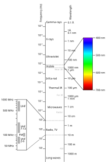
Spectroscopy is the study of the interaction between matter and electromagnetic radiation as a function of the wavelength or frequency of the radiation. In simpler terms, spectroscopy is the precise study of color as generalized from visible light to all bands of the electromagnetic spectrum; indeed, historically, spectroscopy originated as the study of the wavelength dependence of the absorption by gas phase matter of visible light dispersed by a prism. Matter waves and acoustic waves can also be considered forms of radiative energy, and recently gravitational waves have been associated with a spectral signature in the context of the Laser Interferometer Gravitational-Wave Observatory (LIGO).

An optical spectrometer is an instrument used to measure properties of light over a specific portion of the electromagnetic spectrum, typically used in spectroscopic analysis to identify materials. The variable measured is most often the light's intensity but could also, for instance, be the polarization state. The independent variable is usually the wavelength of the light or a unit directly proportional to the photon energy, such as reciprocal centimeters or electron volts, which has a reciprocal relationship to wavelength.

A monochromator is an optical device that transmits a mechanically selectable narrow band of wavelengths of light or other radiation chosen from a wider range of wavelengths available at the input. The name is from the Greek roots mono-, "single", and chroma, "colour", and the Latin suffix -ator, denoting an agent.

X-ray spectroscopy is a general term for several spectroscopic techniques for characterization of materials by using x-ray excitation.

The NASA Infrared Telescope Facility is a 3-meter (9.8 ft) telescope optimized for use in infrared astronomy and located at the Mauna Kea Observatory in Hawaii. It was first built to support the Voyager missions and is now the US national facility for infrared astronomy, providing continued support to planetary, solar neighborhood, and deep space applications. The IRTF is operated by the University of Hawaii under a cooperative agreement with NASA. According to the IRTF's time allocation rules, at least 50% of the observing time is devoted to planetary science.

Spectral imaging is imaging that uses multiple bands across the electromagnetic spectrum. While an ordinary camera captures light across three wavelength bands in the visible spectrum, red, green, and blue (RGB), spectral imaging encompasses a wide variety of techniques that go beyond RGB. Spectral imaging may use the infrared, the visible spectrum, the ultraviolet, x-rays, or some combination of the above. It may include the acquisition of image data in visible and non-visible bands simultaneously, illumination from outside the visible range, or the use of optical filters to capture a specific spectral range. It is also possible to capture hundreds of wavelength bands for each pixel in an image.

In imaging spectroscopy each pixel of an image acquires many bands of light intensity data from the spectrum, instead of just the three bands of the RGB color model. More precisely, it is the simultaneous acquisition of spatially coregistered images in many spectrally contiguous bands.

Hyperspectral imaging, like other spectral imaging, collects and processes information from across the electromagnetic spectrum. The goal of hyperspectral imaging is to obtain the spectrum for each pixel in the image of a scene, with the purpose of finding objects, identifying materials, or detecting processes. There are three general branches of spectral imagers. There are push broom scanners and the related whisk broom scanners, which read images over time, band sequential scanners, which acquire images of an area at different wavelengths, and snapshot hyperspectral imaging, which uses a staring array to generate an image in an instant.

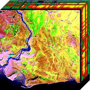
An imaging spectrometer is an instrument used in hyperspectral imaging and imaging spectroscopy to acquire a spectrally-resolved image of an object or scene, often referred to as a datacube due to the three-dimensional representation of the data. Two axes of the image corresponds to vertical and horizontal distance and the third to wavelength. The principle of operation is the same as that of the simple spectrometer, but special care is taken to avoid optical aberrations for better image quality.
Chemical imaging is the analytical capability to create a visual image of components distribution from simultaneous measurement of spectra and spatial, time information. Hyperspectral imaging measures contiguous spectral bands, as opposed to multispectral imaging which measures spaced spectral bands.

An echelle grating is a type of diffraction grating characterised by a relatively low groove density, but a groove shape which is optimized for use at high incidence angles and therefore in high diffraction orders. Higher diffraction orders allow for increased dispersion (spacing) of spectral features at the detector, enabling increased differentiation of these features. Echelle gratings are, like other types of diffraction gratings, used in spectrometers and similar instruments. They are most useful in cross-dispersed high resolution spectrographs, such as HARPS, PARAS, and numerous other astronomical instruments.

Airborne Real-time Cueing Hyperspectral Enhanced Reconnaissance, also known by the acronym ARCHER, is an aerial imaging system that produces ground images far more detailed than plain sight or ordinary aerial photography can. It is the most sophisticated unclassified hyperspectral imaging system available, according to U.S. Government officials. ARCHER can automatically scan detailed imaging for a given signature of the object being sought, for abnormalities in the surrounding area, or for changes from previous recorded spectral signatures.
Electro-optical MASINT is a subdiscipline of Measurement and Signature Intelligence, (MASINT) and refers to intelligence gathering activities which bring together disparate elements that do not fit within the definitions of Signals Intelligence (SIGINT), Imagery Intelligence (IMINT), or Human Intelligence (HUMINT).

Fourier-transform infrared spectroscopy (FTIR) is a technique used to obtain an infrared spectrum of absorption or emission of a solid, liquid or gas. An FTIR spectrometer simultaneously collects high-resolution spectral data over a wide spectral range. This confers a significant advantage over a dispersive spectrometer, which measures intensity over a narrow range of wavelengths at a time.
An integral field spectrograph, or a spectrograph equipped with an integral field unit (IFU), is an optical instrument combining spectrographic and imaging capabilities, used to obtain spatially resolved spectra in astronomy and other fields of research such as bio-medical science and earth observation.
Snapshot hyperspectral imaging is a method for capturing hyperspectral images during a single integration time of a detector array. No scanning is involved with this method and the lack of moving parts means that motion artifacts should be avoided. This instrument typically features detector arrays with a high number of pixels.

The computed tomography imaging spectrometer (CTIS) is a snapshot imaging spectrometer conceived separately by Takayuki Okamoto and Ichirou Yamaguchi at Riken (Japan), and by F. Bulygin and G. Vishnakov in Moscow (Russia). The concept was subsequently further developed by Michael Descour, at the time a PhD student at the University of Arizona, under the direction of Prof. Eustace Dereniak.
Video spectroscopy combines spectroscopic measurements with video technique. This technology has resulted from recent developments in hyperspectral imaging. A video capable imaging spectrometer can work like a camcorder and provide full frame spectral images in real-time that enables advanced mobility and hand-held imaging spectroscopy. Unlike hyperspectral line scanners, a video spectrometer can spectrally capture randomly and quickly moving objects and processes. The product of a conventional hyperspectral line scanner has typically been called a hyperspectral data cube. A video spectrometer produces a spectral image data series at much higher speeds (1 ms) and frequencies (25 Hz) that is called a hyperspectral video. This technology can initiate novel solutions and challenges in spectral tracking, field spectroscopy, spectral mobile mapping, real-time spectral monitoring and many other applications.

The NIRSpec is one of the four scientific instruments which will be flown on the James Webb Space Telescope (JWST). The JWST is the follow-on mission to the Hubble Space Telescope (HST) and is developed to receive more information about the origins of the universe by observing infrared light from the first stars and galaxies. In comparison to HST, its instruments will allow looking further back in time and will study the so-called Dark Ages during which the universe was opaque, about 150 to 800 million years after the Big Bang.
Computational imaging is the process of indirectly forming images from measurements using algorithms that rely on a significant amount of computing. In contrast to traditional imaging, computational imaging systems involve a tight integration of the sensing system and the computation in order to form the images of interest. The ubiquitous availability of fast computing platforms, the advances in algorithms and modern sensing hardware is resulting in imaging systems with significantly enhanced capabilities. Computational Imaging systems cover a broad range of applications include computational microscopy, tomographic imaging, MRI, ultrasound imaging, computational photography, Synthetic Aperture Radar (SAR), seismic imaging etc. The integration of the sensing and the computation in computational imaging systems allows for accessing information which was otherwise not possible. For example:


