| Spitz | |||||||
|---|---|---|---|---|---|---|---|
 Spitz protein | |||||||
| Identifiers | |||||||
| Organism | |||||||
| Symbol | SPI | ||||||
| Entrez | 35253 | ||||||
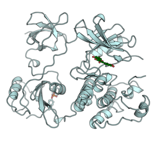
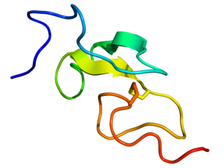
| PDB | 3LTF | ||||||
| RefSeq (Prot) | NM_057561 | ||||||
| UniProt | Q01083 | ||||||
| |||||||
Spitz is a protein in Drosophila species which is the major activator of their epidermal growth factor receptor (EGFR). [1]
| Spitz | |||||||
|---|---|---|---|---|---|---|---|
| Spitz protein | |||||||
| Identifiers | |||||||
| Organism | |||||||
| Symbol | SPI | ||||||
| Entrez | 35253 | ||||||
| PDB | 3LTF | ||||||
| RefSeq (Prot) | NM_057561 | ||||||
| UniProt | Q01083 | ||||||
| |||||||
Spitz is a protein in Drosophila species which is the major activator of their epidermal growth factor receptor (EGFR). [1]
Spitz is produced as a transmembrane protein in the endoplasmic reticulum. There it associates with a cargo receptor called Star and is trafficked to the Golgi. In the Golgi, Spitz is cleaved by a protease called Rhomboid, which releases Spitz to be trafficked to the cell membrane and released out of the cell. [1] From here it can bind EGFR on the surface of other cells, activating the receptor. Alternatively, Spitz can be bound and inactivated by Argos, inhibiting EGFR activation. [2]
Spitz is responsible for activating signaling of the Drosophila epidermal growth factor receptor (DER) and is involved in the development of the embryos, eyes, and wings of fruit flies. Spitz can be sequestered and prevented from binding to DER by the protein Argos (Aos) which then inhibits the epidermal growth factor receptor pathway. [3] Over-expression of epidermal growth factor receptors contribute to human cancers, so the sequestering of activating ligands may be useful in developing ways to diminish EGFR ligands for cancer treatment. [2]


The protein Spitz is structurally similar to transforming growth factor-α (TGF-α) and is a homologue of TGF-α, along with other proteins found in Drosophila such as Gurken and Keren. [1] These proteins are processed by Star, a transmembrane protein, and Rhomboid (Rho), a protease. Spitz binds to and regulates the epidermal growth factor receptor. [4]
EGFR exists as a dimer, and two Spitz proteins bind to the receptor to regulate its function. The dimer is referred to as two subunits: the left hand subunit and the right hand subunit. [5] The binding of the two Spitz proteins on either subunit are not identical, as different amino acid residues participate in the binding on either side. On the right hand side, Arg21 and Arg26 of EGFR interact with residues Asn50 and Ile51 of Spitz. On the left hand side, Arg21 and Arg26 of the other part of the dimer interact with Glu53 and Ile51 of another Spitz protein. The Spitz protein can wedge apart the I and III domains of one of the EGFR subunits, subsequently relocating domain I away from domain III and causing new interactions between side chains of the EGFR subunit. [5] Research has shown that there is negative cooperativity in the binding of the Spitz ligands to EGFR, where the binding event of the second ligand to the dimer decreases the affinity of the receptors for one another. The specificity, autophosphorylation, mechanism, and other behaviors of the dimer can change significantly when it is weakened by the double-occupancy of the Spitz proteins. [5]

Integrins are transmembrane receptors that help cell-cell and cell-extracellular matrix (ECM) adhesion. Upon ligand binding, integrins activate signal transduction pathways that mediate cellular signals such as regulation of the cell cycle, organization of the intracellular cytoskeleton, and movement of new receptors to the cell membrane. The presence of integrins allows rapid and flexible responses to events at the cell surface.

A protein kinase is a kinase which selectively modifies other proteins by covalently adding phosphates to them (phosphorylation) as opposed to kinases which modify lipids, carbohydrates, or other molecules. Phosphorylation usually results in a functional change of the target protein (substrate) by changing enzyme activity, cellular location, or association with other proteins. The human genome contains about 500 protein kinase genes and they constitute about 2% of all human genes. There are two main types of protein kinase. The great majority are serine/threonine kinases, which phosphorylate the hydroxyl groups of serines and threonines in their targets. Most of the others are tyrosine kinases, although additional types exist. Protein kinases are also found in bacteria and plants. Up to 30% of all human proteins may be modified by kinase activity, and kinases are known to regulate the majority of cellular pathways, especially those involved in signal transduction.
A tyrosine kinase is an enzyme that can transfer a phosphate group from ATP to the tyrosine residues of specific proteins inside a cell. It functions as an "on" or "off" switch in many cellular functions.

Paracrine signaling is a form of cell signaling, a type of cellular communication in which a cell produces a signal to induce changes in nearby cells, altering the behaviour of those cells. Signaling molecules known as paracrine factors diffuse over a relatively short distance, as opposed to cell signaling by endocrine factors, hormones which travel considerably longer distances via the circulatory system; juxtacrine interactions; and autocrine signaling. Cells that produce paracrine factors secrete them into the immediate extracellular environment. Factors then travel to nearby cells in which the gradient of factor received determines the outcome. However, the exact distance that paracrine factors can travel is not certain.

The insulin receptor (IR) is a transmembrane receptor that is activated by insulin, IGF-I, IGF-II and belongs to the large class of receptor tyrosine kinase. Metabolically, the insulin receptor plays a key role in the regulation of glucose homeostasis; a functional process that under degenerate conditions may result in a range of clinical manifestations including diabetes and cancer. Insulin signalling controls access to blood glucose in body cells. When insulin falls, especially in those with high insulin sensitivity, body cells begin only to have access to lipids that do not require transport across the membrane. So, in this way, insulin is the key regulator of fat metabolism as well. Biochemically, the insulin receptor is encoded by a single gene INSR, from which alternate splicing during transcription results in either IR-A or IR-B isoforms. Downstream post-translational events of either isoform result in the formation of a proteolytically cleaved α and β subunit, which upon combination are ultimately capable of homo or hetero-dimerisation to produce the ≈320 kDa disulfide-linked transmembrane insulin receptor.

Gefitinib, sold under the brand name Iressa, is a medication used for certain breast, lung and other cancers. Gefitinib is an EGFR inhibitor, like erlotinib, which interrupts signaling through the epidermal growth factor receptor (EGFR) in target cells. Therefore, it is only effective in cancers with mutated and overactive EGFR, but resistances to gefitinib can arise through other mutations. It is marketed by AstraZeneca and Teva.
Epidermal growth factor (EGF) is a protein that stimulates cell growth and differentiation by binding to its receptor, EGFR. Human EGF is 6-kDa and has 53 amino acid residues and three intramolecular disulfide bonds.

The epidermal growth factor receptor is a transmembrane protein that is a receptor for members of the epidermal growth factor family of extracellular protein ligands.

Transforming growth factor beta (TGF-β) is a multifunctional cytokine belonging to the transforming growth factor superfamily that includes three different mammalian isoforms and many other signaling proteins. TGFB proteins are produced by all white blood cell lineages.

Receptor tyrosine kinases (RTKs) are the high-affinity cell surface receptors for many polypeptide growth factors, cytokines, and hormones. Of the 90 unique tyrosine kinase genes identified in the human genome, 58 encode receptor tyrosine kinase proteins. Receptor tyrosine kinases have been shown not only to be key regulators of normal cellular processes but also to have a critical role in the development and progression of many types of cancer. Mutations in receptor tyrosine kinases lead to activation of a series of signalling cascades which have numerous effects on protein expression. Receptor tyrosine kinases are part of the larger family of protein tyrosine kinases, encompassing the receptor tyrosine kinase proteins which contain a transmembrane domain, as well as the non-receptor tyrosine kinases which do not possess transmembrane domains.
The transforming growth factor beta (TGFB) signaling pathway is involved in many cellular processes in both the adult organism and the developing embryo including cell growth, cell differentiation, cell migration, apoptosis, cellular homeostasis and other cellular functions. The TGFB signaling pathways are conserved. In spite of the wide range of cellular processes that the TGFβ signaling pathway regulates, the process is relatively simple. TGFβ superfamily ligands bind to a type II receptor, which recruits and phosphorylates a type I receptor. The type I receptor then phosphorylates receptor-regulated SMADs (R-SMADs) which can now bind the coSMAD SMAD4. R-SMAD/coSMAD complexes accumulate in the nucleus where they act as transcription factors and participate in the regulation of target gene expression.

Transforming growth factor alpha (TGF-α) is a protein that in humans is encoded by the TGFA gene. As a member of the epidermal growth factor (EGF) family, TGF-α is a mitogenic polypeptide. The protein becomes activated when binding to receptors capable of protein kinase activity for cellular signaling.
Zalutumumab is a fully human IgG1 monoclonal antibody (mAb) directed towards the epidermal growth factor receptor (EGFR). It is a product developed by Genmab in Utrecht, the Netherlands. Specifically, zalutumumab is designed for the treatment of squamous cell carcinoma of the head and neck (SCCHN), a type of cancer.
The ErbB family of proteins contains four receptor tyrosine kinases, structurally related to the epidermal growth factor receptor (EGFR), its first discovered member. In humans, the family includes Her1, Her2 (ErbB2), Her3 (ErbB3), and Her4 (ErbB4). The gene symbol, ErbB, is derived from the name of a viral oncogene to which these receptors are homologous: erythroblastic leukemia viral oncogene. Insufficient ErbB signaling in humans is associated with the development of neurodegenerative diseases, such as multiple sclerosis and Alzheimer's disease, while excessive ErbB signaling is associated with the development of a wide variety of types of solid tumor.
Argos is a secreted protein that is an inhibitor of the epidermal growth factor receptor (EGFR) pathway in Drosophila melanogaster. Argos inhibits the EGFR pathway by sequestering the EGFR ligand Spitz. Argos binds the epidermal growth factor domain of Spitz, preventing interaction between Spitz and EGFR. Argos does not directly interact with EGFR. Argos represents the first example of ligand sequestration as a mechanism of inhibition in the ErbB (EGFR) family.
Faint little ball (flb) is a Drosophila gene that encodes the Drosophila epidermal growth factor receptor (DER) homolog. The gene is also called torpedo and Ellipse. The gene is located at 3-26 of the Drosophila melanogaster genome. It is named faint little ball because when the gene is mutated the embryo forms a ball of dorsal hypoderm. flb is necessary for several processes to occur during embryonic development, specifically in central nervous system development. It is expressed as quickly as 4 hours after fertilization of the egg. The peak of expression of the flb gene is between 4–8 hours into development. In all processes that are facilitated by flb the same signal transduction pathway is used. Drosophila EGF receptor is involved in the development of embryos as well as larvae/pupae's wings, eyes, legs and ovaries.

Receptor tyrosine-protein kinase erbB-3, also known as HER3, is a membrane bound protein that in humans is encoded by the ERBB3 gene.

Cell surface receptors are receptors that are embedded in the plasma membrane of cells. They act in cell signaling by receiving extracellular molecules. They are specialized integral membrane proteins that allow communication between the cell and the extracellular space. The extracellular molecules may be hormones, neurotransmitters, cytokines, growth factors, cell adhesion molecules, or nutrients; they react with the receptor to induce changes in the metabolism and activity of a cell. In the process of signal transduction, ligand binding affects a cascading chemical change through the cell membrane.

Autophosphorylation is a type of post-translational modification of proteins. It is generally defined as the phosphorylation of the kinase by itself. In eukaryotes, this process occurs by the addition of a phosphate group to serine, threonine or tyrosine residues within protein kinases, normally to regulate the catalytic activity. Autophosphorylation may occur when a kinases' own active site catalyzes the phosphorylation reaction, or when another kinase of the same type provides the active site that carries out the chemistry. The latter often occurs when kinase molecules dimerize. In general, the phosphate groups introduced are gamma phosphates from nucleoside triphosphates, most commonly ATP.
The transforming growth factor beta (TGFβ) receptors are a family of serine/threonine kinase receptors involved in TGF beta signaling pathway. These receptors bind growth factor and cytokine signaling proteins in the TGF-beta family such as TGFβs, bone morphogenetic proteins (BMPs), growth differentiation factors (GDFs), activin and inhibin, myostatin, anti-Müllerian hormone (AMH), and NODAL.