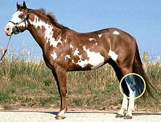
Splints is an ailment of the horse or pony, characterized by a hard, bony swelling, usually on the inside of a front leg, lying between the splint and cannon bone or on the splint bone itself. It may be "hot," meaning that it occurred recently and is still painful; or "cold," meaning that the splint has completely recovered and there is no longer any pain associated with it. Bucked shins are sometimes called 'shin splints,' which involve small stress fractures of the dorsal cannon bone, often seen in race training, and discussed elsewhere.
The splint bones, (metacarpal or metatarsal II and IV), which are remnants of two of the five toes of prehistoric horses, run down either side of the cannon bone. They narrow as they go from the carpal or tarsal joint down, and form a "button" at the bottom or their length, a few inches above the fetlock. Splint bones are attached to the cannon by interosseous ligaments, providing some mobility in the young horse. As the horse ages, the interosseous ligaments are typically replaced by bone. In some older horses, the cannon and splint bones may become completely fused.
Direct trauma, such as from an interference injury or a kick from another horse, is a common causes of splints. The periosteum is damaged by the trauma, and the horse's body lays down new bone in the injured area. Splints caused by trauma are more commonly seen lower down the leg than ones caused by strain. The splints may occur in a front leg or hind, in one leg or both. Severe enough trauma can fracture the splint bone. If minimally displaced, and in the lower portion, some heal well. Others may need surgical removal of a portion of the damaged splint bone. [1]
Concussion is another cause of splints. Concussive forces run from the carpus or tarsus into the splint bones. Working a horse on hard surfaces increases the concussion received by the interosseous ligament, which causes tearing. Splints caused by concussion are usually found on both front legs, most commonly on the inside of the leg a few inches below the knee.
Overworking young or unfit horses at speed or in tight circles may cause splints. The uneven loading of the limb in tight circles places excessive force on the medial splint, which can cause it to move excessively relative to the cannon bone, causing tears in the interosseous ligament and periosteal reaction.
Bench-kneed conformation causes excess loading of the medial splint bone, which can lead to splints. [2]
Because the splint bone does have some mobility independent of the cannon bone, it can cause tension and strain on the periosteum of the splint bone where the interosseous ligament attaches. The horse will then lay down new bone and the area will become inflamed. "Blind splints" are named because the bony reaction happens on the inside border between the splint bone and cannon bone, where it can not be seen, and is usually not palpable. Besides causing pain as any active splint reaction can, the swelling can impinge on the suspensory ligament. This condition is difficult to diagnose, but ultrasound is generally diagnostic. [3] MRI and CT also show these well. [4]
Splints usually cause mild lameness (a grade of 1–2 out of 5). The injured area is hot, painful, and inflamed with a small bony swelling. However, splints do not always cause lameness, especially once "cold". More severe lameness is sometimes associated with a fractured splint bone, or soft tissue injury adjacent to the splints.
"Blind splints" usually produce mild lameness that is difficult to pinpoint because there is no obvious swelling, pain, or bony changes related to the exterior of the splint bone. At times, bone proliferation on the axial border of the splint bone can be seen radiographically, but ultrasound is much more sensitive for detecting blind splints.
The body will eventually absorb some of the bone it placed down in the splint, flattening out the splint over several months and possibly making it completely disappear.
The horse should have a reduced workload for 1–3 weeks. If a trainer does not decrease the workload sufficiently, and the splint bone continues to receive concussion, the injury is likely to continue or worsen. Light exercise on soft ground is best for a horse with splints, as work can help encourage the needed bone growth to heal the splint. Those trainers concerned with the cosmetic appearance of their horse usually prefer to hand-walk twice daily and keep the animal stalled until the splint is resolved, eliminating the chance that the splint will accidentally be knocked during work and the swelling increased.
Several days of cold therapy, sweats, and NSAIDs can help a "hot" splint. NSAIDs can help reduce the inflammation and help the bone growth by doing so. However, none of these treatments are incredibly effective. The most important factor is time. Counter-irritants, which increase inflammation, only hinder the formation of bone and can actually prolong the healing process.
Surgery to remove the fractured end of the splint bone, particularly in the lower third, is typically successful. However, surgical removal of the bone growth in large splints, performed by chiseling it away, usually does not produce satisfying results. Often, bone growth is stimulated by the surgery, and the size of the splint is increased. Only about a third of the time is surgery at all successful.
Prognosis is excellent in uncomplicated cases. The horse will be able to return to full work once the inflammation and pain ceases. Although the horse usually recovers quite quickly, horses with "blind splints" may take longer because there may be impingement on the suspensory ligament. The ossification of the splint is usually a permanent blemish, though over a period of many years, the excess ossification may be remodeled to varying degrees, occasionally to the point that the splint is no longer visible.

A bone fracture is a medical condition in which there is a partial or complete break in the continuity of any bone in the body. In more severe cases, the bone may be broken into several fragments, known as a comminuted fracture. A bone fracture may be the result of high force impact or stress, or a minimal trauma injury as a result of certain medical conditions that weaken the bones, such as osteoporosis, osteopenia, bone cancer, or osteogenesis imperfecta, where the fracture is then properly termed a pathologic fracture.
The hock, tarsus or uncommonly gambrel, is the region formed by the tarsal bones connecting the tibia and metatarsus of a digitigrade or unguligrade quadrupedal mammal, such as a horse, cat, or dog. This joint may include articulations between tarsal bones and the fibula in some species, while in others the fibula has been greatly reduced and is only found as a vestigial remnant fused to the distal portion of the tibia. It is the anatomical homologue of the ankle of the human foot. While homologous joints occur in other tetrapods, the term is generally restricted to mammals, particularly long-legged domesticated species.
A soft tissue injury is the damage of muscles, ligaments and tendons throughout the body. Common soft tissue injuries usually occur from a sprain, strain, a one-off blow resulting in a contusion or overuse of a particular part of the body. Soft tissue injuries can result in pain, swelling, bruising and loss of function.

Laminitis is a disease that affects the feet of ungulates and is found mostly in horses and cattle. Clinical signs include foot tenderness progressing to inability to walk, increased digital pulses, and increased temperature in the hooves. Severe cases with outwardly visible clinical signs are known by the colloquial term founder, and progression of the disease will lead to perforation of the coffin bone through the sole of the hoof or being unable to stand up, requiring euthanasia.
Navicular syndrome, often called navicular disease, is a syndrome of lameness problems in horses. It most commonly describes an inflammation or degeneration of the navicular bone and its surrounding tissues, usually on the front feet. It can lead to significant and even disabling lameness.
Bone spavin is a bony growth within the lower hock joint of horse or cattle. It is caused by osteoarthritis, and the degree of lameness that results can be serious enough to end a horse's competitive career.
Tendinitis/tendonitis is inflammation of a tendon, often involving torn collagen fibers. A bowed tendon is a horseman's term for a tendon after a horse has sustained an injury that causes swelling in one or more tendons creating a "bowed" appearance.

Equine conformation evaluates a horse's bone structure, musculature, and its body proportions in relation to each other. Undesirable conformation can limit the ability to perform a specific task. Although there are several faults with universal disadvantages, a horse's conformation is usually judged by what its intended use may be. Thus "form to function" is one of the first set of traits considered in judging conformation. A horse with poor form for a Grand Prix show jumper could have excellent conformation for a World Champion cutting horse, or to be a champion draft horse. Every horse has good and bad points of its conformation and many horses excel even with conformation faults.

Ringbone is exostosis in the pastern or coffin joint of a horse. In severe cases, the growth can encircle the bones, giving ringbone its name. It has been suggested by some authors that such a colloquial term, whilst commonly used, might be misleading and that it would be better to refer to this condition as osteoarthritis of the inter-phalangeal joints in ungulates.

Arthrodesis, also known as artificial ankylosis or syndesis, is the artificial induction of joint ossification between two bones by surgery. This is done to relieve intractable pain in a joint which cannot be managed by pain medication, splints, or other normally indicated treatments. The typical causes of such pain are fractures which disrupt the joint, severe sprains, and arthritis. It is most commonly performed on joints in the spine, hand, ankle, and foot. Historically, knee and hip arthrodeses were also performed as pain-relieving procedures, but with the great successes achieved in hip and knee arthroplasty, arthrodesis of these large joints has fallen out of favour as a primary procedure, and now is only used as a procedure of last resort in some failed arthroplasties.
Infantile cortical hyperostosis (ICH) is a self-limited inflammatory disorder of infants that causes bone changes, soft tissue swelling and irritability. The disease may be present at birth or occur shortly thereafter. The cause is unknown. Both familial and sporadic forms occur. It is also known as Caffey disease or Caffey's disease.

The Lisfranc ligament is one of several ligaments which connects the medial cuneiform bone to the second metatarsal. Sometimes, the term Lisfranc ligament refers specifically to the ligament that connects the superior, lateral surface of the medial cuneiform to the superior, medial surface of the base of the second metatarsal.
Osselet is arthritis in the fetlock joint of a horse, caused by trauma. Osselets usually occur in the front legs of the horse, because there is more strain and concussion on the fetlock there than in the hind legs. The arthritis will occur at the joint between the cannon bone and large pastern bone, at the front of the fetlock.
Equine lymphangitis is an inflammation or swelling associated with impairment of the lymphatic system, particularly in a limb, in horses. It is most commonly a bacterial infection, although bacterial culture may be negative.

The skeletal system of the horse is a skeletal system of a horse that has three major functions in the body. It protects vital organs, provides framework, and supports soft parts of the body. Horses typically have 205 bones. The pelvic limb typically contains 19 bones, while the thoracic limb contains 20 bones.
Lameness is an abnormal gait or stance of an animal that is the result of dysfunction of the locomotor system. In the horse, it is most commonly caused by pain, but can be due to neurologic or mechanical dysfunction. Lameness is a common veterinary problem in racehorses, sport horses, and pleasure horses. It is one of the most costly health problems for the equine industry, both monetarily for the cost of diagnosis and treatment, and for the cost of time off resulting in loss-of-use.
Curb is defined in older literature as enlargement secondary to inflammation and thickening of the long plantar ligament in horses. However, with the widespread use of diagnostic ultrasonography in equine medicine, curb has been redefined as a collection of soft tissue injuries of the distal plantar hock region. Curb is a useful descriptive term when describing swelling in this area.
Racehorse injuries and fatalities are a side effect of the training and competition of horse racing. Racehorse injuries are considered especially difficult to treat, as they frequently result in the death of the horse. A 2005 study by the United States Department of Agriculture found that injuries are the second leading cause of death in horses, second only to old age.
The limbs of the horse are structures made of dozens of bones, joints, muscles, tendons, and ligaments that support the weight of the equine body. They include two apparatuses: the suspensory apparatus, which carries much of the weight, prevents overextension of the joint and absorbs shock, and the stay apparatus, which locks major joints in the limbs, allowing horses to remain standing while relaxed or asleep. The limbs play a major part in the movement of the horse, with the legs performing the functions of absorbing impact, bearing weight, and providing thrust. In general, the majority of the weight is borne by the front legs, while the rear legs provide propulsion. The hooves are also important structures, providing support, traction and shock absorption, and containing structures that provide blood flow through the lower leg. As the horse developed as a cursorial animal, with a primary defense mechanism of running over hard ground, its legs evolved to the long, sturdy, light-weight, one-toed form seen today.
The treatment of equine lameness is a complex subject. Lameness in horses has a variety of causes, and treatment must be tailored to the type and degree of injury, as well as the financial capabilities of the owner. Treatment may be applied locally, systemically, or intralesionally, and the strategy for treatment may change as healing progresses. The end goal is to reduce the pain and inflammation associated with injury, to encourage the injured tissue to heal with normal structure and function, and to ultimately return the horse to the highest level of performance possible following recovery.