
A microscope is a laboratory instrument used to examine objects that are too small to be seen by the naked eye. Microscopy is the science of investigating small objects and structures using a microscope. Microscopic means being invisible to the eye unless aided by a microscope.
A biosensor is an analytical device, used for the detection of a chemical substance, that combines a biological component with a physicochemical detector. The sensitive biological element, e.g. tissue, microorganisms, organelles, cell receptors, enzymes, antibodies, nucleic acids, etc., is a biologically derived material or biomimetic component that interacts with, binds with, or recognizes the analyte under study. The biologically sensitive elements can also be created by biological engineering. The transducer or the detector element, which transforms one signal into another one, works in a physicochemical way: optical, piezoelectric, electrochemical, electrochemiluminescence etc., resulting from the interaction of the analyte with the biological element, to easily measure and quantify. The biosensor reader device connects with the associated electronics or signal processors that are primarily responsible for the display of the results in a user-friendly way. This sometimes accounts for the most expensive part of the sensor device, however it is possible to generate a user friendly display that includes transducer and sensitive element. The readers are usually custom-designed and manufactured to suit the different working principles of biosensors.

Two-photon excitation microscopy is a fluorescence imaging technique that is particularly well-suited to image scattering living tissue of up to about one millimeter in thickness. Unlike traditional fluorescence microscopy, where the excitation wavelength is shorter than the emission wavelength, two-photon excitation requires simultaneous excitation by two photons with longer wavelength than the emitted light. The laser is focused onto a specific location in the tissue and scanned across the sample to sequentially produce the image. Due to the non-linearity of two-photon excitation, mainly fluorophores in the micrometer-sized focus of the laser beam are excited, which results in the spatial resolution of the image. This contrasts with confocal microscopy, where the spatial resolution is produced by the interaction of excitation focus and the confined detection with a pinhole.

William Esco Moerner, also known as W. E. Moerner, is an American physical chemist and chemical physicist with current work in the biophysics and imaging of single molecules. He is credited with achieving the first optical detection and spectroscopy of a single molecule in condensed phases, along with his postdoc, Lothar Kador. Optical study of single molecules has subsequently become a widely used single-molecule experiment in chemistry, physics and biology. In 2014, he was awarded the Nobel Prize in Chemistry.

Stefan Walter Hell is a Romanian-German physicist and one of the directors of the Max Planck Institute for Multidisciplinary Sciences in Göttingen, and of the Max Planck Institute for Medical Research in Heidelberg, both of which are in Germany. He received the Nobel Prize in Chemistry in 2014 "for the development of super-resolved fluorescence microscopy", together with Eric Betzig and William Moerner.
Chemical imaging is the analytical capability to create a visual image of components distribution from simultaneous measurement of spectra and spatial, time information. Hyperspectral imaging measures contiguous spectral bands, as opposed to multispectral imaging which measures spaced spectral bands.

A molecular sensor or chemosensor is a molecular structure that is used for sensing of an analyte to produce a detectable change or a signal. The action of a chemosensor, relies on an interaction occurring at the molecular level, usually involves the continuous monitoring of the activity of a chemical species in a given matrix such as solution, air, blood, tissue, waste effluents, drinking water, etc. The application of chemosensors is referred to as chemosensing, which is a form of molecular recognition. All chemosensors are designed to contain a signalling moiety and a recognition moiety, that is connected either directly to each other or through a some kind of connector or a spacer. The signalling is often optically based electromagnetic radiation, giving rise to changes in either the ultraviolet and visible absorption or the emission properties of the sensors. Chemosensors may also be electrochemically based. Small molecule sensors are related to chemosensors. These are traditionally, however, considered as being structurally simple molecules and reflect the need to form chelating molecules for complexing ions in analytical chemistry. Chemosensors are synthetic analogues of biosensors, the difference being that biosensors incorporate biological receptors such as antibodies, aptamers or large biopolymers.

Supercritical angle fluorescence microscopy (SAF) is a technique to detect and characterize fluorescent species and their behaviour close or even adsorbed or linked at surfaces. The method is able to observe molecules in a distance of less than 100 to 0 nanometer from the surface even in presence of high concentrations of fluorescent species around. Using an aspheric lens for excitation of a sample with laser light, fluorescence emitted by the specimen is collected above the critical angle of total internal reflection selectively and directed by a parabolic optics onto a detector. The method was invented in 1998 in the laboratories of Stefan Seeger at University of Regensburg/Germany and later at University of Zurich/Switzerland.
Christoph Cremer is a German physicist and emeritus at the Ruprecht-Karls-University Heidelberg, former honorary professor at the University of Mainz and was a former group leader at Institute of Molecular Biology (IMB) at the Johannes Gutenberg University of Mainz, Germany, who has successfully overcome the conventional limit of resolution that applies to light based investigations by a range of different methods. In the meantime, according to his own statement, Christoph Cremer is a member of the Max Planck Institute for Chemistry and the Max Planck Institute for Polymer Research.
Photothermal optical microscopy / "photothermal single particle microscopy" is a technique that is based on detection of non-fluorescent labels. It relies on absorption properties of labels, and can be realized on a conventional microscope using a resonant modulated heating beam, non-resonant probe beam and lock-in detection of photothermal signals from a single nanoparticle. It is the extension of the macroscopic photothermal spectroscopy to the nanoscopic domain. The high sensitivity and selectivity of photothermal microscopy allows even the detection of single molecules by their absorption. Similar to Fluorescence Correlation Spectroscopy (FCS), the photothermal signal may be recorded with respect to time to study the diffusion and advection characteristics of absorbing nanoparticles in a solution. This technique is called photothermal correlation spectroscopy (PhoCS).

Biophysical chemistry is a physical science that uses the concepts of physics and physical chemistry for the study of biological systems. The most common feature of the research in this subject is to seek an explanation of the various phenomena in biological systems in terms of either the molecules that make up the system or the supra-molecular structure of these systems. Apart from the biological applications, recent research showed progress in the medical field as well.
Xiaowei Zhuang is a Chinese-American biophysicist who is the David B. Arnold Jr. Professor of Science, Professor of Chemistry and Chemical Biology, and Professor of Physics at Harvard University, and an Investigator at the Howard Hughes Medical Institute. She is best known for her work in the development of Stochastic Optical Reconstruction Microscopy (STORM), a super-resolution fluorescence microscopy method, and the discoveries of novel cellular structures using STORM. She received a 2019 Breakthrough Prize in Life Sciences for developing super-resolution imaging techniques that get past the diffraction limits of traditional light microscopes, allowing scientists to visualize small structures within living cells. She was elected a Member of the American Philosophical Society in 2019 and was awarded a Vilcek Foundation Prize in Biomedical Science in 2020.

Martin Gruebele is a German-born American physical chemist and biophysicist who is currently James R. Eiszner Professor of Chemistry, Professor of Physics, Professor of Biophysics and Computational Biology at the University of Illinois Urbana-Champaign, where he is the principal investigator of the Gruebele Group.

Andreas Hierlemann is a German chemist and professor of Biosystems Engineering at ETH Zurich. He is known for his work in the field of CMOS-based chemical and biomicrosensors and high-density microelectrode arrays.

Robert Eric Betzig is an American physicist who works as a professor of physics and professor of molecular and cell biology at the University of California, Berkeley. He is also a senior fellow at the Janelia Farm Research Campus in Ashburn, Virginia.
Jürgen P. Rabe is a German physicist and nanoscientist.
Brian T. Cunningham is an American engineer, researcher and academic. He is a Donald Biggar Willett Professor of Engineering at University of Illinois at Urbana-Champaign. He is a professor of Electrical and Computer Engineering, and a professor of bioengineering.

Debabrata Goswami FInstP FRSC, is an Indian chemist and the Prof. S. Sampath Chair Professor of Chemistry, at the Indian Institute of Technology Kanpur. He is also a professor of The Department of Chemistry and The Center for Lasers & Photonics at the same Institute. Goswami is an associate editor of the open-access journal Science Advances. He is also an Academic Editor for PLOS One and PeerJ Chemistry. He has contributed to the theory of Quantum Computing as well as nonlinear optical spectroscopy. His work is documented in more than 200 research publications. He is an elected Fellow of the Royal Society of Chemistry, Fellow of the Institute of Physics, the SPIE, and The Optical Society. He is also a Senior Member of the IEEE, has been awarded a Swarnajayanti Fellowship for Chemical Sciences, and has held a Wellcome Trust Senior Research Fellowship. He is the third Indian to be awarded the International Commission for Optics Galileo Galilei Medal for excellence in optics.
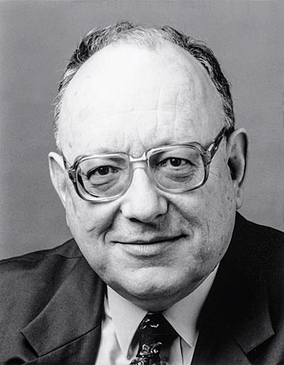
Urs Paul Rolf Wild was a Swiss chemist. He became known for his pioneering works in single molecule detection.

MicroRNA (miRNA) biosensors are analytical devices that involve interactions between the target miRNA strands and recognition element on a detection platform to produce signals that can be measured to indicate levels or the presence of the target miRNA. Research into miRNA biosensors shows shorter readout times, increased sensitivity and specificity of miRNA detection and lower fabrication costs than conventional miRNA detection methods.
