Subconjunctival injection

Last updated
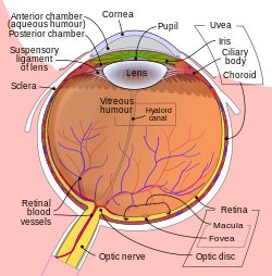
Schematic diagram of the human eye showing. Schematic diagram of the human eye en.svg
Schematic diagram of the human eye showing.

Subconjunctival injection is a type of periocular route of injection for ocular drug administration by administration of a medication either under the conjunctiva or underneath the conjunctiva lining the eyelid.

Using the subconjunctival injection bypasses the fatty layers of the bulbous conjunctiva and putting medications adjacent to sclera that is permeable to water, this will increase the penetration of the water-soluble drug into the eye. [1]

This route is indicated for treatment of different lesions, such as in the cornea, sclera, anterior uvea and vitreous.

Antibiotics and corticosteroids [2] can be administered by this route.

Related Research Articles

<span class="mw-page-title-main">Pterygium (eye)</span> Pinkish, triangular tissue growth on the cornea of the eye

A pterygium of the eye is a pinkish, roughly triangular tissue growth of the conjunctiva onto the cornea of the eye. It typically starts on the cornea near the nose. It may slowly grow but rarely grows so large that it covers the pupil and impairs vision. Often both eyes are involved.

<span class="mw-page-title-main">Route of administration</span> Path by which a drug, fluid, poison, or other substance is taken into the body

In pharmacology and toxicology, a route of administration is the way by which a drug, fluid, poison, or other substance is taken into the body.

<span class="mw-page-title-main">Eye surgery</span> Surgery performed on the eye or its adnexa

Eye surgery, also known as ophthalmic surgery or ocular surgery, is surgery performed on the eye or its adnexa. Eye surgery is part of ophthalmology and is performed by an ophthalmologist or eye surgeon. The eye is a fragile organ, and requires due care before, during, and after a surgical procedure to minimize or prevent further damage. An eye surgeon is responsible for selecting the appropriate surgical procedure for the patient, and for taking the necessary safety precautions. Mentions of eye surgery can be found in several ancient texts dating back as early as 1800 BC, with cataract treatment starting in the fifth century BC. It continues to be a widely practiced class of surgery, with various techniques having been developed for treating eye problems.

<span class="mw-page-title-main">Conjunctiva</span> Part of the eye; protective outer layer covering the sclera

In the anatomy of the eye, the conjunctiva is a thin mucous membrane that lines the inside of the eyelids and covers the sclera. It is composed of non-keratinized, stratified squamous epithelium with goblet cells, stratified columnar epithelium and stratified cuboidal epithelium. The conjunctiva is highly vascularised, with many microvessels easily accessible for imaging studies.

<span class="mw-page-title-main">Subconjunctival bleeding</span> Bleeding within the whites of the eye

Subconjunctival bleeding, also known as subconjunctival hemorrhage or subconjunctival haemorrhage, is bleeding from a small blood vessel over the whites of the eye. It results in a red spot in the white of the eye. There is generally little to no pain and vision is not affected. Generally only one eye is affected.

<span class="mw-page-title-main">Red eye (medicine)</span> Eye that appears red due to illness or injury

A red eye is an eye that appears red due to illness or injury. It is usually injection and prominence of the superficial blood vessels of the conjunctiva, which may be caused by disorders of these or adjacent structures. Conjunctivitis and subconjunctival hemorrhage are two of the less serious but more common causes.

<span class="mw-page-title-main">Scleritis</span> Medical condition

Scleritis is a serious inflammatory disease that affects the white outer coating of the eye, known as the sclera. The disease is often contracted through association with other diseases of the body, such as granulomatosis with polyangiitis or rheumatoid arthritis. There are three types of scleritis: diffuse scleritis, nodular scleritis, and necrotizing scleritis. Scleritis may be the first symptom of onset of connective tissue disease.

<span class="mw-page-title-main">Anterior ciliary arteries</span> Small arteries in the eye-socket

The anterior ciliary arteries are seven arteries in each eye-socket that arise from muscular branches of the ophthalmic artery and supply the conjunctiva, sclera, rectus muscles, and the ciliary body. The arteries end by anastomosing with branches of the long posterior ciliary arteries to form the circulus arteriosus major.

<span class="mw-page-title-main">Intravitreal administration</span>

Intravitreal administration is a route of administration of a drug, or other substance, in which the substance is delivered into the vitreous humor of the eye. "Intravitreal" literally means "inside an eye". Intravitreal injections were first introduced in 1911 when Ohm gave an injection of air into the vitreous humor to repair a detached retina. In the mid-1940s, intravitreal injections became a standard way to administer drugs to treat endophthalmitis and cytomegalovirus retinitis.

<span class="mw-page-title-main">Trabeculectomy</span> Surgical procedure used in the treatment of glaucoma

Trabeculectomy is a surgical procedure used in the treatment of glaucoma to relieve intraocular pressure by removing part of the eye's trabecular meshwork and adjacent structures. It is the most common glaucoma surgery performed and allows drainage of aqueous humor from within the eye to underneath the conjunctiva where it is absorbed. This outpatient procedure was most commonly performed under monitored anesthesia care using a retrobulbar block or peribulbar block or a combination of topical and subtenon anesthesia. Due to the higher risks associated with bulbar blocks, topical analgesia with mild sedation is becoming more common. Rarely general anesthesia will be used, in patients with an inability to cooperate during surgery.

<span class="mw-page-title-main">Episcleritis</span> Medical condition

Episcleritis is a benign, self-limiting inflammatory disease affecting part of the eye called the episclera. The episclera is a thin layer of tissue that lies between the conjunctiva and the connective tissue layer that forms the white of the eye (sclera). Episcleritis is a common condition, and is characterized by the abrupt onset of painless eye redness.

<span class="mw-page-title-main">Intraocular hemorrhage</span> Medical condition

Intraocular hemorrhage is bleeding inside the eye. Bleeding can occur from any structure of the eye where there is vasculature or blood flow, including the anterior chamber, vitreous cavity, retina, choroid, suprachoroidal space, or optic disc.

<span class="mw-page-title-main">Accessory visual structures</span> External parts of the eye including eyebrow, eyelid, and lacrimal apparatus

The accessory visual structures are the protecting and supporting structures (adnexa) of the eye, including the eyebrow, eyelids, and lacrimal apparatus. The eyebrows, eyelids, eyelashes, lacrimal gland and drainage apparatus all play a crucial role with regards to globe protection, lubrication, and minimizing the risk of ocular infection. The adnexal structures also help to keep the cornea moist and clean.

<span class="mw-page-title-main">Oral administration</span> Route of administration where a substance is taken through the mouth

Oral administration is a route of administration whereby a substance is taken through the mouth, swallowed, and then processed via the digestive system. This is a common route of administration for many medications.

<span class="mw-page-title-main">Conjunctivochalasis</span> Medical condition

Conjunctivochalasis, also known as mechanical dry eye (MDE), is a common eye surface condition characterized by the presence of excess folds of the conjunctiva located between the globe of the eye and the eyelid margin.

<span class="mw-page-title-main">Acute hemorrhagic conjunctivitis</span> Medical condition

Acute hemorrhagic conjunctivitis (AHC) is a derivative of the highly contagious conjunctivitis virus, otherwise known as pink eye. Symptoms include excessively red, swollen eyes as well as subconjunctival hemorrhaging. Currently, there is no known treatment and patients are required to merely endure the symptoms while the virus runs its five- to seven-day course. While it was first identified in Ghana, the virus has now been seen in China, India, Egypt, Cuba, Singapore, Taiwan, Japan, Pakistan, Thailand, and the United States.

Micro-invasive glaucoma surgery (MIGS) is the latest advance in surgical treatment for glaucoma, which aims to reduce intraocular pressure by either increasing outflow of aqueous humor or reducing its production. MIGS comprises a group of surgical procedures which share common features. MIGS procedures involve a minimally invasive approach, often with small cuts or micro-incisions through the cornea that causes the least amount of trauma to surrounding scleral and conjunctival tissues. The techniques minimize tissue scarring, allowing for the possibility of traditional glaucoma procedures such as trabeculectomy or glaucoma valve implantation to be performed in the future if needed.

<span class="mw-page-title-main">Intravitreal injection</span> Method of administration of drugs into the eye by injection with a fine needle

Intravitreal injection is the method of administration of drugs into the eye by injection with a fine needle. The medication will be directly applied into the vitreous humor. It is used to treat various eye diseases, such as age-related macular degeneration (AMD), diabetic retinopathy, and infections inside the eye such as endophthalmitis. As compared to topical administration, this method is beneficial for a more localized delivery of medications to the targeted site, as the needle can directly pass through the anatomical eye barrier and dynamic barrier. It could also minimize adverse drug effects on other body tissues via the systemic circulation, which could be a possible risk for intravenous injection of medications. Although there are risks of infections or other complications, with suitable precautions throughout the injection process, chances for these complications could be lowered.

<span class="mw-page-title-main">Suprachoroidal drug delivery</span>

Suprachoroidal drug delivery is an ocular route of drug administration. It involves using a microneedle to provide a minimally invasive method and injecting particles of a medication into the suprachoroidal space (SCS) between the sclera and choroid in the eye.

<span class="mw-page-title-main">Sub-Tenon injection</span> Ocular route of drug administration

Sub-Tenon injection is an ocular route of drug administration. It involves administration of a medication to the area between the sclera and the Tenon's capsule.

References

  1. Eye: drug administration - subconjunctival injection
  2. Koff, Raphael; Rome, SOL; Kasper, Richard; Commons, Robert R.; Button, Richard; Starr, Paul (1950). "Subconjunctival Injection of Cortisone in Iritis". Journal of the American Medical Association. 144 (15): 1259. doi:10.1001/jama.1950.62920150004009a. PMID   14778735.