Related Research Articles

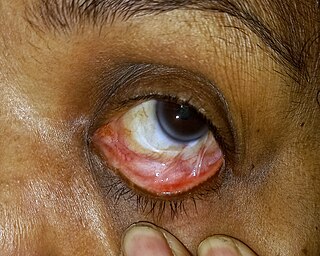
Conjunctivitis, also known as pink eye, is inflammation of the outermost layer of the white part of the eye and the inner surface of the eyelid. It makes the eye appear pink or reddish. Pain, burning, scratchiness, or itchiness may occur. The affected eye may have increased tears or be "stuck shut" in the morning. Swelling of the white part of the eye may also occur. Itching is more common in cases due to allergies. Conjunctivitis can affect one or both eyes.

Cherry eye is a disorder of the nictitating membrane (NM), also called the third eyelid, present in the eyes of dogs and cats. Cherry eye is most often seen in young dogs under the age of two. Common misnomers include adenitis, hyperplasia, adenoma of the gland of the third eyelid; however, cherry eye is not caused by hyperplasia, neoplasia, or primary inflammation. In many species, the third eyelid plays an essential role in vision by supplying oxygen and nutrients to the eye via tear production. Normally, the gland can turn inside-out without detachment. Cherry eye results from a defect in the retinaculum which is responsible for anchoring the gland to the periorbita. This defect causes the gland to prolapse and protrude from the eye as a red fleshy mass. Problems arise as sensitive tissue dries out and is subjected to external trauma Exposure of the tissue often results in secondary inflammation, swelling, or infection. If left untreated, this condition can lead to dry eye syndrome and other complications.

The conjunctiva is a tissue that lines the inside of the eyelids and covers the sclera. It is composed of non-keratinized, stratified squamous epithelium with goblet cells, stratified columnar epithelium and stratified cuboidal epithelium. The conjunctiva is highly vascularised, with many microvessels easily accessible for imaging studies.

Trachoma is an infectious disease caused by bacterium Chlamydia trachomatis. The infection causes a roughening of the inner surface of the eyelids. This roughening can lead to pain in the eyes, breakdown of the outer surface or cornea of the eyes, and eventual blindness. Untreated, repeated trachoma infections can result in a form of permanent blindness when the eyelids turn inward.
Trichiasis is a medical term for abnormally positioned eyelashes that grow back toward the eye, touching the cornea or conjunctiva. This can be caused by infection, inflammation, autoimmune conditions, congenital defects, eyelid agenesis and trauma such as burns or eyelid injury. It is the leading cause of infectious blindness in the world.
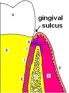
In biological morphology and anatomy, a sulcus is a furrow or fissure. It may be a groove in the surface of a limb or an organ, notably in the surface of the brain, but also in the lungs, certain muscles, as well as in bones, and elsewhere. Many sulci are the product of a surface fold or junction, such as in the gums, where they fold around the neck of the tooth.
Ocular melanosis (OM) is a blue-gray and/or brown lesion of the conjunctiva that can be separated into benign conjunctival epithelial melanosis (BCEM) and primary acquired melanosis (PAM), of which the latter is considered a risk factor for uveal melanoma. The disease is caused by an increase of melanocytes in the iris, choroid, and surrounding structures. Overproduction of pigment by these cells can block the trabecular meshwork through which fluid drains from the eye. The increased fluid in the eye leads to increased pressure, which can lead to glaucoma. In humans, this is sometimes known as pigment dispersion syndrome.

A distichia is an eyelash that arises from an abnormal part of the eyelid. This abnormality, attributed to a genetic mutation, is known to affect dogs and humans. Distichiae usually exit from the duct of the meibomian gland at the eyelid margin. They are usually multiple, and sometimes more than one arises from a duct. They can affect either the upper or lower eyelid and are usually bilateral. The lower eyelids of dogs usually have no eyelashes.

Superior limbic keratoconjunctivitis is a disease of the eye characterized by episodes of recurrent inflammation of the superior cornea and limbus, as well as of the superior tarsal and bulbar conjunctiva. It was first described by F. H. Théodore in 1963.

The lacrimal artery is an artery of the orbit around the eye. It arises close to the optic foramen, and is a branch of the ophthalmic artery. It accompanies the lacrimal nerve along the upper border of the lateral rectus muscle. It supplies the lacrimal gland, two rectus muscles of the eye, the eyelids, and the conjunctiva.

The superior tarsal muscle is a smooth muscle adjoining the levator palpebrae superioris muscle that helps to raise the upper eyelid.

The plica semilunaris is a small fold of bulbar conjunctiva on the medial canthus of the eye. It functions during movement of the eye, to help maintain tear drainage via the lacrimal lake, and to permit greater rotation of the globe, for without the plica the conjunctiva would attach directly to the eyeball, restricting movement. It is the vestigial remnant of the nictitating membrane which is drawn across the eye for protection, and is present in other animals such as birds, reptiles, and fish, but is rare in mammals, mainly found in monotremes and marsupials. Its associated muscles are also vestigial. It is loose, thus eye movements are not restricted by it. Only one species of primate, the Calabar angwantibo, is known to have a functioning nictitating membrane.

Carl Ferdinand Ritter von Arlt was an Austrian ophthalmologist born in Ober-Graupen, a village near Teplitz (Teplice) in Bohemia.
Krause's glands are small, mucous accessory lacrimal glands that are found underneath the eyelid where the upper and lower conjunctivae meet. Their ducts unite into a rather long sinus which open into the fornix conjunctiva. There are approximately forty Krause glands in the region of the upper eyelid, and around 6 to 8 in the region of the lower lid. The function of these glands are to produce tears which are secreted onto the surface of the conjunctiva.
A symblepharon is a partial or complete adhesion of the palpebral conjunctiva of the eyelid to the bulbar conjunctiva of the eyeball. It results either from disease or trauma. Cicatricial pemphigoid and, in severe cases, rosacea may cause symblepharon. It is rarely congenital. Its treatment is symblepharectomy.

The accessory visual structures are the protecting and supporting structures (adnexa) of the eye, including the eyebrow, eyelids, and lacrimal apparatus. The eyebrows, eyelids, eyelashes, lacrimal gland and drainage apparatus all play a crucial role with regards to globe protection, lubrication, and minimizing the risk of ocular infection. The adnexal structures also help to keep the cornea moist and clean.

Conjunctivochalasis is a common eye surface condition characterized by the presence of excess folds of the conjunctiva located between the globe of the eye and the eyelid margin.
Arlt's line is a thick band of scar tissue in the conjunctiva of the eye, near the lid margin, that is associated with eye infections. Arlt's line is a characteristic finding of trachoma, an infection of the eye caused by Chlamydia trachomatis. The line runs horizontally, parallel to eyelid, and is found at the junction of the anterior one third and posterior two thirds of the conjunctiva.
Ankyloblepharon is defined as the adhesion of the edges of the upper eyelid with the lower eyelid. Ankyloblepharon must be differentiated from blepharophimosis, in which palpebral aperture is reduced and there is telecanthus, but the eyelid margins are normal. Another condition similar to Ankyloblepharon is Symblepharon where palpebral conjunctiva is attached to bulbar conjunctiva. Recognition of ankyloblepharon necessitates systemic examination to detect associated abnormalities such as genitourinary, cardiac, and syndactyly.
Exposure keratopathy is medical condition affecting the cornea of eyes. It can lead to corneal ulceration and permanent loss of vision due to corneal opacity.
References
- ↑ "Conjunctiva". Online Resources of Ophthalmology. Retrieved 19 June 2013.
- ↑ Khurana, A. K. (2008). Comprehensive Ophthalmology (4 ed.). Kent, UK: Anshan Publishers. p. 65. ISBN 978-1905740789.
| | This article about the eye is a stub. You can help Wikipedia by expanding it. |