Symphysis pubis dysfunction

Last updated
Symphysis pubis dysfunction
Symphysis Pubis.png
Location of pubic symphysis
Specialty OB/Gyn

Symphysis pubis dysfunction (SPD), commonly known as pubic symphysis dysfunction or lightning crotch, [1] is a condition that causes excessive movement of the pubic symphysis, either anterior or lateral, as well as associated pain, possibly because of a misalignment of the pelvis. Most commonly associated with pregnancy and childbirth,[ citation needed ] it is diagnosed in approximately 1 in 300 pregnancies, although some estimates of incidence are as high as 1 in 50. [2]

Contents

SPD is associated with pelvic girdle pain and the names are often used interchangeably.

Symptoms

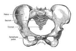
The main symptom is usually pain or discomfort in the pelvic region, usually centered on the joint at the front of the pelvis (the pubic symphysis). Some sufferers report being able to hear and feel the pubic symphysis and/or sacroiliac, clicking or popping in and out as they walk or change position. Sufferers frequently also experience pain in the lower back, hips, groin, lower abdomen, and legs. The severity of the pain can range from mild discomfort to extreme pain that interferes with routine activities, family, social and professional life, and sleep. [3] There have been links between SPD and depression due to the associated physical discomfort. [4] [5] Sufferers may walk with a characteristic side-to-side gait and have difficulty climbing stairs, problems with leg abduction and adduction, pain when carrying out weight bearing activities, difficulties carrying out everyday activities, and difficulties standing. [4]

Diagnosis

A diagnosis is usually made from the symptoms, history, and physical exam alone. After pregnancy, MRI scans, X-rays and ultrasound scanning are sometimes used. Patients typically initially report symptoms to a midwife, chiropractor, obstetrician, general practitioner, physiotherapist or an osteopath. On seeing a health professional, patients should expect to receive a thorough physical examination to rule out other lumbar spine problems, such as a prolapsed disc or pelvic and or pubis joint misalignment, or other conditions such as iliopsoas muscle spasms, urinary tract infections and Braxton Hicks contractions.

Unnecessary radiation from medical imaging is avoided during pregnancy, so in most cases a physical examination and history are considered sufficient to refer to physical therapy.

Treatment

A promising treatment for chronic or post natal dysfunction is prolotherapy. [6] Other treatments include the use of elbow crutches, pelvic support devices and prescribed pain relief. The majority of problems will resolve spontaneously after delivery. [7] There are two case studies that show reduction of pain and dysfunction with conservative chiropractic care. [8]

Physical therapists—especially those specializing in pelvic floor physical therapy—can assist with pain relief techniques, provide manual therapy to alleviate related muscle spasms, and manage exercise protocols.

While most pregnancy-related cases are reported to resolve postpartum, definitive diagnosis and treatment are still appropriate in order to optimize comfort and function and ensure a good course of recovery.

Long-term complications can develop without proper care. Postpartum follow-up in cases of pregnancy-related SPD may include radiologic imaging, evaluation by a specialist such as an orthopedist or physiatrist, ongoing pelvic floor physical therapy, and assessment for any underlying or related musculoskeletal issues.

In extreme cases that do not resolve with conservative management, surgery is considered after pregnancy to stabilise the pelvis, but success rates are very poor. [4]

Everyday living

Typical advice usually given to people with SPD includes avoiding strenuous exercise, prolonged standing, repetitive reaching movements, lunges, stretching exercises and squatting. Patients are also frequently advised to:

If the pain is very severe, using a walker or crutches will help take the weight off the pelvis and assist with mobility. Alternatively, for more extreme cases a wheelchair may be considered advisable.

Pharmacological interventions

It is not usually considered advisable to take anti-inflammatory medication in pregnancy, which makes SPD a particularly difficult condition to manage. Acetaminophen may be a safer option. Of note, opiates are considered high risk with a more addictive nature, and carry a risk of depressed respiration in the newborn baby if taken near the time of birth, if taken at all. Therefore, it is considered advisable to discuss any pain relief medications with a physician, and cease taking any opiates 2–4 weeks before the estimated due date, as advised by a medical professional.

See also

Related Research Articles

<span class="mw-page-title-main">Levator ani</span> Broad, thin muscle group, situated on either side of the pelvis

The levator ani is a broad, thin muscle group, situated on either side of the pelvis. It is formed from three muscle components: the pubococcygeus, the iliococcygeus, and the puborectalis.

<span class="mw-page-title-main">Acetabulum</span> Cavity where the thigh bone (femur) articulates with the pelvis

The acetabulum, also called the cotyloid cavity, is a concave surface of the pelvis. The head of the femur meets with the pelvis at the acetabulum, forming the hip joint.

Dyspareunia is painful sexual intercourse due to medical or psychological causes. The term dyspareunia covers both female dyspareunia and male dyspareunia, but many discussions that use the term without further specification concern the female type, which is more common than the male type. In females, the pain can primarily be on the external surface of the genitalia, or deeper in the pelvis upon deep pressure against the cervix. Medically, dyspareunia is a pelvic floor dysfunction and is frequently underdiagnosed. It can affect a small portion of the vulva or vagina or be felt all over the surface. Understanding the duration, location, and nature of the pain is important in identifying the causes of the pain.

<span class="mw-page-title-main">Pubic symphysis</span> Cartilaginous joint between the front of the left and right hip bones

The pubic symphysis is a secondary cartilaginous joint between the left and right superior rami of the pubis of the hip bones. It is in front of and below the urinary bladder. In males, the suspensory ligament of the penis attaches to the pubic symphysis. In females, the pubic symphysis is attached to the suspensory ligament of the clitoris. In most adults, it can be moved roughly 2 mm and with 1 degree rotation. This increases for women at the time of childbirth.

<span class="mw-page-title-main">Pelvic floor dysfunction</span> Medical condition

Pelvic floor dysfunction is a term used for a variety of disorders that occur when pelvic floor muscles and ligaments are impaired. The condition affects up to 50 percent of women who have given birth. Although this condition predominantly affects women, up to 16 percent of men are affected as well. Symptoms can include pelvic pain, pressure, pain during sex, urinary incontinence (UI), overactive bladder, bowel incontinence, incomplete emptying of feces, constipation, myofascial pelvic pain and pelvic organ prolapse. When pelvic organ prolapse occurs, there may be visible organ protrusion or a lump felt in the vagina or anus. Research carried out in the UK has shown that symptoms can restrict everyday life for women. However, many people found it difficult to talk about it and to seek care, as they experienced embarrassment and stigma.

<span class="mw-page-title-main">Rectus abdominis muscle</span> Paired straight muscle

The rectus abdominis muscle, also known as the "abdominal muscle" or simply the "abs", is a pair of segmented skeletal muscle on the ventral aspect of a person's abdomen. The paired muscle is separated at the midline by a band of dense connective tissue called the linea alba, and the connective tissue defining each lateral margin of the rectus abdominus is the linea semilunaris. The muscle extends from the pubic symphysis, pubic crest and pubic tubercle inferiorly, to the xiphoid process and costal cartilages of the 5th–7th ribs superiorly.

<span class="mw-page-title-main">Shoulder dystocia</span> Birthing obstruction complication

Shoulder dystocia is when, after vaginal delivery of the head, the baby's anterior shoulder gets caught above the mother's pubic bone. Signs include retraction of the baby's head back into the vagina, known as "turtle sign". Complications for the baby may include brachial plexus injury, or clavicle fracture. Complications for the mother may include vaginal or perineal tears, postpartum bleeding, or uterine rupture.

<span class="mw-page-title-main">Osteitis pubis</span> Medical condition

Osteitis pubis is a noninfectious inflammation of the pubis symphysis, causing varying degrees of lower abdominal and pelvic pain. Osteitis pubis was first described in patients who had undergone suprapubic surgery, and it remains a well-known complication of invasive procedures about the pelvis. It may also occur as an inflammatory process in athletes. The incidence and cause of osteitis pubis as an inflammatory process versus an infectious process continues to fuel debate among physicians when confronted by a patient who presents complaining of abdominal pain or pelvic pain and overlapping symptoms. It was first described in 1924.

<span class="mw-page-title-main">Abdomen</span> Part of the body between the chest and pelvis

The abdomen is the part of the body between the thorax (chest) and pelvis, in humans and in other vertebrates. The abdomen is the front part of the abdominal segment of the torso. The area occupied by the abdomen is called the abdominal cavity. In arthropods, it is the posterior tagma of the body; it follows the thorax or cephalothorax.

<span class="mw-page-title-main">Pubis (bone)</span> Most forward-facing of the three main regions making up the os coxa

In vertebrates, the pubis or pubic bone forms the lower and anterior part of each side of the hip bone. The pubis is the most forward-facing of the three bones that make up the hip bone. The left and right pubic bones are each made up of three sections; a superior ramus, an inferior ramus, and a body.

<span class="mw-page-title-main">Pelvic fracture</span> Broken bone in nonacetabular portions of pelvis

A pelvic fracture is a break of the bony structure of the pelvis. This includes any break of the sacrum, hip bones, or tailbone. Symptoms include pain, particularly with movement. Complications may include internal bleeding, injury to the bladder, or vaginal trauma.

<span class="mw-page-title-main">Pubic symphysis diastasis</span> Medical condition

Pubic symphysis diastasis is the separation of normally joined pubic bones, as in the dislocation of the bones, without a fracture that measures radiologically more than 10 mm. Separation of the symphysis pubis is a rare pathology associated with childbirth and has an incidence of 1 in 300 to 1 in 30,000 births. It is usually noticed after delivery but can be observed up to six months postpartum. Risk factors associated with this injury include cephalopelvic disproportion, rapid second stage of labor, epidural anesthesia, severe abduction of the thighs during delivery, or previous trauma to the pelvis. Common signs and symptoms include symphyseal pain aggravated by weight-bearing and walking, a waddling gait, pubic tenderness, and a palpable interpubic gap. Treatment for pubic symphysis diastasis is largely conservative, with treatment modalities including pelvic bracing, bed rest, analgesia, physical therapy, and in some severe cases, surgery.

Pelvic girdle pain can be described as a pregnancy discomfort for some women and a severe disability for others. PGP can cause pain, instability and limitation of mobility and functioning in any of the three pelvic joints. PGP has a long history of recognition, mentioned by Hippocrates and later described in medical literature by Snelling.

The affection appears to consist of relaxation of the pelvic articulations, becoming apparent suddenly after parturition or gradually during pregnancy and permitting a degree of mobility of the pelvic bones which effectively hinders locomotion and gives rise to the most peculiar and alarming sensations.

The following outline is provided as an overview of and topical guide to obstetrics:

Round ligament pain (RLP) is pain associated with the round ligament of the uterus, usually during pregnancy. RLP is one of the most common discomforts of pregnancy and usually starts at the second trimester of gestation and continues until delivery. It usually resolves completely after delivery although cases of postpartum RLP have been reported. RLP also occurs in nonpregnant women.

In surgery, a surgical incision is a cut made through the skin and soft tissue to facilitate an operation or procedure. Often, multiple incisions are possible for an operation. In general, a surgical incision is made as small and unobtrusive as possible to facilitate safe and timely operating conditions.

<span class="mw-page-title-main">Sacroiliac joint dysfunction</span> Medical condition

The term sacroiliac joint dysfunction refers to abnormal motion in the sacroiliac joint, either too much motion or too little motion, that causes pain in this region.

<span class="mw-page-title-main">Pelvis</span> Lower torso of the human body

The pelvis is the lower part of the trunk, between the abdomen and the thighs, together with its embedded skeleton.

<span class="mw-page-title-main">Vaginal support structures</span> Structures that maintain the position of the vagina within the pelvic cavity

The vaginal support structures are those muscles, bones, ligaments, tendons, membranes and fascia, of the pelvic floor that maintain the position of the vagina within the pelvic cavity and allow the normal functioning of the vagina and other reproductive structures in the female. Defects or injuries to these support structures in the pelvic floor leads to pelvic organ prolapse. Anatomical and congenital variations of vaginal support structures can predispose a woman to further dysfunction and prolapse later in life. The urethra is part of the anterior wall of the vagina and damage to the support structures there can lead to incontinence and urinary retention.

<span class="mw-page-title-main">Hard flaccid syndrome</span> Medical condition

Hard flaccid syndrome (HFS), also known as hard flaccid (HF), is a chronic painful condition characterized by a semi-rigid penis at the flaccid state, a soft glans at the erect state (cold glans syndrome), pelvic pain, low libido, erectile dysfunction, erectile pain, pain on ejaculation, penile sensory changes (numbness or coldness), lower urinary tract symptoms, contraction of the pelvic floor muscles, and psychological distress. Other complaints include rectal and perineal discomfort, cold hands and feet, and a hollow or detached feeling inside the penile shaft. The majority of HFS patients are in their 20s–30s and symptoms significantly affect one's quality of life.

References

  1. Murkoff, Heidi (31 May 2016). What to Expect when You're Expecting. Workman. ISBN   9780761187486.
  2. "Symphysis Pubis Dysfunction (SPD) in Pregnancy".
  3. "The Pelvic Partnership". www.pelvicpartnership.org.uk. Archived from the original on 2002-09-29.
  4. 1 2 3 Pregnancy Related Pelvic Girdle Pain For mothers to be and new mothers. Pelvic Obstetric and Gynaecological Physiotherapy. 2018.
  5. Jain, Smita; Eedarapalli, Padma; Jamjute, Pradumna; Sawdy, Robert (July 2006). "Symphysis pubis dysfunction: a practical approach to management". The Obstetrician & Gynaecologist. 8 (3): 153–158. doi: 10.1576/toag.8.3.153.27250 . S2CID   72701799.
  6. Hauser, Ross A.; Lackner, Johanna B.; Steilen-Matias, Danielle; Harris, David K. (7 July 2016). "A Systematic Review of Dextrose Prolotherapy for Chronic Musculoskeletal Pain". Clinical Medicine Insights. Arthritis and Musculoskeletal Disorders. 9: 139–159. doi:10.4137/CMAMD.S39160. PMC   4938120 . PMID   27429562.
  7. "Antenatal care (NICE clinical guideline 62)". Royal College of Obstetricians & Gynaecologists.
  8. Howell, Emily R (June 2012). "Pregnancy-related symphysis pubis dysfunction management and postpartum rehabilitation: two case reports". The Journal of the Canadian Chiropractic Association. 56 (2): 102–11. PMC   3364059 . PMID   22675223.

Further reading