Synchysis scintillans

Last updated
Synchysis scintillans
Schematic diagram of the human eye en.svg
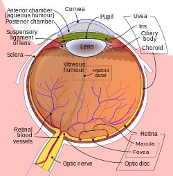
Human eye (vitreous humor in the middle)
Specialty Ophthalmology

Synchysis scintillans is a degenerative condition of the eye resulting in liquefied vitreous humor and the accumulation of cholesterol crystals within the vitreous. It is also known as cholesterolosis bulbi. The vitreous liquifies in a process known as syneresis. Synchysis scintillans appears as small white floaters that freely move in the posterior part of the eye, giving a snow globe effect. It is most commonly seen in eyes that have suffered from a degenerative disease and are end-stage. [1]

The condition is seen rarely. Associated with the advanced stages of diabetic retinopathy, but the exact pathogenesis is unknown.

The condition is symptomless and untreatable. In ophthalmoscopic examination it appears as small, flat, yellow, highly refractive crystals of cholesterol floating freely in the vitreous. These will settle, due to gravity, if the eye is immobilized.

Related Research Articles

Floater Deposits within the eyes vitreous humour

Floaters or eye floaters are sometimes visible deposits within the eye's vitreous humour, which is normally transparent, or between the vitreous and retina. They can become particularly noticeable when looking at a blank surface or an open monochromatic space, such as blue sky. Each floater can be measured by its size, shape, consistency, refractive index, and motility. They are also called muscae volitantes, or mouches volantes. The vitreous usually starts out transparent, but imperfections may gradually develop as one ages. The common type of floater, present in most people's eyes, is due to these degenerative changes of the vitreous. The perception of floaters, which may be annoying or problematic to some people, is known as myodesopsia, or, less commonly, as myodaeopsia, myiodeopsia, or myiodesopsia. It is not often treated, except in severe cases, where vitrectomy (surgery), laser vitreolysis, and medication may be effective.

Vitreous body Gel in eyeballs

The vitreous body is the clear gel that fills the space between the lens and the retina of the eyeball in humans and other vertebrates. It is often referred to as the vitreous humor or simply "the vitreous". Vitreous fluid or "liquid vitreous" is the liquid component of the vitreous gel, found after a vitreous detachment. It is not to be confused with the aqueous humor, the other fluid in the eye that is found between the cornea and lens.

Moore's lightning streaks are lightning type streaks (photopsia) due to sudden head or eye movement in the dark. They are generally caused by shock waves in the vitreous humor hitting the retina or traction on the retina from fibers in the vitreous humor. The implication is that the vitreous is softer than normal, generally this is not a cause for alarm provided they are momentary, occur only in the dark, are due to sudden head movements (acceleration) and do not occur along with many new tiny black floating specks in the vision. Professional advice should be sought in cases of doubt, as retinal detachment, a serious condition, also can cause flashes in the eye.

Accommodation (eye) Focusing ability of eye

Accommodation is the process by which the vertebrate eye changes optical power to maintain a clear image or focus on an object as its distance varies. In this, distances vary for individuals from the far point—the maximum distance from the eye for which a clear image of an object can be seen, to the near point—the minimum distance for a clear image. Accommodation usually acts like a reflex, including part of the accommodation-vergence reflex, but it can also be consciously controlled. Mammals, birds and reptiles vary their eyes' optical power by changing the form of the elastic lens using the ciliary body. Fish and amphibians vary the power by changing the distance between a rigid lens and the retina with muscles.

Entoptic phenomena are visual effects whose source is within the human eye itself.

Hyaloid artery

The hyaloid artery is a branch of the ophthalmic artery, which is itself a branch of the internal carotid artery. It is contained within the optic stalk of the eye and extends from the optic disc through the vitreous humor to the lens. Usually fully regressed before birth, its purpose is to supply nutrients to the developing lens in the growing fetus.

Posterior vitreous detachment Eye condition

A posterior vitreous detachment (PVD) is a condition of the eye in which the vitreous membrane separates from the retina. It refers to the separation of the posterior hyaloid membrane from the retina anywhere posterior to the vitreous base.

Milky seas effect Luminous phenomenon in the ocean

Milky seas, also called mareel, is a luminous phenomenon in the ocean in which large areas of seawater appear to glow translucently. Such occurrences glow brightly enough at night to be visible from satellites orbiting Earth.

Photopsia Presence of perceived flashes of light in ones field of vision

Photopsia is the presence of perceived flashes of light in the field of vision.

Ectopia lentis Medical condition

Ectopia lentis is a displacement or malposition of the eye's crystalline lens from its normal location. A partial dislocation of a lens is termed lens subluxation or subluxated lens; a complete dislocation of a lens is termed lens luxation or luxated lens.

Asteroid hyalosis Medical condition

Asteroid hyalosis is a degenerative condition of the eye involving small white opacities in the vitreous humor. It is known to occur in humans, dogs, cats, horses, and chinchillas. Clinically, these opacities are quite refractile, giving the appearance of stars shining in the night sky—except that ocular asteroids are often quite mobile. Ocular asteroids must be distinguished from the more common typical vitreous floaters, which are usually fibrillar or cellular condensates. The cause of asteroid hyalosis is unknown, but it has been associated with diabetes mellitus, hypertension, hypercholesterolemia, and, in certain animals, tumors of the ciliary body. In dogs, asteroid hyalosis is considered to be an age related change. The asteroid bodies are made up of hydroxylapatite, which in turn consists of calcium and phosphates or phospholipids. While asteroid hyalosis does not usually severely affect vision, the floating opacities can be quite annoying, and may interfere significantly with visualization and testing of the retina. While treatment of asteroid hyalosis is usually unnecessary, vitrectomy may occasionally be indicated, for both diagnostic and therapeutic purposes.

Eales disease is a type of obliterative vasculopathy, also known as angiopathia retinae juvenilis, periphlebitis retinae, primary perivasculitis of the retina. It was first described by the British ophthalmologist Henry Eales (1852–1913) in 1880 and is a rare ocular disease characterized by inflammation and possible blockage of retinal blood vessels, abnormal growth of new blood vessels (neovascularization), and recurrent retinal and vitreal hemorrhages. This disease is identified by its three characteristic steps: vasculitis, occlusion, and retinal neovascularization, leading to recurrent vitreous hemorrhages and vision loss. Eales Disease with a characteristic clinical picture, fluorescein angiographic finding, and natural course is considered a specific disease entity. The exact cause of this disease is unknown but it appears to affect individuals that are from Asian subcontinents. This disease tends to begin between the ages of 20-30 years and begins with unilateral peripheral retinal perivasculitis and peripheral retinal capillary non-perfusion. These are not normally recognized until the inflammation results in vitreous hemorrhage. This disease has been found to affect the second eye 50-90% of the time so there is a large chance that both eyes will begin to show signs of the disease.

Coats disease Human eye disease causing full or partial blindness

Coats' disease is a rare congenital, nonhereditary eye disorder, causing full or partial blindness, characterized by abnormal development of blood vessels behind the retina. Coats' disease can also fall under glaucoma.

Intermediate uveitis Medical condition

Intermediate uveitis is a form of uveitis localized to the vitreous and peripheral retina. Primary sites of inflammation include the vitreous of which other such entities as pars planitis, posterior cyclitis, and hyalitis are encompassed. Intermediate uveitis may either be an isolated eye disease or associated with the development of a systemic disease such as multiple sclerosis or sarcoidosis. As such, intermediate uveitis may be the first expression of a systemic condition. Infectious causes of intermediate uveitis include Epstein-Barr virus infection, Lyme disease, HTLV-1 virus infection, cat scratch disease, and hepatitis C.

Epiretinal membrane Eye disease

Epiretinal membrane or macular pucker is a disease of the eye in response to changes in the vitreous humor or more rarely, diabetes. Sometimes, as a result of immune system response to protect the retina, cells converge in the macular area as the vitreous ages and pulls away in posterior vitreous detachment (PVD). PVD can create minor damage to the retina, stimulating exudate, inflammation, and leucocyte response. These cells can form a transparent layer gradually and, like all scar tissue, tighten to create tension on the retina which may bulge and pucker, or even cause swelling or macular edema. Often this results in distortions of vision that are clearly visible as bowing and blurring when looking at lines on chart paper within the macular area, or central 1.0 degree of visual arc. Usually it occurs in one eye first, and may cause binocular diplopia or double vision if the image from one eye is too different from the image of the other eye. The distortions can make objects look different in size, especially in the central portion of the visual field, creating a localized or field dependent aniseikonia that cannot be fully corrected optically with glasses. Partial correction often improves the binocular vision considerably though. In the young, these cells occasionally pull free and disintegrate on their own; but in the majority of those affected the condition is permanent. The underlying photoreceptor cells, rod cells and cone cells, are usually not damaged unless the membrane becomes quite thick and hard; so usually there is no macular degeneration.

Acute retinal necrosis (ARN) is a medical inflammatory condition of the eye. The condition presents itself as a necrotizing retinitis. The inflammation onset is due to certain herpes viruses, varicella zoster virus (VZV), herpes simplex virus and Epstein–Barr virus (EBV).

Persistent hyperplastic primary vitreous Medical condition

Persistent hyperplastic primary vitreous (PHPV), also known as persistent fetal vasculature (PFV), is a rare congenital developmental anomaly of the eye that results following failure of the embryological, primary vitreous and hyaloid vasculature to regress. It can be present in three forms: purely anterior, purely posterior and a combination of both. Most examples of PHPV are unilateral and non-hereditary. When bilateral, PHPV may follow an autosomal recessive or autosomal dominant inheritance pattern.

Hyalocytes, also known as vitreous cells, are cells of the vitreous body, which is the clear gel that fills the space between the lens and the retina of the eye. Hyalocytes occur in the peripheral part of the vitreous body, and may produce hyaluronic acid, collagen, fibrils, and hyaluronan. Hyalocytes are star-shaped (stellate) cells with oval nuclei.

Fuchs heterochromic iridocyclitis (FHI) is a chronic unilateral uveitis appearing with the triad of heterochromia, predisposition to cataract and glaucoma, and keratitic precipitates on the posterior corneal surface. Patients are often asymptomatic and the disease is often discovered through investigation of the cause of the heterochromia or cataract. Neovascularisation is possible and any eye surgery, such as cataract surgery, can cause bleeding from the fragile vessels in the atrophic iris causing accumulation of blood in the anterior chamber of the eye, also known as hyphema.

Sickle cell retinopathy can be defined as retinal changes due to blood vessel damage in the eye of a person with a background of sickle cell disease. It can likely progress to loss of vision in late stages due to vitreous hemorrhage or retinal detachment. Sickle cell disease is a structural red blood cell disorder leading to consequences in multiple systems. It is characterized by chronic red blood cell destruction, vascular injury, and tissue ischemia causing damage to the brain, eyes, heart, lungs, kidneys, spleen, and musculoskeletal system.

References

  1. Gelatt, Kirk N. (ed.) (1999). Veterinary Ophthalmology (3rd ed.). Lippincott, Williams & Wilkins. ISBN   0-683-30076-8.{{cite book}}: |author= has generic name (help)