
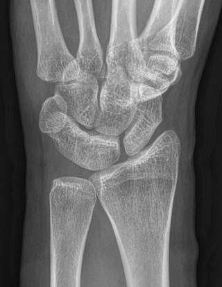
The carpal bones are the eight small bones that make up the wrist (carpus) that connects the hand to the forearm. The terms "carpus" and "carpal" are derived from the Latin carpus and the Greek καρπός (karpós), meaning "wrist". In human anatomy, the main role of the carpal bones is to articulate with the radial and ulnar heads to form a highly mobile condyloid joint, to provide attachments for thenar and hypothenar muscles, and to form part of the rigid carpal tunnel which allows the median nerve and tendons of the anterior forearm muscles to be transmitted to the hand and fingers.

In human anatomy, the wrist is variously defined as (1) the carpus or carpal bones, the complex of eight bones forming the proximal skeletal segment of the hand; (2) the wrist joint or radiocarpal joint, the joint between the radius and the carpus and; (3) the anatomical region surrounding the carpus including the distal parts of the bones of the forearm and the proximal parts of the metacarpus or five metacarpal bones and the series of joints between these bones, thus referred to as wrist joints. This region also includes the carpal tunnel, the anatomical snuff box, bracelet lines, the flexor retinaculum, and the extensor retinaculum.

The ankle, the talocrural region or the jumping bone (informal) is the area where the foot and the leg meet. The ankle includes three joints: the ankle joint proper or talocrural joint, the subtalar joint, and the inferior tibiofibular joint. The movements produced at this joint are dorsiflexion and plantarflexion of the foot. In common usage, the term ankle refers exclusively to the ankle region. In medical terminology, "ankle" can refer broadly to the region or specifically to the talocrural joint.

The scaphoid bone is one of the carpal bones of the wrist. It is situated between the hand and forearm on the thumb side of the wrist. It forms the radial border of the carpal tunnel. The scaphoid bone is the largest bone of the proximal row of wrist bones, its long axis being from above downward, lateralward, and forward. It is approximately the size and shape of a medium cashew nut.

The capitate bone is a bone in the human wrist found in the center of the carpal bone region, located at the distal end of the radius and ulna bones. It articulates with the third metacarpal bone and forms the third carpometacarpal joint. The capitate bone is the largest of the carpal bones in the human hand. It presents, above, a rounded portion or head, which is received into the concavity formed by the scaphoid and lunate bones; a constricted portion or neck; and below this, the body. The bone is also found in many other mammals, and is homologous with the "third distal carpal" of reptiles and amphibians.

The lunate bone is a carpal bone in the human hand. It is distinguished by its deep concavity and crescentic outline. It is situated in the center of the proximal row carpal bones, which lie between the ulna and radius and the hand. The lunate carpal bone is situated between the lateral scaphoid bone and medial triquetral bone.

The radius or radial bone is one of the two large bones of the forearm, the other being the ulna. It extends from the lateral side of the elbow to the thumb side of the wrist and runs parallel to the ulna. The ulna is longer than the radius, but the radius is thicker. The radius is a long bone, prism-shaped and slightly curved longitudinally.

Cleidocranial dysostosis (CCD), also called cleidocranial dysplasia, is a birth defect that mostly affects the bones and teeth. The collarbones are typically either poorly developed or absent, which allows the shoulders to be brought close together. The front of the skull often does not close until later, and those affected are often shorter than average. Other symptoms may include a prominent forehead, wide set eyes, abnormal teeth, and a flat nose. Symptoms vary among people; however, cognitive function is typically unaffected.
The Brunelli Procedure is a surgical procedure that can be used to correct instability in the wrist. Instability in the wrist can be caused by a torn Scapholunate ligament. The Brunelli Procedure does not fix the torn ligament. A hole is drilled through the Scaphoid bone and a part of a tendon taken from the patient is put through this hole and attached to the nearby bones. The procedure usually results in reduced movement of the wrist. Instability in the wrist can, over time, lead to wrist osteoarthritis.

The midcarpal joint is formed by the scaphoid, lunate, and triquetral bones in the proximal row, and the trapezium, trapezoid, capitate, and hamate bones in the distal row. The distal pole of the scaphoid articulates with two trapezial bones as a gliding type of joint. The proximal end of the scaphoid combines with the lunate and triquetrum to form a deep concavity that articulates with the convexity of the combined capitate and hamate in a form of diarthrodial, almost condyloid joint.
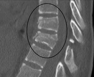
A Chance fracture is a type of vertebral fracture that results from excessive flexion of the spine. Symptoms may include abdominal bruising, or less commonly paralysis of the legs. In around half of cases there is an associated abdominal injury such as a splenic rupture, small bowel injury, pancreatic injury, or mesenteric tear. Injury to the bowel may not be apparent on the first day.

The scapholunate ligament is a ligament of the wrist.
A syndesmophyte is a bony growth originating inside a ligament, commonly seen in the ligaments of the spine, specifically the ligaments in the intervertebral joints leading to fusion of vertebrae. Syndesmophytes are pathologically similar to osteophytes. Ankylosing spondylitis patients are particularly prone to developing syndesmophytes. They are also commonly seen in patients who have had back surgery or other chronic stresses on the ligaments of their spine. Syndesmophytes indicate spine degeneration, similar to osteophytes of spine; however, they bridge across the joint as compared to osteophytes which are non-bridging.

Wrist pain or open wrist is a syndrome inhibiting use of a hand due to pain in anatomical structures of the wrist. It most commonly results from an injury to a ligament. The pain may be sharp from a traumatic injury or from chronic repetitive wrist activities.

Wrist osteoarthritis is gradual loss of articular cartilage and hypertrophic bone changes (osteophytes). While in many joints this is part of normal aging (senescence), in the wrist osteoarthritis usually occurs over years to decades after scapholunate interosseous ligament rupture or an unhealed fracture of the scaphoid. Characteristic symptoms including pain, deformity and stiffness. Pain intensity and incapability are notably variable and do not correspond with arthritis severity on radiographs.

Dorsal intercalated segment instability (DISI) is a deformity of the wrist where the lunate bone angulates to the dorsal side of the hand.

Scapholunate advanced collapse is a type of wrist osteoarthritis. SLAC wrist is the most common type of post-traumatic wrist osteoarthritis and is often the result of an undiagnosed or untreated scapholunate ligament rupture. The condition follows a predictable pattern of development, which was first described by H. Kirk Watson, M.D. and Frederick L. Ballet, M.D. in 1984. Diagnosis of SLAC wrist is made using wrist x-rays, but the diagnosis may be aided using certain provocative tests. Management and treatment of SLAC wrist depends on the stage at the time of diagnosis but includes both non-surgical and surgical options.
Wilkinson's syndrome is a radiographic term which describes a unilaterally enlarged pedicle opposite a contralateral pars defect. The enlarged pedicle may due to stress hypertrophy, and changes may extend into the adjacent lamina and transverse processes.
Srb's anomaly is the clinical condition describing synostosis, or fusion, between the first and second ribs. It may be either a partial or complete fusion between the two ribs to create an entirely indistinguishable new rib.
Carpal coalition is the abnormal fusion of two or more carpal bones when they fail to segment during intrauterine development. First described by Eduard Sandifort in 1779, carpal coalitions are often an isolated issue which connect two carpal bones in the same row of the wrist. These issues are congenital and occur at various rates throughout the population.
