
Human teeth function to mechanically break down items of food by cutting and crushing them in preparation for swallowing and digesting. As such, they are considered part of the human digestive system. Humans have four types of teeth: incisors, canines, premolars, and molars, which each have a specific function. The incisors cut the food, the canines tear the food and the molars and premolars crush the food. The roots of teeth are embedded in the maxilla or the mandible and are covered by gums. Teeth are made of multiple tissues of varying density and hardness.

Cementum is a specialized calcified substance covering the root of a tooth. The cementum is the part of the periodontium that attaches the teeth to the alveolar bone by anchoring the periodontal ligament.

Tooth enamel is one of the four major tissues that make up the tooth in humans and many animals, including some species of fish. It makes up the normally visible part of the tooth, covering the crown. The other major tissues are dentin, cementum, and dental pulp. It is a very hard, white to off-white, highly mineralised substance that acts as a barrier to protect the tooth but can become susceptible to degradation, especially by acids from food and drink. In rare circumstances enamel fails to form, leaving the underlying dentin exposed on the surface.

Dentin or dentine is a calcified tissue of the body and, along with enamel, cementum, and pulp, is one of the four major components of teeth. It is usually covered by enamel on the crown and cementum on the root and surrounds the entire pulp. By volume, 45% of dentin consists of the mineral hydroxyapatite, 33% is organic material, and 22% is water. Yellow in appearance, it greatly affects the color of a tooth due to the translucency of enamel. Dentin, which is less mineralized and less brittle than enamel, is necessary for the support of enamel. Dentin rates approximately 3 on the Mohs scale of mineral hardness. There are two main characteristics which distinguish dentin from enamel: firstly, dentin forms throughout life; secondly, dentin is sensitive and can become hypersensitive to changes in temperature due to the sensory function of odontoblasts, especially when enamel recedes and dentin channels become exposed.

Ameloblasts are cells present only during tooth development that deposit tooth enamel, which is the hard outermost layer of the tooth forming the surface of the crown.

The striae of Retzius are incremental growth lines or bands seen in tooth enamel. They represent the incremental pattern of enamel, the successive apposition of different layers of enamel during crown formation.
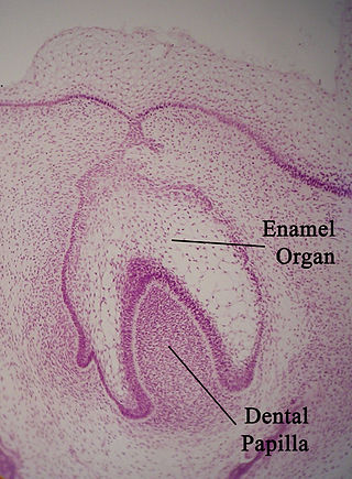
The enamel organ, also known as the dental organ, is a cellular aggregation seen in a developing tooth and it lies above the dental papilla. The enamel organ which is differentiated from the primitive oral epithelium lining the stomodeum. The enamel organ is responsible for the formation of enamel, initiation of dentine formation, establishment of the shape of a tooth's crown, and establishment of the dentoenamel junction.
Interrod enamel is histologically identified on microscopic views of tooth enamel. Because interrod enamel is located around enamel rods, the areas of interrod enamel enhances the "keyhole" appearance of enamel rods by acting as its border. The location where the two areas of enamel meet is known as the rod sheath.

Tooth development or odontogenesis is the complex process by which teeth form from embryonic cells, grow, and erupt into the mouth. For human teeth to have a healthy oral environment, all parts of the tooth must develop during appropriate stages of fetal development. Primary (baby) teeth start to form between the sixth and eighth week of prenatal development, and permanent teeth begin to form in the twentieth week. If teeth do not start to develop at or near these times, they will not develop at all, resulting in hypodontia or anodontia.
Amelogenesis is the formation of enamel on teeth and begins when the crown is forming during the advanced bell stage of tooth development after dentinogenesis forms a first layer of dentin. Dentin must be present for enamel to be formed. Ameloblasts must also be present for dentinogenesis to continue.

In embryology and prenatal development, the dental papilla is a condensation of ectomesenchymal cells called odontoblasts, seen in histologic sections of a developing tooth. It lies below a cellular aggregation known as the enamel organ. The dental papilla appears after 8–10 weeks intra uteral life. The dental papilla gives rise to the dentin and pulp of a tooth.
In vertebrates, an odontoblast is a cell of neural crest origin that is part of the outer surface of the dental pulp, and whose biological function is dentinogenesis, which is the formation of dentin, the substance beneath the tooth enamel on the crown and the cementum on the root.

The dental follicle, also known as dental sac, is made up of mesenchymal cells and fibres surrounding the enamel organ and dental papilla of a developing tooth. It is a vascular fibrous sac containing the developing tooth and its odontogenic organ. The dental follicle (DF) differentiates into the periodontal ligament. In addition, it may be the precursor of other cells of the periodontium, including osteoblasts, cementoblasts and fibroblasts. They develop into the alveolar bone, the cementum with Sharpey's fibers and the periodontal ligament fibers respectively. Similar to dental papilla, the dental follicle provides nutrition to the enamel organ and dental papilla and also have an extremely rich blood supply.

The inner enamel epithelium, also known as the internal enamel epithelium, is a layer of columnar cells located on the rim nearest the dental papilla of the enamel organ in a developing tooth. This layer is first seen during the cap stage, in which these inner enamel epithelium cells are pre-ameloblast cells. These will differentiate into Ameloblasts which are responsible for secretion of enamel during tooth development.
The reduced enamel epithelium, sometimes called reduced dental epithelium, overlies a developing tooth and is formed by two layers: a layer of ameloblast cells and the adjacent layer of cuboidal cells from the dental lamina. As the cells of the reduced enamel epithelium degenerate, the tooth is revealed progressively with its eruption into the mouth. The degeneration of reduced enamel epithelium also mediates the initial epithelial attachment to the tooth, which is called the junctional epithelium.
An odontoblast process is an extension of a cell called an odontoblast, which forms dentin in a tooth. The odontoblast process is located in dentinal tubules. It forms during dentinogenesis and results from a part of the odontoblast staying in its location as the main body of the odontoblast moves toward the center of the tooth's pulp. The odontoblast process causes the secretion of hydroxyapatite crystals and mineralization of the matrix secreted by the odontoblasts.
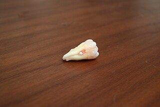
Enamel pearls are developmental variations of teeth that present as beads or nodules of enamel in places where they are not normally observed.
Dental anatomy is a field of anatomy dedicated to the study of human tooth structures. The development, appearance, and classification of teeth fall within its purview. Tooth formation begins before birth, and the teeth's eventual morphology is dictated during this time. Dental anatomy is also a taxonomical science: it is concerned with the naming of teeth and the structures of which they are made, this information serving a practical purpose in dental treatment.
The junctional epithelium (JE) is that epithelium which lies at, and in health also defines, the base of the gingival sulcus. The probing depth of the gingival sulcus is measured by a calibrated periodontal probe. In a healthy-case scenario, the probe is gently inserted, slides by the sulcular epithelium (SE), and is stopped by the epithelial attachment (EA). However, the probing depth of the gingival sulcus may be considerably different from the true histological gingival sulcus depth.
An enamel prism, or enamel rod, is the basic unit of tooth enamel. Measuring 3-6 μm in diameter in primates, enamel prism are tightly packed hydroxyapatite crystals structures. The hydroxyapatite crystals are hexagonal in shape, providing rigidity to the prism and strengthening the enamel. In cross-section, it is best compared to a complex “keyhole” or a “fish-like” shape. The head, which is called the prism core, is oriented toward the tooth’s crown; The tail, which is called the prism sheath, is oriented toward the tooth cervical margin[1][2]. The prism core has tightly packed hydroxyapatite crystals. On the other hand, the prism sheath has its crystals less tightly packed and has more space for organic components. These prism structures can usually be visualised within ground sections and/or with the use of a scanning electron microscope on enamel that has been acid etched[3].