The trabecular bone score is a measure of bone texture correlated with bone microarchitecture and a marker for the risk of osteoporosis. Introduced in 2008,[1] its main projected use is alongside measures of bone density in better predicting fracture risk in people with metabolic bone problems.
To diagnose osteoporosis, despite the inclusion of bone mineral density (BMD), biological markers and clinical factors of fracture risk, many not detected patients are at risk and many fractures are not explained. Bone mineral density is an assessment of the quantity of bone. It does not provide information on bone quality, another important parameter to describe the bone. In addition, clinical risk factors for fracture are at best an indirect assessment of the bone quality. One way to describe the quality of the bone is to assess its microarchitecture. Bone microarchitecture is related to the mechanical strength of bone and hence its greater or lesser risk of fracture. Indeed, for the same quantity of bone, different mechanically resistant bone structures may exist (few large trabeculae or numerous thin trabeculae that are mechanically stronger). Actually, bone loss is often accompanied by a deterioration of bone architecture, resulted in a decreased number of trabeculae, increased inter-trabecular distances, and a loss connectivity of the trabecular meshwork. Moreover, reduction of cortical bone thickness and increased porosity accompany trabecular bone loss, and in particular promote the fragility of the femoral neck. Osteoporotic bone is called "porous".[citation needed]
Technical
Relative Risk of TBS expressed by standard deviation and compared with relative risks of major clinical risk factors for fracture
The trabecular bone score is a textural parameter that can be applied to DEXA, which quantifies the local variations in gray level. TBS is derived from the evaluation of the experimental variogram, obtained from the grayscale DEXA.[citation needed]
It was found that TBS is a reflection of the structural condition of the bone microarchitecture. TBS is strongly correlated with the number of trabeculae and their connectivity and negatively with the space between trabeculae.[2][3] That is to say that a high TBS value means that microarchitecture bone is dense, well connected with little spaces between trabeculaes. Conversely, a low TBS value means that the microarchitecture of bone is incomplete and poorly connected with wide spaces between trabeculae.[4]
in combination with BMD, increase the number of patients at risk (correctly) identified;[5][6][7]
improvement the management of patients with secondary osteoporosis;[8]
follow-up of the evolution of microarchitecture of a patient over time;[4]
monitoring of the effect of anti-resorptive or anabolic.[citation needed]
All these studies have shown that TBS can be used as a clinical risk factor for osteoporotic fracture since it is reversible (with or without treatment), quantitative and independent of BMD. It should therefore be used as such in the same way that taking corticosteroids, rheumatoid arthritis or prevalent fracture after age 50.[citation needed]
The FRAX calculator has an option to include TBS for a TBS adjusted FRAX risk score. The calculated probabilities of fracture have been shown to be more accurate when computed including TBS.[9]
As TBS relies on measurement of soft tissue, it is considered unreliable in individuals with a BMI over 37,[10] or with extremely high waist circumference.[11]
↑ Pothuaud, Laurent; Carceller, Pascal; Hans, Didier (2008). "Correlations between grey-level variations in 2D projection images (TBS) and 3D microarchitecture: Applications in the study of human trabecular bone microarchitecture". Bone. 42 (4): 775–87. doi:10.1016/j.bone.2007.11.018. PMID18234577.
↑ Hans, Didier; Barthe, Nicole; Boutroy, Stephanie; Pothuaud, Laurent; Winzenrieth, Renaud; Krieg, Marc-Antoine (2011). "Correlations Between Trabecular Bone Score, Measured Using Anteroposterior Dual-Energy X-Ray Absorptiometry Acquisition, and 3-Dimensional Parameters of Bone Microarchitecture: An Experimental Study on Human Cadaver Vertebrae". Journal of Clinical Densitometry. 14 (3): 302–12. doi:10.1016/j.jocd.2011.05.005. PMID21724435.
↑ Piveteau, Teddy; Winzenrieth, Renaud; Hans, Didier (2011). "Assessment of correlations between 3D μCT microarchitecture parameters and TBS: Effects of resolution and correlation with TBS DXA measurements". Journal of Clinical Densitometry. 14 (2): 169. doi:10.1016/j.jocd.2011.02.056.
1 2 Pothuaud, Laurent; Barthe, Nicole; Krieg, Marc-Antoine; Mehsen, Nadia; Carceller, Pascal; Hans, Didier (2009). "Evaluation of the Potential Use of Trabecular Bone Score to Complement Bone Mineral Density in the Diagnosis of Osteoporosis: A Preliminary spine BMD–Matched, Case-Control Study". Journal of Clinical Densitometry. 12 (2): 170–6. doi:10.1016/j.jocd.2008.11.006. PMID19181553.
↑ Winzenrieth, Renaud; Dufour, Rémy; Pothuaud, Laurent; Hans, Didier (2009). "A Retrospective Case–Control Study Assessing the Role of Trabecular Bone Score in Postmenopausal Caucasian Women with Osteopenia: Analyzing the Odds of Vertebral Fracture". Calcified Tissue International. 86 (2): 104–9. doi:10.1007/s00223-009-9322-y. PMID19998029. S2CID24345190.
↑ Rio, L. M.; Winzenrieth, R.; Cormier, C.; Gregorio, S. (2012). "Is bone microarchitecture status of the lumbar spine assessed by TBS related to femoral neck fracture? A Spanish case–control study". Osteoporosis International. 24 (3): 991–8. doi:10.1007/s00198-012-2008-8. PMID22581295. S2CID5844264.
Osteoporosis is a systemic skeletal disorder characterized by low bone mass, micro-architectural deterioration of bone tissue leading to bone sterility, and consequent increase in fracture risk. It is the most common reason for a broken bone among the elderly. Bones that commonly break include the vertebrae in the spine, the bones of the forearm, the wrist, and the hip. Until a broken bone occurs there are typically no symptoms. Bones may weaken to such a degree that a break may occur with minor stress or spontaneously. After the broken bone heals, the person may have chronic pain and a decreased ability to carry out normal activities.
Dual-energy X-ray absorptiometry is a means of measuring bone mineral density (BMD) using spectral imaging. Two X-ray beams, with different energy levels, are aimed at the patient's bones. When soft tissue absorption is subtracted out, the bone mineral density (BMD) can be determined from the absorption of each beam by bone. Dual-energy X-ray absorptiometry is the most widely used and most thoroughly studied bone density measurement technology.
Teriparatide, sold under the brand name Forteo, is a form of parathyroid hormone (PTH) consisting of the first (N-terminus) 34 amino acids, which is the bioactive portion of the hormone. It is an effective anabolic agent used in the treatment of some forms of osteoporosis. Teriparatide is a recombinant human parathyroid hormone analog. It has an identical sequence to the 34 N-terminal amino acids of the 84-amino acid human parathyroid hormone.
A trabecula is a small, often microscopic, tissue element in the form of a small beam, strut or rod that supports or anchors a framework of parts within a body or organ. A trabecula generally has a mechanical function, and is usually composed of dense collagenous tissue. It can be composed of other material such as muscle and bone. In the heart, muscles form trabeculae carneae and septomarginal trabeculae. Cancellous bone is formed from groupings of trabeculated bone tissue.
Osteopenia, known as "low bone mass" or "low bone density", is a condition in which bone mineral density is low. Because their bones are weaker, people with osteopenia may have a higher risk of fractures, and some people may go on to develop osteoporosis. In 2010, 43 million older adults in the US had osteopenia. Unlike osteoporosis, osteopenia does not usually cause symptoms, and losing bone density in itself does not cause pain.
Bone density, or bone mineral density, is the amount of bone mineral in bone tissue. The concept is of mass of mineral per volume of bone, although clinically it is measured by proxy according to optical density per square centimetre of bone surface upon imaging. Bone density measurement is used in clinical medicine as an indirect indicator of osteoporosis and fracture risk. It is measured by a procedure called densitometry, often performed in the radiology or nuclear medicine departments of hospitals or clinics. The measurement is painless and non-invasive and involves low radiation exposure. Measurements are most commonly made over the lumbar spine and over the upper part of the hip. The forearm may be scanned if the hip and lumbar spine are not accessible.
Quantitative computed tomography (QCT) is a medical technique that measures bone mineral density (BMD) using a standard X-ray Computed Tomography (CT) scanner with a calibration standard to convert Hounsfield Units (HU) of the CT image to bone mineral density values. Quantitative CT scans are primarily used to evaluate bone mineral density at the lumbar spine and hip.
Strontium ranelate, a strontium(II) salt of ranelic acid, is a medication for osteoporosis marketed as Protelos or Protos by Servier. Studies indicate it can also slow the course of osteoarthritis of the knee. The drug is unusual in that it both increases deposition of new bone by osteoblasts and reduces the resorption of bone by osteoclasts. It is therefore promoted as a "dual action bone agent" (DABA).
Senile osteoporosis has been recently recognized as a geriatric syndrome with a particular pathophysiology. There are different classification of osteoporosis: primary, in which bone loss is a result of aging and secondary, in which bone loss occurs from various clinical and lifestyle factors. Primary, or involuntary osteoporosis, can further be classified into Type I or Type II. Type I refers to postmenopausal osteoporosis and is caused by the deficiency of estrogen. While senile osteoporosis is categorized as an involuntary, Type II, and primary osteoporosis, which affects both men and women over the age of 70 years. It is accompanied by vitamin D deficiency, body's failure to absorb calcium, and increased parathyroid hormone.
Steroid-induced osteoporosis is osteoporosis arising from the use of glucocorticoids analogous to Cushing's syndrome but involving mainly the axial skeleton. The synthetic glucocorticoid prescription drug prednisone is a main candidate after prolonged intake. Bisphosphonates are beneficial in reducing the risk of vertebral fractures. Some professional guidelines recommend prophylactic calcium and vitamin D supplementation in patients who take the equivalent of more than 30 mg hydrocortisone, especially when this is in excess of three months. The use of thiazide diuretics, and gonadal hormone replacement has also been recommended, with the use of calcitonin, bisphosphonates, sodium fluoride or anabolic steroids also suggested in refractory cases. Alternate day use may not prevent this complication.
FRAX is a diagnostic tool used to evaluate the 10-year probability of bone fracture risk. It was developed by the University of Sheffield. FRAX integrates clinical risk factors and bone mineral density at the femoral neck to calculate the 10-year probability of hip fracture and the 10-year probability of a major osteoporotic fracture. The models used to develop the FRAX diagnostic tool were derived from studying patient populations in North America, Europe, Latin America, Asia and Australia.
Abaloparatide, sold under the brand name Tymlos among others, is a parathyroid hormone-related protein (PTHrP) analog medication used to treat osteoporosis. It is an anabolic agent.
Eldecalcitol is an analog of calcitriol, the active form of vitamin D.
The human skeletal system is a complex organ in constant equilibrium with the rest of the body. In addition to support and structure of the body, bone is the major reservoir for many minerals and compounds essential for maintaining a healthy pH balance. The deterioration of the body with age renders the elderly particularly susceptible to and affected by poor bone health. Illnesses like osteoporosis, characterized by weakening of the bone's structural matrix, increases the risk of hip-fractures and other life-changing secondary symptoms. In 2010, over 258,000 people aged 65 and older were admitted to the hospital for hip fractures. Incidence of hip fractures is expected to rise by 12% in America, with a projected 289,000 admissions in the year 2030. Other sources estimate up to 1.5 million Americans will have an osteoporotic-related fracture each year. The cost of treating these people is also enormous, in 1991 Medicare spent an estimated $2.9 billion for treatment and out-patient care of hip fractures, this number can only be expected to rise.
Dual X-ray absorptiometry and laser technique (DXL) in the area of bone density studies for osteoporosis assessment is an improvement to the DXA Technique, adding an exact laser measurement of the thickness of the region scanned. The addition of object thickness adds a third input to the two x-ray energies used by DXA, better solving the equation for bone and excluding more efficiently these soft tissues components.
Single photon absorptiometry is a measuring method for bone density invented by John R. Cameron and James A. Sorenson in 1963.
A bone biopsy is a procedure in which a small bone sample is removed from the outer layers of bone for examination, unlike a bone marrow biopsy, which involves the innermost part of the bone. The bone biopsy sample retains the architecture of bone when seen using histopathological examination slide.
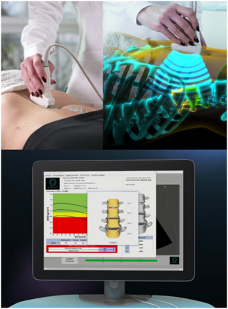
Radiofrequency Echographic Multi Spectrometry (REMS) is a non-ionizing technology for osteoporosis diagnosis and for fracture risk assessment. REMS processes the raw, unfiltered ultrasound signals acquired during an echographic scan of the axial sites, femur and spine. The analysis is performed in the frequency domain. Bone mineral density (BMD) is estimated by comparing the results against reference models.
International Society for Clinical Densitometry (ISCD) is a professional community of physicians with more than 2,700 individual members from over 25 countries. The society advocated an advance in the assessment of musculoskeletal health through education, certification and facility accreditation. The association is established in 1993 and headquartered in Middletown, Connecticut, United States.
Mary Larsen Bouxsein is an American biomechanical engineer and an orthopedic researcher. She is the president of the American Society for Bone and Mineral Research, director of the Centre of Advanced Orthopaedic Studies at Beth Israel Deaconess Medical Center, professor at the department of Orthopedic Surgery at Harvard Medical School. She is known for her work on bone density and the use of imaging to define the factors leading to bone fractures.
This page is based on this Wikipedia article Text is available under the CC BY-SA 4.0 license; additional terms may apply. Images, videos and audio are available under their respective licenses.