Related Research Articles

Temporomandibular joint dysfunction is an umbrella term covering pain and dysfunction of the muscles of mastication and the temporomandibular joints. The most important feature is pain, followed by restricted mandibular movement, and noises from the temporomandibular joints (TMJ) during jaw movement. Although TMD is not life-threatening, it can be detrimental to quality of life; this is because the symptoms can become chronic and difficult to manage.

Jaundice, also known as icterus, is a yellowish or greenish pigmentation of the skin and sclera due to high bilirubin levels. Jaundice in adults is typically a sign indicating the presence of underlying diseases involving abnormal heme metabolism, liver dysfunction, or biliary-tract obstruction. The prevalence of jaundice in adults is rare, while jaundice in babies is common, with an estimated 80% affected during their first week of life. The most commonly associated symptoms of jaundice are itchiness, pale feces, and dark urine.
A syndrome is a set of medical signs and symptoms which are correlated with each other and often associated with a particular disease or disorder. The word derives from the Greek σύνδρομον, meaning "concurrence". When a syndrome is paired with a definite cause this becomes a disease. In some instances, a syndrome is so closely linked with a pathogenesis or cause that the words syndrome, disease, and disorder end up being used interchangeably for them. This substitution of terminology often confuses the reality and meaning of medical diagnoses. This is especially true of inherited syndromes. About one third of all phenotypes that are listed in OMIM are described as dysmorphic, which usually refers to the facial gestalt. For example, Down syndrome, Wolf–Hirschhorn syndrome, and Andersen–Tawil syndrome are disorders with known pathogeneses, so each is more than just a set of signs and symptoms, despite the syndrome nomenclature. In other instances, a syndrome is not specific to only one disease. For example, toxic shock syndrome can be caused by various toxins; another medical syndrome named as premotor syndrome can be caused by various brain lesions; and premenstrual syndrome is not a disease but simply a set of symptoms.

Signs and symptoms are diagnostic indications of an illness, injury, or condition.

Myoclonus is a brief, involuntary, irregular twitching of a muscle, a joint, or a group of muscles, different from clonus, which is rhythmic or regular. Myoclonus describes a medical sign and, generally, is not a diagnosis of a disease. It belongs to the hyperkinetic movement disorders, among tremor and chorea for example. These myoclonic twitches, jerks, or seizures are usually caused by sudden muscle contractions or brief lapses of contraction. The most common circumstance under which they occur is while falling asleep. Myoclonic jerks occur in healthy people and are experienced occasionally by everyone. However, when they appear with more persistence and become more widespread they can be a sign of various neurological disorders. Hiccups are a kind of myoclonic jerk specifically affecting the diaphragm. When a spasm is caused by another person it is known as a provoked spasm. Shuddering attacks in babies fall in this category.
Hypoparathyroidism is decreased function of the parathyroid glands with underproduction of parathyroid hormone (PTH). This can lead to low levels of calcium in the blood, often causing cramping and twitching of muscles or tetany, and several other symptoms. It is a very rare disease. The condition can be inherited, but it is also encountered after thyroid or parathyroid gland surgery, and it can be caused by immune system-related damage as well as a number of rarer causes. The diagnosis is made with blood tests, and other investigations such as genetic testing depending on the results. The primary treatment of hypoparathyroidism is calcium and vitamin D supplementation. Calcium replacement or vitamin D can ameliorate the symptoms but can increase the risk of kidney stones and chronic kidney disease. Additionally, medications such as recombinant human parathyroid hormone or teriparatide may be given by injection to replace the missing hormone.

Primary familial brain calcification (PFBC), also known as familial idiopathic basal ganglia calcification (FIBGC) and Fahr's disease, is a rare, genetically dominant or recessive, inherited neurological disorder characterized by abnormal deposits of calcium in areas of the brain that control movement. Through the use of CT scans, calcifications are seen primarily in the basal ganglia and in other areas such as the cerebral cortex.

Enchondroma is a type of benign bone tumor belonging to the group of cartilage tumors. There may be no symptoms, or it may present typically in the short tubular bones of the hands with a swelling, pain or pathological fracture.

Calcification is the accumulation of calcium salts in a body tissue. It normally occurs in the formation of bone, but calcium can be deposited abnormally in soft tissue, causing it to harden. Calcifications may be classified on whether there is mineral balance or not, and the location of the calcification. Calcification may also refer to the processes of normal mineral deposition in biological systems, such as the formation of stromatolites or mollusc shells.

Valvular heart disease is any cardiovascular disease process involving one or more of the four valves of the heart. These conditions occur largely as a consequence of aging, but may also be the result of congenital (inborn) abnormalities or specific disease or physiologic processes including rheumatic heart disease and pregnancy.

Sturge–Weber syndrome, sometimes referred to as encephalotrigeminal angiomatosis, is a rare congenital neurological and skin disorder. It is one of the phakomatoses and is often associated with port-wine stains of the face, glaucoma, seizures, intellectual disability, and ipsilateral leptomeningeal angioma. Sturge–Weber syndrome can be classified into three different types. Type 1 includes facial and leptomeningeal angiomas as well as the possibility of glaucoma or choroidal lesions. Normally, only one side of the brain is affected. This type is the most common. Type 2 involvement includes a facial angioma with a possibility of glaucoma developing. There is no evidence of brain involvement. Symptoms can show at any time beyond the initial diagnosis of the facial angioma. The symptoms can include glaucoma, cerebral blood flow abnormalities and headaches. More research is needed on this type of Sturge–Weber syndrome. Type 3 has leptomeningeal angioma involvement exclusively. The facial angioma is absent and glaucoma rarely occurs. This type is only diagnosed via brain scan.
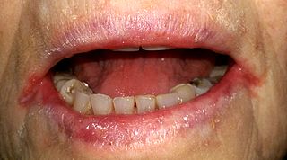
Angular cheilitis (AC) is inflammation of one or both corners of the mouth. Often the corners are red with skin breakdown and crusting. It can also be itchy or painful. The condition can last for days to years. Angular cheilitis is a type of cheilitis.
Pseudohypoparathyroidism is a rare autosomal dominant genetic condition associated primarily with resistance to the parathyroid hormone. Those with the condition have a low serum calcium and high phosphate, but the parathyroid hormone level (PTH) is inappropriately high. Its pathogenesis has been linked to dysfunctional G proteins. Pseudohypoparathyroidism is a very rare disorder, with estimated prevalence between 0.3 and 1.1 cases per 100,000 population depending on geographic location.
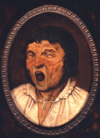
Meige's syndrome is a type of dystonia. It is also known as Brueghel's syndrome and oral facial dystonia. It is actually a combination of two forms of dystonia, blepharospasm and oromandibular dystonia (OMD).
Pseudopseudohypoparathyroidism (PPHP) is an inherited disorder, named for its similarity to pseudohypoparathyroidism in presentation. It is more properly Albright hereditary osteodystrophy, although without resistance of parathyroid hormone (PTH), as frequently seen in that affliction. The term is used to describe a condition where the individual has the phenotypic appearance of pseudohypoparathyroidism type 1a, but has normal labs, including calcium and PTH.

Eagle syndrome is an uncommon condition commonly characterized but not limited to sudden, sharp nerve-like pain in the jaw bone and joint, back of the throat, and base of the tongue, triggered by swallowing, moving the jaw, or turning the neck. First described by American otorhinolaryngologist Watt Weems Eagle in 1937, the condition is caused by an elongated or misshapen styloid process and/or calcification of the stylohyoid ligament, either of which interferes with the functioning of neighboring regions in the body, such as the glossopharyngeal nerve.
Oral and maxillofacial pathology refers to the diseases of the mouth, jaws and related structures such as salivary glands, temporomandibular joints, facial muscles and perioral skin. The mouth is an important organ with many different functions. It is also prone to a variety of medical and dental disorders.

A panoramic radiograph is a panoramic scanning dental X-ray of the upper and lower jaw. It shows a two-dimensional view of a half-circle from ear to ear. Panoramic radiography is a form of focal plane tomography; thus, images of multiple planes are taken to make up the composite panoramic image, where the maxilla and mandible are in the focal trough and the structures that are superficial and deep to the trough are blurred.

Current standards for diagnosing multiple sclerosis (MS) are based on the 2018 revision of McDonald criteria. They rely on MRI detection of demyelinating lesions in the CNS, which are distributed in space (DIS) and in time (DIT). It is also a requirement that any possible known disease that produces demyelinating lesions is ruled out before applying McDonald's criteria.

Ground-glass opacity (GGO) is a finding seen on chest x-ray (radiograph) or computed tomography (CT) imaging of the lungs. It is typically defined as an area of hazy opacification (x-ray) or increased attenuation (CT) due to air displacement by fluid, airway collapse, fibrosis, or a neoplastic process. When a substance other than air fills an area of the lung it increases that area's density. On both x-ray and CT, this appears more grey or hazy as opposed to the normally dark-appearing lungs. Although it can sometimes be seen in normal lungs, common pathologic causes include infections, interstitial lung disease, and pulmonary edema.
References
- ↑ Gunderman RB. Essential radiology. 2nd ed. Thieme: New York.
- ↑ Kaplan, Bernard S.; Meyers, Kevin E. C. (2004). Pediatric nephrology and urology: the requisites in pediatrics. Elsevier Health Sciences. pp. 151–. ISBN 978-0-323-01841-8 . Retrieved 30 July 2011.
- ↑ Ramina, Ricardo; Aguiar, Paulo Henrique Pires; Tatagiba, Marcos (2007-11-29). Samii's Essentials in Neurosurgery. Springer. pp. 85–. ISBN 978-3-540-49249-8 . Retrieved 30 July 2011.
| | This medical sign article is a stub. You can help Wikipedia by expanding it. |