
Apocrine is a term used to classify the mode of secretion of exocrine glands. In apocrine secretion, secretory cells accumulate material at their apical ends, often forming blebs or "snouts", and this material then buds off from the cells, forming extracellular vesicles. The secretory cells therefore lose part of their cytoplasm in the process of secretion.

Hyperplasia, or hypergenesis, is an enlargement of an organ or tissue caused by an increase in the amount of organic tissue that results from cell proliferation. It may lead to the gross enlargement of an organ, and the term is sometimes confused with benign neoplasia or benign tumor.

Metaplasia is the transformation of a cell type to another cell type. The change from one type of cell to another may be part of a normal maturation process, or caused by some sort of abnormal stimulus. In simplistic terms, it is as if the original cells are not robust enough to withstand their environment, so they transform into another cell type better suited to their environment. If the stimulus causing metaplasia is removed or ceases, tissues return to their normal pattern of differentiation. Metaplasia is not synonymous with dysplasia, and is not considered to be an actual cancer. It is also contrasted with heteroplasia, which is the spontaneous abnormal growth of cytologic and histologic elements. Today, metaplastic changes are usually considered to be an early phase of carcinogenesis, specifically for those with a history of cancers or who are known to be susceptible to carcinogenic changes. Metaplastic change is thus often viewed as a premalignant condition that requires immediate intervention, either surgical or medical, lest it lead to cancer via malignant transformation.

A seminoma is a germ cell tumor of the testicle or, more rarely, the mediastinum or other extra-gonadal locations. It is a malignant neoplasm and is one of the most treatable and curable cancers, with a survival rate above 95% if discovered in early stages.

A Hürthle cell is a cell in the thyroid that is often associated with Hashimoto's thyroiditis as well as benign and malignant tumors. This version is a relatively rare form of differentiated thyroid cancer, accounting for only 3-10% of all differentiated thyroid cancers. Oncocytes in the thyroid are often called Hürthle cells. Although the terms oncocyte, oxyphilic cell, and Hürthle cell are used interchangeably, Hürthle cell is used only to indicate cells of thyroid follicular origin.

Hürthle cell neoplasm is a rare tumor of the thyroid, typically seen in women between the ages of 70 and 80 years old. When benign, it is called a Hürthle cell adenoma, and when malignant it is called a Hürthle cell carcinoma. Hürthle cell adenoma is characterized by a mass of benign Hürthle cells. Typically such a mass is removed because it is not easy to predict whether it will transform into the malignant counterpart of Hürthle cell carcinoma, which is a subtype of follicular thyroid cancer.

Microcalcifications are tiny deposits of calcium salts that are too small to be felt but can be detected by imaging.

A ranula is a mucus extravasation cyst involving a sublingual gland and is a type of mucocele found on the floor of the mouth. Ranulae present as a swelling of connective tissue consisting of collected mucin from a ruptured salivary gland caused by local trauma. If small and asymptomatic further treatment may not be needed, otherwise minor oral surgery may be indicated.

Fibrocystic breast changes is a condition of the breasts where there may be pain, breast cysts, and breast masses. The breasts may be described as "lumpy" or "doughy". Symptoms may worsen during certain parts of the menstrual cycle due to hormonal stimulation. These are normal breast changes, not associated with cancer.

A mucinous neoplasm is an abnormal and excessive growth of tissue (neoplasia) with associated mucin. It arises from epithelial cells that line certain internal organs and skin, and produce mucin. A malignant mucinous neoplasm is called a mucinous carcinoma. For example, for ovarian mucinous tumors, approximately 75% are benign, 10% are borderline and 15% are malignant.

High-grade prostatic intraepithelial neoplasia (HGPIN) is an abnormality of prostatic glands and believed to precede the development of prostate adenocarcinoma.

Visual artifacts are anomalies apparent during visual representation as in digital graphics and other forms of imagery, especially photography and microscopy.

An oncocyte is an epithelial cell characterized by an excessive number of mitochondria, resulting in an abundant acidophilic, granular cytoplasm. Oncocytes can be benign or malignant.

Atypical ductal hyperplasia (ADH) is the term used for a benign lesion of the breast that indicates an increased risk of breast cancer.
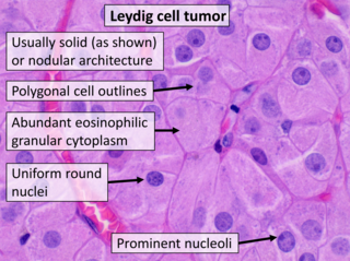
Leydig cell tumour, also Leydig cell tumor, (testicular) interstitial cell tumour and (testicular) interstitial cell tumor, is a member of the sex cord-stromal tumour group of ovarian and testicular cancers. It arises from Leydig cells. While the tumour can occur at any age, it occurs most often in young adults.

A solid pseudopapillary tumour is a low-grade malignant neoplasm of the pancreas of papillary architecture that typically afflicts young women.
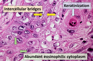
Squamous-cell carcinoma (SCC), also known as epidermoid carcinoma, comprises a number of different types of cancer that begin in squamous cells. These cells form on the surface of the skin, on the lining of hollow organs in the body, and on the lining of the respiratory and digestive tracts.

A fundic gland polyp is a type of polyp, found in the fundus of the stomach. Fundic gland polyps are found in 0.8 to 1.9% of patients who undergo esophagogastroduodenoscopy, and are more common in middle-aged women.

Mucinous carcinoma of the breast is a form of mucinous carcinoma and a breast cancer type. Rare cases of this carcinoma have been diagnosed in men.
Tubular carcinoma is a subtype of invasive ductal carcinoma of the breast. More rarely, tubular carcinomas may arise in the pancreas or kidney. Most tubular carcinomas begin in the milk duct of the breast and spread to healthy tissue around it.

