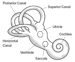
The inner ear is the innermost part of the vertebrate ear. In vertebrates, the inner ear is mainly responsible for sound detection and balance. In mammals, it consists of the bony labyrinth, a hollow cavity in the temporal bone of the skull with a system of passages comprising two main functional parts:
Ménière's disease (MD) is a disease of the inner ear that is characterized by potentially severe and incapacitating episodes of vertigo, tinnitus, hearing loss, and a fullness in the ear. Typically, only one ear is affected initially, but over time, both ears may become involved. Episodes generally last from 20 minutes to a few hours. The time between episodes varies. The hearing loss and ringing in the ears can become constant over time.

The vestibulocochlear nerve or auditory vestibular nerve, also known as the eighth cranial nerve, cranial nerve VIII, or simply CN VIII, is a cranial nerve that transmits sound and equilibrium (balance) information from the inner ear to the brain. Through olivocochlear fibers, it also transmits motor and modulatory information from the superior olivary complex in the brainstem to the cochlea.
The saccule is a bed of sensory cells in the inner ear. It translates head movements into neural impulses for the brain to interpret. The saccule detects linear accelerations and head tilts in the vertical plane. When the head moves vertically, the sensory cells of the saccule are disturbed and the neurons connected to them begin transmitting impulses to the brain. These impulses travel along the vestibular portion of the eighth cranial nerve to the vestibular nuclei in the brainstem.

The vestibular system, in vertebrates, is a sensory system that creates the sense of balance and spatial orientation for the purpose of coordinating movement with balance. Together with the cochlea, a part of the auditory system, it constitutes the labyrinth of the inner ear in most mammals.
Ototoxicity is the property of being toxic to the ear (oto-), specifically the cochlea or auditory nerve and sometimes the vestibular system, for example, as a side effect of a drug. The effects of ototoxicity can be reversible and temporary, or irreversible and permanent. It has been recognized since the 19th century. There are many well-known ototoxic drugs used in clinical situations, and they are prescribed, despite the risk of hearing disorders, for very serious health conditions. Ototoxic drugs include antibiotics, loop diuretics, and platinum-based chemotherapy agents. A number of nonsteroidal anti-inflammatory drugs (NSAIDS) have also been shown to be ototoxic. This can result in sensorineural hearing loss, dysequilibrium, or both. Some environmental and occupational chemicals have also been shown to affect the auditory system and interact with noise.

Labyrinthitis is inflammation of the labyrinth – a maze of fluid-filled channels in the inner ear. Vestibular neuritis is inflammation of the vestibular nerve – the nerve in the inner ear that sends messages related to motion and position to the brain. Both conditions involve inflammation of the inner ear.labyrinths that house the vestibular system, senses changes in the head's position or the head's motion. Inflammation of these inner ear parts results in a sensation of the world spinning and also possible hearing loss or ringing in the ears. It can occur as a single attack, a series of attacks, or a persistent condition that diminishes over three to six weeks. It may be associated with nausea, vomiting, and eye nystagmus.

Sensorineural hearing loss (SNHL) is a type of hearing loss in which the root cause lies in the inner ear or sensory organ or the vestibulocochlear nerve. SNHL accounts for about 90% of reported hearing loss. SNHL is usually permanent and can be mild, moderate, severe, profound, or total. Various other descriptors can be used depending on the shape of the audiogram, such as high frequency, low frequency, U-shaped, notched, peaked, or flat.
Pendred syndrome is a genetic disorder leading to congenital bilateral sensorineural hearing loss and goitre with euthyroid or mild hypothyroidism. There is no specific treatment, other than supportive measures for the hearing loss and thyroid hormone supplementation in case of hypothyroidism. It is named after Vaughan Pendred (1869–1946), the British doctor who first described the condition in an Irish family living in Durham in 1896. It accounts for 7.5% to 15% of all cases of congenital deafness.

Vertigo is a condition where a person has the sensation of movement or of surrounding objects moving when they are not. Often it feels like a spinning or swaying movement. This may be associated with nausea, vomiting, sweating, or difficulties walking. It is typically worse when the head is moved. Vertigo is the most common type of dizziness.
Nonsyndromic deafness is hearing loss that is not associated with other signs and symptoms. In contrast, syndromic deafness involves hearing loss that occurs with abnormalities in other parts of the body. Genetic changes are related to the following types of nonsyndromic deafness.

The ampullary cupula, or cupula, is a structure in the vestibular system, providing the sense of spatial orientation.

The otolithic membrane is a fibrous structure located in the vestibular system of the inner ear. It plays a critical role in the brain's interpretation of equilibrium. The membrane serves to determine if the body or the head is tilted, in addition to the linear acceleration of the body. The linear acceleration could be in the horizontal direction as in a moving car or vertical acceleration such as that felt when an elevator moves up or down.

The crista ampullaris is the sensory organ of rotation. They are found in the ampullae of each of the semicircular canals of the inner ear, meaning that there are three pairs in total. The function of the crista ampullaris is to sense angular acceleration and deceleration.

Wolframin is a protein that in humans is encoded by the WFS1 gene.
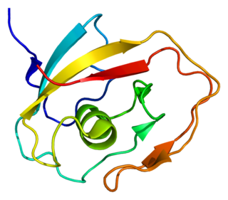
Cochlin is a protein that in humans is encoded by the COCH gene. It is an extracellular matrix (ECM) protein highly abundant in the cochlea and vestibule of the inner ear, constituting the major non-collagen component of the ECM of the inner ear. The protein is highly conserved in human, mouse, and chicken, showing 94% and 79% amino acid identity of human to mouse and chicken sequences, respectively.

Alpha-tectorin is a protein that in humans is encoded by the TECTA gene.
Hearing loss has multiple causes, including ageing, genetics, perinatal problems and acquired causes like noise and disease. For some kinds of hearing loss the cause may be classified as of unknown cause.
CAPOS syndrome is a rare genetic neurological disorder which is characterized by abnormalities of the feet, eyes and brain which affect their normal function. These symptoms occur episodically when a fever-related infection is present within the body.
Inner ear decompression sickness, (IEDCS) or audiovestibular decompression sickness is a medical condition of the inner ear caused by the formation of gas bubbles in the tissues of the inner ear. Generally referred to as a form of decompression sickness, it can also occur at constant pressure due to inert gas counterdiffusion effects. Usually only one side is affected, and the most common symptoms are vertigo with nystagmus, loss of balance, and nausea. First aid is breathing the highest practicable concentration of normobaric oxygen. Definitive treatment is recompression with hyperbaric oxygen therapy. Anti-vertigo and anti-nausea drugs are usually effective at suppressing symptoms, but do not reduce the tissue damage. Hyperbaric oxygen may be effective for reducing oedema and ischaemia even after the most effective period for reducing the injury has passed. IEDCS is often associated with relatively deep diving, relatively long periods of decompression obligation, and breathing gas switches involving changes in inert gas type and concentration. Onset may occur during the dive or afterwards. The symptoms are similar to those caused by some other diving injuries and differential diagnosis can be complicated and uncertain if several possible causes for the symptoms coexist. IEDCS is a relatively uncommonn manifestation of decompression sickness, occurring in about 5 to 6% of cases. The most commonly used decompression models do not appear to accurately model IEDCS, and therefore dive computers based on those models alone are not particularly effective at predicting it, or avoiding it. There are a few rule of thumb methods which have been reasonably effective for avoidance, but they have not been tested under controlled conditions.