Related Research Articles

Microscopy is the technical field of using microscopes to view objects and areas of objects that cannot be seen with the naked eye. There are three well-known branches of microscopy: optical, electron, and scanning probe microscopy, along with the emerging field of X-ray microscopy.

Video is an electronic medium for the recording, copying, playback, broadcasting, and display of moving visual media. Video was first developed for mechanical television systems, which were quickly replaced by cathode ray tube (CRT) systems which were later replaced by flat panel displays of several types.

The optical microscope, also referred to as a light microscope, is a type of microscope that commonly uses visible light and a system of lenses to generate magnified images of small objects. Optical microscopes are the oldest design of microscope and were possibly invented in their present compound form in the 17th century. Basic optical microscopes can be very simple, although many complex designs aim to improve resolution and sample contrast.

Medical ultrasound is a diagnostic imaging technique, or therapeutic application of ultrasound. It is used to create an image of internal body structures such as tendons, muscles, joints, blood vessels, and internal organs. Its aim is often to find a source of a disease or to exclude pathology. The practice of examining pregnant women using ultrasound is called obstetric ultrasound, and was an early development and application of clinical ultrasonography.

A panorama is any wide-angle view or representation of a physical space, whether in painting, drawing, photography, film, seismic images or a three-dimensional model. The word was originally coined in the 18th century by the English painter Robert Barker to describe his panoramic paintings of Edinburgh and London. The motion-picture term panning is derived from panorama.

Stereoscopy is a film (non-digital) technique for creating or enhancing the illusion of depth in an image by means of stereopsis for binocular vision. The word stereoscopy derives from Greek στερεός (stereos), meaning 'firm, solid', and σκοπέω (skopeō), meaning 'to look, to see'. Any stereoscopic image is called a stereogram. Originally, stereogram referred to a pair of stereo images which could be viewed using a stereoscope.

An image scanner—often abbreviated to just scanner, is a device that optically scans images, printed text, handwriting or an object and converts it to a digital image. Commonly used in offices are variations of the desktop flatbed scanner where the document is placed on a glass window for scanning. Hand-held scanners, where the device is moved by hand, have evolved from text scanning "wands" to 3D scanners used for industrial design, reverse engineering, test and measurement, orthotics, gaming and other applications. Mechanically driven scanners that move the document are typically used for large-format documents, where a flatbed design would be impractical.
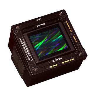
A digital camera back is a device that attaches to the back of a camera in place of the traditional negative film holder and contains an electronic image sensor. This lets cameras that were designed to use film take digital photographs. These camera backs are generally expensive by consumer standards and are primarily built to be attached on medium- and large-format cameras used by professional photographers.

3D scanning is the process of analyzing a real-world object or environment to collect data on its shape and possibly its appearance. The collected data can then be used to construct digital 3D models.

Scanography, more commonly referred to as scanner photography, is the process of capturing digitized images of objects for the purpose of creating printable art using a flatbed "photo" scanner with a CCD array capturing device. Fine art scanography differs from traditional document scanning by using atypical objects, often three-dimensional, as well as from photography, due to the nature of the scanner's operation.
Virtual microscopy is a method of posting microscope images on, and transmitting them over, computer networks. This allows independent viewing of images by large numbers of people in diverse locations. It involves a synthesis of microscopy technologies and digital technologies. The use of virtual microscopes can transform traditional teaching methods by removing the reliance on physical space, equipment, and specimens to a model that is solely dependent upon computer-internet access. This increases the convenience of accessing the slide sets and making the slides available to a broader audience. Digitized slides can have a high resolution and are resistant to being damaged or broken over time.
The Northern Ireland Virtual Tissue Archive (NIVTA) is an initiative intending to establish a European wide network for virtual tissue archiving, aimed at supporting clinical trials, biomarker research, tissue microarray analysis and virtual slide-based education.
Digital pathology is a sub-field of pathology that focuses on data management based on information generated from digitized specimen slides. Through the use of computer-based technology, digital pathology utilizes virtual microscopy. Glass slides are converted into digital slides that can be viewed, managed, shared and analyzed on a computer monitor. With the practice of Whole-Slide Imaging (WSI), which is another name for virtual microscopy, the field of digital pathology is growing and has applications in diagnostic medicine, with the goal of achieving efficient and cheaper diagnoses, prognosis, and prediction of diseases.
Teledermatology is a subspecialty in the medical field of dermatology and probably one of the most common applications of telemedicine and e-health. In teledermatology, telecommunication technologies are used to exchange medical information over a distance using audio, visual and data communication. Applications comprise health care management such as diagnoses, consultation and treatment as well as (continuous) education.
Telepathology is the practice of pathology at a distance. It uses telecommunications technology to facilitate the transfer of image-rich pathology data between distant locations for the purposes of diagnosis, education, and research. Performance of telepathology requires that a pathologist selects the video images for analysis and the rendering of diagnoses. The use of "television microscopy", the forerunner of telepathology, did not require that a pathologist have physical or virtual "hands-on" involvement in the selection of microscopic fields-of-view for analysis and diagnosis.

A panoramic radiograph is a panoramic scanning dental X-ray of the upper and lower jaw. It shows a two-dimensional view of a half-circle from ear to ear. Panoramic radiography is a form of focal plane tomography; thus, images of multiple planes are taken to make up the composite panoramic image, where the maxilla and mandible are in the focal trough and the structures that are superficial and deep to the trough are blurred.
Computer-generated imagery (CGI) is the application of computer graphics to create or contribute to images in art, printed media, video games, films, television programs, shorts, commercials, videos, and simulators. The visual scenes may be dynamic or static and may be two-dimensional (2D), though the term "CGI" is most commonly used to refer to 3D computer graphics used for creating scenes or special effects in films and television. Additionally, the use of 2D CGI is often mistakenly referred to as "traditional animation", most often in the case when dedicated animation software such as Adobe Flash or Toon Boom is not used or the CGI is hand drawn using a tablet and mouse.
The Virtual Boy hardware includes the console as well as a variety of accessories. The Virtual Boy is Nintendo's first 32-bit system. The PPU is 16-bit, and the hardware control unit is 8-bit.
3Scan, Inc. is an American biotechnology company based in San Francisco, California. It offers automated microscopy services using a coordinated combination of both hardware and software for the 3D analysis of cells, tissues, and organs. The company was founded in 2011 by Todd Huffman, Megan Klimen, Matthew Goodman, and Cody Daniel. The 3Scan technology is based on the Knife Edge Scanning Microscope developed in the late 1990s by Bruce McCormick, founder of the Brain Networks Lab at Texas A&M University.
Applied Spectral Imaging or ASI is a multinational biomedical company that develops and manufactures microscopy imaging and digital diagnostics solutions for hospitals, service laboratories and research centers. The company provides cytogenetic, pathology and research laboratories with brightfield, fluorescence and spectral imaging clinical applications. Sample slides are scanned, captured, reviewed, analyzed and archived on ASI system platforms in order to automate the workflow process and reduce human error in the identification and classification of chromosomal disorders, various oncological malignancies, among other diseases.
References
- ↑ Home. Some companies manufacturing digital slide scanners for pathology include Zeiss, Aperio, and Olympus
- ↑ Molnar, B; Berczi, I.; Diczhazy, C.; Tagscherer, A.; Varga, S.V.; Szende, B.; Tulassay, Z. (2003-06-01). "Digital slide and virtual microscopy based routine and telepathology evaluation of routine gastrointestinal biopsy specimens". Journal of Clinical Pathology. 56 (6): 433–438. doi:10.1136/jcp.56.6.433. PMC 1769977 . PMID 12783970.
| This bioinformatics-related article is a stub. You can help Wikipedia by expanding it. |