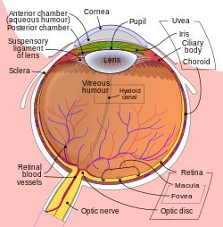
Vitreomacular adhesion

Last updated
Vitreomacular adhesion
Schematic diagram of the human eye en.svg
Schematic diagram of the human eye.

Vitreomacular adhesion (VMA) is a human medical condition where the vitreous gel (or simply vitreous, AKA vitreous humour) of the human eye adheres to the retina in an abnormally strong manner. As the eye ages, it is common for the vitreous to separate from the retina. But if this separation is not complete, i.e. there is still an adhesion, this can create pulling forces on the retina that may result in subsequent loss or distortion of vision. The adhesion in of itself is not dangerous, but the resulting pathological vitreomacular traction (VMT) can cause severe ocular damage.

Contents

The current standard of care for treating these adhesions is pars plana vitrectomy (PPV), which involves surgically removing the vitreous from the eye. A biological agent for non-invasive treatment of adhesions called ocriplasmin has been approved by the FDA on October 17, 2012.

Symptom and signs

Traction caused by VMA is the underlying pathology of an eye disease called symptomatic VMA. There is evidence that symptomatic VMA can contribute to the development of several well-known eye disorders, such as macular hole and macular pucker, that can cause visual impairment, including blindness. It may also be associated with age-related macular degeneration (AMD), diabetic macular edema (DME), retinal vein occlusion, and diabetic retinopathy (DR).[ citation needed ]

Pathology

Over time, it is common for the vitreous within the human eye to liquify and collapse in processes known as syneresis and synchisis respectively. This creates fluid-filled areas that can combine to form pockets of vitreous gel that are mostly liquid with very small concentrations of collagen. If these liquid pockets are close enough to the interface between the vitreous gel and the retina, they can cause complete separation of the vitreous from the retina in a normally occurring process in older humans called posterior vitreous detachment (PVD). PVD in of itself is not dangerous and a natural process.[ citation needed ]

If the separation of the vitreous from the retina is not complete, areas of focal attachment or adhesions can occur, i.e. a VMA. The pulling forces or traction from this adhesion on the retinal surface can sometimes cause edema within the retina, damage to retinal blood vessels causing bleeding, or damage to the optic nerve causing disruption in the nerve signals sent to the brain for visual processing. It is important to note that while the VMA itself is not dangerous, the resultant pulling on the retina called vitreomacular traction (VMT) causes the above damage. The size and strength of the VMA determine the variety of resulting pathologies or symptoms. [1]

VMA can also lead to the development of VMT/traction-related complications such as macular puckers and macular holes leading to distorted vision or metamorphopsia; epiretinal membrane; tractional macular oedema; myopic macular retinoschisis; visual impairment; blindness. The incidence of VMA is reported as high as 84% for patients with macular hole, 100% for patients with vitreomacular traction syndrome, and 56% in idiopathic epimacular membrane. [2]

Diagnosis

Careful eye examination by an ophthalmologist or optometrist is critical for diagnosing symptomatic VMA. Imaging technologies such as optical coherence tomography (OCT) have significantly improved the accuracy of diagnosing symptomatic VMA.[ citation needed ]

Treatment

A new FDA approved drug was released on the market late 2013. Jetrea (Brand name) or Ocriplasmin (Generic name) is the first drug of its kind used to treat vitreomacular adhesion.[ citation needed ]

Mechanism of Action: Ocriplasmin is a truncated human plasmin with proteolytic activity against protein components of the vitreous body and vitreoretinal interface. It dissolves the protein matrix responsible for the vitreomacular adhesion. Adverse drug reactions: Decreased vision, potential for lens subluxation, dyschromatopsia (yellow vision), eye pain, floaters, blurred vision. New Drug comparison Rating gave Jetea a 5 indicating an important advance. Previously, no recommended treatment was available for the patient with mild symptomatic VMA. In symptomatic VMA patients with more significant vision loss, the standard of care is pars plana vitrectomy (PPV), which involves surgically removing the vitreous from the eye, thereby surgically releasing the symptomatic VMA. In other words, vitrectomy induces PVD to release the traction/adhesion on the retina. An estimated 850,000 vitrectomy procedures are performed globally on an annual basis with 250,000 in the United States alone.

A standard PPV procedure can lead to serious complications [3] [4] [5] [6] including small-gauge PPV. [7] [8] [9] [10] [11] Complications can include retinal detachment, retinal tears, endophthalmitis, and postoperative cataract formation. Additionally, PPV may result in incomplete separation, and it may potentially leave a nidus for vasoactive and vasoproliferative substances, or it may induce development of fibrovascular membranes. As with any invasive surgical procedure, PPV introduces trauma to the vitreous and surrounding tissue. [12] [13]

There are data showing that nonsurgical induction of PVD using ocriplasmin (a recombinant protease with activity against fibronectin and laminin) [14] can offer the benefits of successful PVD while eliminating the risks associated with a surgical procedure, i.e. vitrectomy. Pharmacologic vitreolysis is an improvement over invasive surgery as it induces complete separation, creates a more physiologic state of the vitreomacular interface, prevents the development of fibrovascular membranes, is less traumatic to the vitreous, and is potentially prophylactic. [15] [16] As of 2012, ThromboGenics is still developing the ocriplasmin biological agent. Ocriplasmin is approved recently under the name Jetrea for use in the United States by the FDA.view. [17]

An experimental test of injections of perfluoropropane (C3F8) on 15 symptomatic eyes of 14 patients showed that vitreomacular traction resolved in 6 eyes within 1 month and resolved in 3 more eyes within 6 months. [18] A systematic review found high-certainty evidence that Ocriplasmin compared to no treatment increases the chance of resolution and improves vision in people with symptomatic VMA. [19] Ocriplasmin probably reduces the need for surgery but there were more adverse events in eyes treated with ocriplasmin compared to control.

Related Research Articles

Diabetic retinopathy Medical condition

Diabetic retinopathy, also known as diabetic eye disease (DED), is a medical condition in which damage occurs to the retina due to diabetes mellitus. It is a leading cause of blindness in developed countries.

Floater Deposits within the eyes vitreous humour

Floaters or eye floaters are sometimes visible deposits - e.g. the shadows of tiny structures of protein or other cell debris projected onto the retina - within the eye's vitreous humour, which is normally transparent, or between the vitreous and retina. They can become particularly noticeable when looking at a blank surface or an open monochromatic space, such as blue sky. Each floater can be measured by its size, shape, consistency, refractive index, and motility. They are also called muscae volitantes, or mouches volantes. The vitreous usually starts out transparent, but imperfections may gradually develop as one ages. The common type of floater, present in most people's eyes, is due to these degenerative changes of the vitreous. The perception of floaters, which may be annoying or problematic to some people, is known as myodesopsia, or, less commonly, as myodaeopsia, myiodeopsia, or myiodesopsia. It is not often treated, except in severe cases, where vitrectomy (surgery), laser vitreolysis, and medication may be effective.

Vitrectomy Type of eye surgery

Vitrectomy is a surgery to remove some or all of the vitreous humor from the eye.

Macular edema Medical condition

Macular edema occurs when fluid and protein deposits collect on or under the macula of the eye and causes it to thicken and swell (edema). The swelling may distort a person's central vision, because the macula holds tightly packed cones that provide sharp, clear, central vision to enable a person to see detail, form, and color that is directly in the centre of the field of view.

Eye surgery Surgery performed on the eye or its adnexa

Eye surgery, also known as ocular surgery, is surgery performed on the eye or its adnexa, typically by an ophthalmologist. The eye is a very fragile organ, and requires extreme care before, during, and after a surgical procedure to minimise or prevent further damage. An expert eye surgeon is responsible for selecting the appropriate surgical procedure for the patient, and for taking the necessary safety precautions. Mentions of eye surgery can be found in several ancient texts dating back as early as 1800 BC, with cataract treatment starting in the fifth century BC. Today it continues to be a widely practiced type of surgery, with various techniques having been developed for treating eye problems.

Retinal detachment is a disorder of the eye in which the retina peels away from its underlying layer of support tissue. Initial detachment may be localized, but without rapid treatment the entire retina may detach, leading to vision loss and blindness. It is a surgical emergency.

Posterior vitreous detachment Eye condition

A posterior vitreous detachment (PVD) is a condition of the eye in which the vitreous membrane separates from the retina. It refers to the separation of the posterior hyaloid membrane from the retina anywhere posterior to the vitreous base.

Intravitreal administration

Intravitreal is a route of administration of a drug, or other substance, in which the substance is delivered into the vitreous humor of the eye. "Intravitreal" literally means "inside an eye". Intravitreal injections were first introduced in 1911 when Ohm gave an injection of air into the vitreous humor to repair a detached retina. In the mid-1940s, intravitreal injections became a standard way to administer drugs to treat endophthalmitis and cytomegalovirus retinitis.

Endophthalmitis Medical condition

Endophthalmitis is inflammation of the interior cavity of the eye, usually caused by infection. It is a possible complication of all intraocular surgeries, particularly cataract surgery, and can result in loss of vision or loss of the eye itself. Infection can be caused by bacteria or fungi, and is classified as exogenous, or endogenous. Other non-infectious causes include toxins, allergic reactions, and retained intraocular foreign bodies. Intravitreal injections are a rare cause, with an incidence rate usually less than .05%.

Intermediate uveitis Medical condition

Intermediate uveitis is a form of uveitis localized to the vitreous and peripheral retina. Primary sites of inflammation include the vitreous of which other such entities as pars planitis, posterior cyclitis, and hyalitis are encompassed. Intermediate uveitis may either be an isolated eye disease or associated with the development of a systemic disease such as multiple sclerosis or sarcoidosis. As such, intermediate uveitis may be the first expression of a systemic condition. Infectious causes of intermediate uveitis include Epstein-Barr virus infection, Lyme disease, HTLV-1 virus infection, cat scratch disease, and hepatitis C.

Epiretinal membrane Eye disease

Epiretinal membrane or macular pucker is a disease of the eye in response to changes in the vitreous humor or more rarely, diabetes. Sometimes, as a result of immune system response to protect the retina, cells converge in the macular area as the vitreous ages and pulls away in posterior vitreous detachment (PVD). PVD can create minor damage to the retina, stimulating exudate, inflammation, and leucocyte response. These cells can form a transparent layer gradually and, like all scar tissue, tighten to create tension on the retina which may bulge and pucker, or even cause swelling or macular edema. Often this results in distortions of vision that are clearly visible as bowing and blurring when looking at lines on chart paper within the macular area, or central 1.0 degree of visual arc. Usually it occurs in one eye first, and may cause binocular diplopia or double vision if the image from one eye is too different from the image of the other eye. The distortions can make objects look different in size, especially in the central portion of the visual field, creating a localized or field dependent aniseikonia that cannot be fully corrected optically with glasses. Partial correction often improves the binocular vision considerably though. In the young, these cells occasionally pull free and disintegrate on their own; but in the majority of sufferers the condition is permanent. The underlying photoreceptor cells, rod cells and cone cells, are usually not damaged unless the membrane becomes quite thick and hard; so usually there is no macular degeneration.

Optic pit Medical condition

Optic pit, optic nerve pit, or optic disc pit is a congenital excavation of the optic disc, resulting from a malformation during development of the eye. Optic pits are important because they are associated with posterior vitreous detachments (PVD) and even serous retinal detachments.

Macular hole Medical condition

A macular hole is a small break in the macula, located in the center of the eye's light-sensitive tissue called the retina.

Ron P. Gallemore is a registered ophthalmologist with the American Academy of Ophthalmology involved in research and treatment of diseases of the macula and retina.

Gholam A. Peyman

Gholam A. Peyman is an ophthalmologist, retina surgeon, and inventor. He is best known for his invention of LASIK eye surgery, a vision correction procedure designed to allow people to see clearly without glasses. He was awarded the first US patent for the procedure in 1989.

Bascom Palmer Eye Institute, of the University of Miami Leonard M. Miller School of Medicine, is a center for ophthalmic care, research, and education. Faculty and staff treat patients from around the world in facilities in Miami-Dade County, Broward County, Palm Beach County, and Collier County. The institute's full-time faculty and researchers encompass many ophthalmic sub-specialties and has been consistently ranked as the #1 eye hospital and vision research center in the country by US News & World Report.

Branch retinal vein occlusion Medical condition

Branch retinal vein occlusion is a common retinal vascular disease of the elderly. It is caused by the occlusion of one of the branches of central retinal vein.

Ocriplasmin is a recombinant protease with activity against fibronectin and laminin, components of the vitreoretinal interface. It is used for treatment of symptomatic vitreomacular adhesion, for which it received FDA approval on 17 October 2012. It works by dissolving the proteins that link the vitreous to the macula, resulting in posterior detachment of the vitreous from the retina.

Intravitreal injection

Intravitreal injection is the method of administration of drugs into the eye by injection with a fine needle. The medication will be directly applied into the vitreous humor. It is used to treat various eye diseases, such as age-related macular degeneration (AMD), diabetic retinopathy, and infections inside the eye such as endophthalmitis. As compared to topical administration, this method is beneficial for a more localized delivery of medications to the targeted site, as the needle can directly pass through the anatomical eye barrier and dynamic barrier. It could also minimize adverse drug effect to other body tissues via the systemic circulation, which could be a possible risk for intravenous injection of medications. Although there are risks of infections or other complications, with suitable precautions throughout the injection process, chances for these complications could be lowered.

Sickle cell retinopathy can be defined as retinal changes due to blood vessel damage in the eye of a person with a background of sickle cell disease. It can likely progress to loss of vision in late stages due to vitreous hemorrhage or retinal detachment. Sickle cell disease is a structural red blood cell disorder leading to consequences in multiple systems. It is characterized by chronic red blood cell destruction, vascular injury, and tissue ischemia causing damage to the brain, eyes, heart, lungs, kidneys, spleen, and musculoskeletal system.

References

  1. Paolo Carpineto, Luca Di Antonio, Agbeanda Aharrh-Gnama, Vincenzo Ciciarelli, Leonardo Mastropasqua. Diagnosing and Treating Vitreomacular Adhesion. European Ophthalmic Review, 2011,5(1):69-73
  2. Koerner F, Garweg J. [Diseases of the vitreo-macular interface]. Klin Monbl Augenheilkd. 1999;214(5):305-310.
  3. Mojana F, Cheng L, Bartsch DU, et al. The role of abnormal vitreomacular adhesion in age-related macular degeneration: spectral optical coherence tomography and surgical results. Am J Ophthalmol. 2008;146(2):218-227.
  4. Doft BH, Wisniewski SR, Kelsey SF, Groer-Fitzgerald S; Endophthalmitis Vitrectomy Study Group. Diabetes and postcataract extraction endophthalmitis. Curr Opin Ophthalmol. 2002;13(3):147-151.
  5. Doft BM, Kelsey SF, Wisniewski SR. Retinal detachment in the endophthalmitis vitrectomy study. Arch Ophthalmol. 2000;118(12):1661-1665.
  6. Wisniewski SR, Capone A, Kelsey SF, et al. Characteristics after cataract extraction or secondary lens implantation among patients screened for the Endophthalmitis Vitrectomy Study. Ophthalmology. 2000;107(7):1274-1282.
  7. Gupta OP, Weichel ED, Regillo CD, et al. Postoperative complications associated with 25-gauge pars plana vitrectomy. Ophthalmic Surg Lasers Imaging. 2007;38(4):270–275.
  8. Liu DT, Chan CK, Fan DS, Lam SW, Lam DS, Chan WM. Choroidal folds after 25 gauge transconjunctival sutureless vitrectomy. Eye. 2005;19(7):825–827.
  9. Scott IU, Flynn HW Jr, Dev S, et al. Endophthalmitis after 25-gauge and 20-gauge pars plana vitrectomy: incidence and outcomes. Retina. 2008;28(1):138–142.
  10. Kunimoto DY, Kaiser RS; Wills Eye Retina Service. Incidence of endophthalmitis after 20- and 25- gauge vitrectomy. Ophthalmology. 2007;114(12):2133–2137.
  11. Kaiser RS. Complications of sutureless vitrectomy and the findings of the Micro-Surgical Safety Task Force. Paper presented at: Retina Subspecialty Day, Annual Meeting of the American Academy of Ophthalmology; November 7–8, 2008; Atlanta, GA.
  12. de Smet MD, Gandorfer A, Stalmans P, et al. Microplasmin intravitreal administration in patients with vitreomacular traction scheduled for vitrectomy: the MIVI I trial. Ophthalmology. 2009;116(7):1349-1355.
  13. Goldenberg DT, Trese MT. Pharmacologic vitreodynamics and molecular flux. Dev Ophthalmol. 2009;44:31-36.
  14. Stalmans P, Benz MS, Gandorfer A, Kampik A, Girach A, Pakola S, Haller JA; MIVI-TRUST Study Group. Enzymatic vitreolysis with ocriplasmin for vitreomacular traction and macular holes. N Engl J Med. 2012 Aug 16;367(7):606-15.
  15. de Smet MD, Gandorfer A, Stalmans P, et al. Microplasmin intravitreal administration in patients with vitreomacular traction scheduled for vitrectomy: the MIVI I trial. Ophthalmology. 2009;116(7):1349-1355.
  16. Goldenberg DT, Trese MT. Pharmacologic vitreodynamics and molecular flux. Dev Ophthalmol. 2009;44:31-36.
  17. "Retina Today - May 2012".
  18. Rodrigues IA; Stangos AN; McHugh DA; Jackson TL (2013). "Intravitreal injection of expansile perfluoropropane (C3F8) for the treatment of vitreomacular traction". American Journal of Ophthalmology. 155 (2)): 270–276. doi:10.1016/j.ajo.2012.08.018. PMID   23164159.
  19. Neffendorf, James E; Kirthi, Varo; Pringle, Edward; Jackson, Timothy L (2017-10-17). "Ocriplasmin for symptomatic vitreomacular adhesion". Cochrane Database of Systematic Reviews. 10: CD011874. doi:10.1002/14651858.cd011874.pub2. ISSN   1465-1858. PMC   6485716 . PMID   29040800.
Classification
D