Related Research Articles

The cell cycle, or cell-division cycle, is the series of events that take place in a cell that cause it to divide into two daughter cells. These events include the duplication of its DNA and some of its organelles, and subsequently the partitioning of its cytoplasm and other components into two daughter cells in a process called cell division.

In cell biology, mitosis is a part of the cell cycle in which replicated chromosomes are separated into two new nuclei. Cell division by mitosis gives rise to genetically identical cells in which the total number of chromosomes is maintained. Therefore, mitosis is also known as equational division. In general, mitosis is preceded by S phase of interphase and is often followed by telophase and cytokinesis; which divides the cytoplasm, organelles and cell membrane of one cell into two new cells containing roughly equal shares of these cellular components. The different stages of mitosis altogether define the mitotic (M) phase of an animal cell cycle—the division of the mother cell into two daughter cells genetically identical to each other.

Cell division is the process by which a parent cell divides, when a mother cell divides into two or more daughter cells. Cell division usually occurs as part of a larger cell cycle. In eukaryotes, there are two distinct types of cell division; a vegetative division, whereby each daughter cell is genetically identical to the parent cell (mitosis), and a reproductive cell division, whereby the number of chromosomes in the daughter cells is reduced by half to produce haploid gametes (meiosis). In cell biology, mitosis (/maɪˈtoʊsɪs/) is a part of the cell cycle, in which, replicated chromosomes are separated into two new nuclei. Cell division gives rise to genetically identical cells in which the total number of chromosomes is maintained. In general, mitosis is preceded by the S stage of interphase and is often followed by telophase and cytokinesis; which divides the cytoplasm, organelles, and cell membrane of one cell into two new cells containing roughly equal shares of these cellular components. The different stages of mitosis all together define the mitotic (M) phase of animal cell cycle—the division of the mother cell into two genetically identical daughter cells. Meiosis results in four haploid daughter cells by undergoing one round of DNA replication followed by two divisions. Homologous chromosomes are separated in the first division, and sister chromatids are separated in the second division. Both of these cell division cycles are used in the process of sexual reproduction at some point in their life cycle. Both are believed to be present in the last eukaryotic common ancestor.
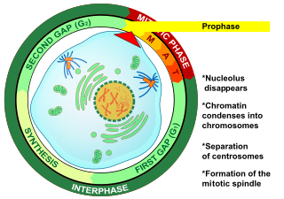
Prophase is the first stage of cell division in both mitosis and meiosis. Beginning after interphase, DNA has already been replicated when the cell enters prophase. The main occurrences in prophase are the condensation of the chromatin reticulum and the disappearance of the nucleolus.

In cell biology, the spindle apparatus refers to the cytoskeletal structure of eukaryotic cells that forms during cell division to separate sister chromatids between daughter cells. It is referred to as the mitotic spindle during mitosis, a process that produces genetically identical daughter cells, or the meiotic spindle during meiosis, a process that produces gametes with half the number of chromosomes of the parent cell.
Baylor College of Medicine (BCM) is a private, independent health sciences center in Houston, Texas within the Texas Medical Center, the world's largest medical center. BCM is composed of four academic components: the School of Medicine, the Graduate School of Biomedical Sciences; the School of Health Professions, and the National School of Tropical Medicine.
The microtubule-organizing center (MTOC) is a structure found in eukaryotic cells from which microtubules emerge. MTOCs have two main functions: the organization of eukaryotic flagella and cilia and the organization of the mitotic and meiotic spindle apparatus, which separate the chromosomes during cell division. The MTOC is a major site of microtubule nucleation and can be visualized in cells by immunohistochemical detection of γ-tubulin. The morphological characteristics of MTOCs vary between the different phyla and kingdoms. In animals, the two most important types of MTOCs are 1) the basal bodies associated with cilia and flagella and 2) the centrosome associated with spindle formation.
A kinetochore is a disc-shaped protein structure associated with duplicated chromatids in eukaryotic cells where the spindle fibers attach during cell division to pull sister chromatids apart. The kinetochore assembles on the centromere and links the chromosome to microtubule polymers from the mitotic spindle during mitosis and meiosis. The term kinetochore was first used in a footnote in a 1934 Cytology book by Lester W. Sharp and commonly accepted in 1936. Sharp's footnote reads: "The convenient term kinetochore has been suggested to the author by J. A. Moore", likely referring to John Alexander Moore who had joined Columbia University as a freshman in 1932.

Aurora kinase B is a protein that functions in the attachment of the mitotic spindle to the centromere.

A mitotic inhibitor is a drug that inhibits mitosis, or cell division. These drugs disrupt microtubules, which are structures that pull the chromosomes apart when a cell divides. Mitotic inhibitors are used in cancer treatment, because cancer cells are able to grow and eventually spread through the body (metastasize) through continuous mitotic division. Thus, cancer cells are more sensitive to inhibition of mitosis than normal cells. Mitotic inhibitors are also used in cytogenetics, where they stop cell division at a stage where chromosomes can be easily examined.

Mitotic checkpoint serine/threonine-protein kinase BUB1 also known as BUB1 is an enzyme that in humans is encoded by the BUB1 gene.

Aurora kinase C, also Serine/threonine-protein kinase 13 is an enzyme that in humans is encoded by the AURKC gene.

An aster is a cellular structure shaped like a star, consisting of a centrosome and its associated microtubules during the early stages of mitosis in an animal cell. Asters do not form during mitosis in plants. Astral rays, composed of microtubules, radiate from the centrosphere and look like a cloud. Astral rays are one variant of microtubule which comes out of the centrosome; others include kinetochore microtubules and polar microtubules.
Angelika Amon was an Austrian American molecular and cell biologist, and the Kathleen and Curtis Marble Professor in Cancer Research at the Massachusetts Institute of Technology (MIT) in Cambridge, Massachusetts, United States. Amon's research centered on how chromosomes are regulated, duplicated, and partitioned in the cell cycle. Amon was elected to the American Academy of Arts and Sciences in 2017.
In cell biology, Meiomitosis is an aberrant cellular division pathway that combines normal mitosis pathways with ectopically expressed meiotic machinery resulting in genomic instability.
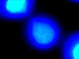
Chromatin bridge is a mitotic occurrence that forms when telomeres of sister chromatids fuse together and fail to completely segregate into their respective daughter cells. Because this event is most prevalent during anaphase, the term anaphase bridge is often used as a substitute. After the formation of individual daughter cells, the DNA bridge connecting homologous chromosomes remains fixed. As the daughter cells exit mitosis and re-enter interphase, the chromatin bridge becomes known as an interphase bridge. These phenomena are usually visualized using the laboratory techniques of staining and fluorescence microscopy.
Conly Leroy Rieder is a cancer researcher in the field of mitotic cellular division. The bulk of his research between 1980 and 2011, was funded through NIH grants and conducted at the Wadsworth Center in the New York State Department of Health in Albany, New York. He has published on the subjects of chromosome motility, spindle assembly and mitotic checkpoints. His research has contributed to the growing understanding of the process of cell division and the pathology of cancer.

Jonathon Noë Joseph Pines is Head of the Cancer Biology Division at the Institute of Cancer Research in London. He was formerly a senior group leader at the Gurdon Institute at the University of Cambridge.
J. Richard McIntosh is a Distinguished Professor Emeritus in Molecular, Cellular, and Developmental Biology at the University of Colorado Boulder. McIntosh first graduated from Harvard with a BA in Physics in 1961, and again with a Ph.D. in Biophysics in 1968. He began his teaching career at Harvard but has spent most of his career at the University of Colorado Boulder. At the University of Colorado Boulder, McIntosh taught biology courses at both the undergraduate and graduate levels. Additionally, he created an undergraduate course in the biology of cancer towards the last several years of his teaching career. McIntosh's research career looks at a variety of things, including different parts of mitosis, microtubules, and motor proteins.

Tim J. Yen is an American molecular biologist and cancer biologist. Yen is currently director of the Biological Imaging Facility at Fox Chase Cancer Center in Philadelphia, Pennsylvania. Yen is known for pioneering work in the field of mitosis.
References
- 1 2 http://www.researchamerica.org/brinkley_william Archived 2008-04-24 at the Wayback Machine Research America biography profile
- ↑ "Friends Honor Alumnus With Scholarship". Communications Office, Sam Houston State University, (SHSU). 2011. Retrieved 2018-11-19.
- 1 2 Heald, Rebecca (2007). "Brinkley-Fest of Mitosis". Developmental Cell. 13 (2): 168–76. doi: 10.1016/j.devcel.2007.07.010 . PMID 17681129.
- ↑ Garrison, Howard H.; Masters, Bettie Sue; Bond, Judith (2021-03-30). "William R. Brinkley (1936–2020)". FASEB BioAdvances. 3 (4): 280–281. doi:10.1096/fba.2021-00034. ISSN 2573-9832. PMC 8019260 .
- ↑ Fleischman, John (2014). "Giants of the Cytoskeleton Win E.B. Wilson Medal". American Society for Cell Biology. Retrieved May 10, 2017.