Actinobacillus pleuropneumoniae

Last updated

Actinobacillus pleuropneumoniae
Scientific classification OOjs UI icon edit-ltr.svg
Domain: Bacteria
Phylum: Pseudomonadota
Class: Gammaproteobacteria
Order: Pasteurellales
Family: Pasteurellaceae
Genus: Actinobacillus
Species:
A. pleuropneumoniae
Binomial name
Actinobacillus pleuropneumoniae
(Pohl, 1983)

Actinobacillus pleuropneumoniae (previously Haemophilus pleuropneumoniae), is a Gram-negative, facultative anaerobic, respiratory pathogen found in pigs. It was first reported in 1957, and was formally declared to be the causative agent of porcine pleuropneumonia in 1964. [1] [2] It was reclassified in 1983 after DNA studies showed it was more closely related to A. lignieresii. [3]

Contents

Microbiology

Actinobacillus pleuropneumoniae is a nonmotile, Gram-negative, encapsulated coccobacillus bacterium found in the family Pasteurellaceae. [4] [5] It exhibits β-hemolysis activity, [6] thus explaining its growth on chocolate or blood agar, but must be supplemented with NAD ('V factor') to facilitate growth for one of its biological variants (biovar 1). [3] As a facultative anaerobic pathogen, A. pleuropneumoniae may need CO2 to grow. [3] Depending on the biovar, the bacteria may or may not be positive for urease; both biovars are positive for porphyrin. [3]

Porcine pleuropneumonia

Actinobacillus pleuropneumoniae was found to be the causative agent for up to 20% of all bacterial pneumonia cases in swine. [3] The main disease associated with this bacterium is porcine pleuropneumonia, a highly contagious respiratory disease, affecting primarily young pigs (usually less than 6 months). [5] All of the symptoms and signs of porcine pleuropneumonia can be attributed to its virulence factors. The symptoms include respiratory distress, bloodstained discharge (usually frothy) from the mouth, fever, anorexia, mild diarrhea, cyanosis, lethargy, and spontaneous abortion in sows. [4] [5] The most common sign for a pig farmer is the sudden death of several pigs over a short period of time. [4] Peak mortality is usually reached when pigs are 10–16 weeks old. [1] Not uncommonly,r mortality rates can reach 20-80% in fattening pigs, with similarly high morbidity. [1] Pigs that do survive the disease remain as carriers and spread the bacterium to other swine. [4] Several bacterial combinations are seen in vivo , the most common simultaneous infection being Pasteurella multocida . [3] Treatment must be immediate and continuous. Antibiotics used include ceftiofur, tetracycline, synthetic penicillins, tylosin, and sulfonamides. [7]

Serotypes

Nineteen different serotype variants (serovars) have been recognized for A. pleuropneumoniae, [8] based on the different capsular polysaccharides exhibited. [9] Two different biovars exist, with biovar 1 having 13 different serovars and biovar 2 having two serovars. [4] Differences in virulence potential, immunogenicity, and worldwide geographical distribution contribute to the diversity of the A. pleuropneumoniae serotypes. [9] All 15 serotypes can cause disease, with one serotype usually predominating in a particular herd. [10] The main difference between the serotypes is the expression of Apx toxins and other virulence factors.

Pathophysiology

The bacterium rapidly colonizes the host and attaches to the epithelial cells of the tonsils, moving down to the respiratory tract using type IV fimbriae. [4] [10] As the bacteria replicate, they release cytotoxins (in the form of Apx toxins), hemolysins and the lipopolysaccharides (LPSs)on their outer membranes. [4] The subsequent lysis of macrophages causes a release of lysozymes, which in turn cause the tissue damage seen in porcine pleuropneumonia. [4] Members of the family Pasteurellaceae routinely change the cellular processes of the infected cell. [11] In particular, A. pleuropneumoniae activates the creation of various cytokines such as interleukin 1β (IL-1β), IL-8, and tumor necrosis factor-alpha (TNF-α). [11] IL-8 is itself a chemical signal used to attract neutrophils to the infection site. [11]

The typical presentation of A. pleuropneumoniae in pigs is the characteristic demarcated lesions in the middle, cranial, and caudal lobes of the lungs. [3] Areas of severe pneumonic growth are dark and consolidated. [3] In the case of chronically infected pigs, pleural adhesions and abscesses are normally found. [3] Histological studies of infected lung tissue normally showcase lung necrosis, neutrophil infiltration, macrophage and platelet activation, and an exudate. [3] Severe hemolysis or hemorrhaging is also present.

Several virulence factors account for the remarkable pathogenicity of A. pleuropneumoniae. [9] The more important ones include the production and release of the Apx toxins, the ability to produce a biofilm, its LPS layer, capsule polysaccharides, and its ability to survive within an iron-limited environment. [9] Of these, the most important are its capsule and Apx toxin production. [9]

The Apx toxin, a member of the RTX toxin family, is subdivided into four types: ApxI through ApxIV. [10] As a pore-forming exotoxin, Apx toxin lyses alveolar epithelial cells, endothelial cells, red blood cells, neutrophils, and macrophages. [10] Each serotype expresses different levelss of the four Apx toxins. [9] The most virulent combination known to exist, ApxI and ApxII, is expressed by serovars 1, 5, 9, and 11. [9] The ApxII and ApxIII combination is of medium virulence and is expressed by serovars 2, 3, 4, 6, 8, and 15. [9]

Epidemiology

The bacteria are usually spread through direct nose-to-nose contact. [1] It is species-specific, as its Apx toxin only affects pigs and other swine. [12] Overcrowding in pigpens, co-infections of other respiratory pathogens, and unusual stress all contribute to the spread of the disease. [1] A. pleuropneumoniae must have a host to survive, and does not survive for a significant time outside a host. [1] This bacterium is found worldwide, with different serotypes prevailing in different locations. Serotypes 1, 3, 5, and 7 are most commonly found in North America. [1]

Economic impact

A. pleuropneumoniae has a profound economic impact on pork production and pig farmers. In 1995, Its infections cost the U.S. economy about $30 million. [13] Such losses usually result from medication and veterinary expenses, increased mortality of pigs, extra labor, and other factors, such as reduced weight gain. [13]

Related Research Articles

<i>Salmonella</i> Genus of prokaryotes

Salmonella is a genus of rod-shaped (bacillus) gram-negative bacteria of the family Enterobacteriaceae. The two known species of Salmonella are Salmonella enterica and Salmonella bongori. S. enterica is the type species and is further divided into six subspecies that include over 2,600 serotypes. Salmonella was named after Daniel Elmer Salmon (1850–1914), an American veterinary surgeon.

<i>Francisella tularensis</i> Species of bacterium

Francisella tularensis is a pathogenic species of Gram-negative coccobacillus, an aerobic bacterium. It is nonspore-forming, nonmotile, and the causative agent of tularemia, the pneumonic form of which is often lethal without treatment. It is a fastidious, facultative intracellular bacterium, which requires cysteine for growth. Due to its low infectious dose, ease of spread by aerosol, and high virulence, F. tularensis is classified as a Tier 1 Select Agent by the U.S. government, along with other potential agents of bioterrorism such as Yersinia pestis, Bacillus anthracis, and Ebola virus. When found in nature, Francisella tularensis can survive for several weeks at low temperatures in animal carcasses, soil, and water. In the laboratory, F. tularensis appears as small rods, and is grown best at 35–37 °C.

<span class="mw-page-title-main">Serotype</span> Distinct variation within a species of bacteria or virus or among immune cells

A serotype or serovar is a distinct variation within a species of bacteria or virus or among immune cells of different individuals. These microorganisms, viruses, or cells are classified together based on their surface antigens, allowing the epidemiologic classification of organisms to a level below the species. A group of serovars with common antigens is called a serogroup or sometimes serocomplex.

<i>Chlamydia psittaci</i> Species of bacterium

Chlamydia psittaci is a lethal intracellular bacterial species that may cause endemic avian chlamydiosis, epizootic outbreaks in other mammals, and respiratory psittacosis in humans. Potential hosts include feral birds and domesticated poultry, as well as cattle, pigs, sheep, and horses. C. psittaci is transmitted by inhalation, contact, or ingestion among birds and to mammals. Psittacosis in birds and in humans often starts with flu-like symptoms and becomes a life-threatening pneumonia. Many strains remain quiescent in birds until activated by stress. Birds are excellent, highly mobile vectors for the distribution of chlamydia infection, because they feed on, and have access to, the detritus of infected animals of all sorts.

<i>Brucella suis</i> Bacterium that causes swine brucellosis

Brucella suis is a bacterium that causes swine brucellosis, a zoonosis that affects pigs. The disease typically causes chronic inflammatory lesions in the reproductive organs of susceptible animals or orchitis, and may even affect joints and other organs. The most common symptom is abortion in pregnant susceptible sows at any stage of gestation. Other manifestations are temporary or permanent sterility, lameness, posterior paralysis, spondylitis, and abscess formation. It is transmitted mainly by ingestion of infected tissues or fluids, semen during breeding, and suckling infected animals.

Mycoplasma hyopneumoniae is a species of bacteria known to cause the disease porcine enzootic pneumonia, a highly contagious and chronic disease affecting pigs. As with other mollicutes, M. hyopneumoniae is small in size (400–1200 nm), has a small genome and lacks a cell wall. It is difficult to grow in laboratories due to its complex nutritional requirements and the high chances of contamination associated with mycoplasma culture. To successfully grow the bacterium, an environment of 5–10% carbon dioxide is required, and the medium should demonstrate an acid colour shift.

<i>Shigella flexneri</i> Species of bacterium

Shigella flexneri is a species of Gram-negative bacteria in the genus Shigella that can cause diarrhea in humans. Several different serogroups of Shigella are described; S. flexneri belongs to group B. S. flexneri infections can usually be treated with antibiotics, although some strains have become resistant. Less severe cases are not usually treated because they become more resistant in the future. Shigella are closely related to Escherichia coli, but can be differentiated from E.coli based on pathogenicity, physiology and serology.

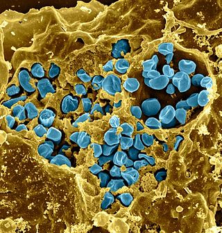
Betaarterivirus suid 1, commonly Porcine reproductive and respiratory syndrome virus (PRRSV), is a virus that causes a disease of pigs, called porcine reproductive and respiratory syndrome (PRRS), also known as blue-ear pig disease. This economically important, panzootic disease causes reproductive failure in breeding stock and respiratory tract illness in young pigs.

<i>Bordetella pertussis</i> Species of bacterium causing pertussis or whooping cough

Bordetella pertussis is a Gram-negative, aerobic, pathogenic, encapsulated coccobacillus of the genus Bordetella, and the causative agent of pertussis or whooping cough. Like B. bronchiseptica, B. pertussis can express a flagellum-like structure, even if it has been historically categorized as a nonmotile bacteria. Its virulence factors include pertussis toxin, adenylate cyclase toxin, filamentous hæmagglutinin, pertactin, fimbria, and tracheal cytotoxin.

Aujeszky's disease, usually called pseudorabies in the United States, is a viral disease in swine that is endemic in most parts of the world. It is caused by Suid herpesvirus 1 (SuHV-1). Aujeszky's disease is considered to be the most economically important viral disease of swine in areas where classical swine fever has been eradicated. Other mammals, such as cattle, sheep, goats, cats, dogs, and raccoons, are also susceptible. The disease is usually fatal in these animal species.

Adenylate cyclase toxin is a virulence factor produced by some members of the genus Bordetella. Together with the pertussis toxin it is the most important virulence factor of the causative agent of whooping cough, Bordetella pertussis. Bordetella bronchiseptica and Bordetella parapertussis, also able to cause pertussis-like symptoms, also produce adenylate cyclase toxin. It is a toxin secreted by the bacteria to influence the host immune system.

<i>Pasteurella multocida</i> Species of bacterium

Pasteurella multocida is a Gram-negative, nonmotile, penicillin-sensitive coccobacillus of the family Pasteurellaceae. Strains of the species are currently classified into five serogroups based on capsular composition and 16 somatic serovars (1–16). P. multocida is the cause of a range of diseases in mammals and birds, including fowl cholera in poultry, atrophic rhinitis in pigs, and bovine hemorrhagic septicemia in cattle and buffalo. It can also cause a zoonotic infection in humans, which typically is a result of bites or scratches from domestic pets. Many mammals and birds harbor it as part of their normal respiratory microbiota.

Porphyromonas gingivalis belongs to the phylum Bacteroidota and is a nonmotile, Gram-negative, rod-shaped, anaerobic, pathogenic bacterium. It forms black colonies on blood agar.

Aggregatibacter actinomycetemcomitans is a Gram-negative, facultative anaerobe, nonmotile bacterium that is often found in association with localized aggressive periodontitis, a severe infection of the periodontium. It is also suspected to be involved in chronic periodontitis. Less frequently, A. actinomycetemcomitans is associated with nonoral infections such as endocarditis. Its role in aggressive periodontitis was first discovered by Danish-born periodontist Jørgen Slots, a professor of dentistry and microbiology at the University of Southern California School of Dentistry.

<i>Rhodococcus equi</i> Species of bacterium

Rhodococcus equi is a Gram-positive coccobacillus bacterium. The organism is commonly found in dry and dusty soil and can be important for diseases of domesticated animals. The frequency of infection can reach near 60%. R. equi is an important pathogen causing pneumonia in foals. Since 2008, R. equi has been known to infect wild boar and domestic pigs. R. equi can infect humans. At-risk groups are immunocompromised people, such as HIV-AIDS patients or transplant recipients. Rhodococcus infection in these patients resemble clinical and pathological signs of pulmonary tuberculosis. It is facultative intracellular.

<i>Bacillus anthracis</i> Species of bacterium

Bacillus anthracis is a gram-positive and rod-shaped bacterium that causes anthrax, a deadly disease to livestock and, occasionally, to humans. It is the only permanent (obligate) pathogen within the genus Bacillus. Its infection is a type of zoonosis, as it is transmitted from animals to humans. It was discovered by a German physician Robert Koch in 1876, and became the first bacterium to be experimentally shown as a pathogen. The discovery was also the first scientific evidence for the germ theory of diseases.

Feline coronavirus (FCoV) is a positive-stranded RNA virus that infects cats worldwide. It is a coronavirus of the species Alphacoronavirus 1, which includes canine coronavirus (CCoV) and porcine transmissible gastroenteritis coronavirus (TGEV). FCoV has two different forms: feline enteric coronavirus (FECV), which infects the intestines, and feline infectious peritonitis virus (FIPV), which causes the disease feline infectious peritonitis (FIP).

<i>Salmonella enterica <span style="font-style:normal;">subsp.</span> enterica</i> Subspecies of bacterium

Salmonella enterica subsp. enterica is a subspecies of Salmonella enterica, the rod-shaped, flagellated, aerobic, Gram-negative bacterium. Many of the pathogenic serovars of the S. enterica species are in this subspecies, including that responsible for typhoid.

<i>Staphylococcus hyicus</i> Species of bacterium

Staphylococcus hyicus is a Gram-positive, facultatively anaerobic bacterium in the genus Staphylococcus. It consists of clustered cocci and forms white circular colonies when grown on blood agar. S. hyicus is a known animal pathogen. It causes disease in poultry, cattle, horses, and pigs. Most notably, it is the agent that causes porcine exudative epidermitis, also known as greasy pig disease, in piglets. S. hyicus is generally considered to not be zoonotic, however it has been shown to be able to cause bacteremia and sepsis in humans.

Actinobacillus equuli is a gram-negative, non-motile rod bacteria from the family Pasteurellaceae.

References

  1. 1 2 3 4 5 6 7 "Actinobacillus pleuropneumoniae". Iowa State University College of Veterinary Medicine. Retrieved 3 September 2018.
  2. Shope, RE (1964). "Porcine Contagious Pleuropnemonia: 1. Experimental Transmission, Etiology, and Pathology". Journal of Experimental Medicine. 119 (3): 357–368. doi:10.1084/jem.119.3.357. PMC   2137882 . PMID   14129707.
  3. 1 2 3 4 5 6 7 8 9 10 Marsteller, TA; Fenwick B (1999). "Actinobacillus pleuropneumoniae disease and serology". Swine Health and Production. 7 (4): 161–165.
  4. 1 2 3 4 5 6 7 8 Brownfield, B. "Actinobacillus pleuropneumoniae in swine". Purdue University-Animal Disease Diagnostic Laboratory. Retrieved 20 April 2013.
  5. 1 2 3 Xu, Z; Chen X; Li L; et al. (2010). "Comparative Genomic Characterization of Actinobacillus pleuropneumoniae". Journal of Bacteriology. 192 (21): 5625–5636. doi:10.1128/JB.00535-10. PMC   2953695 . PMID   20802045.
  6. Gillaspy, A. "Actinobacillus pleuropneumoniae". Laboratory for Molecular Biology and Cytometry Research-University of Oklahoma Health Sciences Center. Archived from the original on 13 October 2002. Retrieved 20 April 2013.
  7. Dee, S. "Pleuropneumonia in Pigs". Merck Veterinary Manual. Retrieved 20 April 2013.
  8. Stringer, O; Bosse J; Lacouture S (2021). "Proposal of Actinobacillus pleuropneumoniae serovar 19, and reformulation of previous multiplex PCRs for capsule-specific typing of all known serovars". Veterinary Microbiology. 255: 109021. doi:10.1016/j.vetmic.2021.109021. hdl: 10044/1/86195 . PMID   33667982. S2CID   232132425.
  9. 1 2 3 4 5 6 7 8 Klitgaard, K; Friis C; Angen O; et al. (2010). "Comparative profiling of the transcriptional response to iron restriction in six serotypes of Actinobacillus pleuropneumoniae with different virulence potential". BMC Genomics. 11: 698. doi: 10.1186/1471-2164-11-698 . PMC   3091793 . PMID   21143895.
  10. 1 2 3 4 Chiers, K; De Waele T; Masmans F; et al. (2010). "Virulence factors of Actinobacillus pleuropneumoniae involved in colonization, persistence and induction of lesions in its porcine host". Veterinary Research. 41 (65): 65. doi:10.1051/vetres/2010037. PMC   2899255 . PMID   20546697.
  11. 1 2 3 Auger, E; Deslandes V; Ramjeet M; et al. (2009). "Host-pathogen interactions of Actinobacillus pleuropneumoniae with porcine lung and tracheal epithelial cells". Infection and Immunity. 77 (4): 1426–1441. doi:10.1128/IAI.00297-08. PMC   2663157 . PMID   19139196.
  12. Vanden Bergh, PGAC; Zecchinon LM; Fett T; et al. (2009). "Porcine CD18 mediates Actinobacillus pleuropneumoniae ApxIII species-specific toxicity". Veterinary Research. 40 (33): 33. doi:10.1051/vetres/2009016. PMC   2701182 . PMID   19356397.
  13. 1 2 Losinger, WC (2005). "Economic impacts of reduced pork production associated with the diagnosis of Actinobacillus pleuropneumoniae on grower/finisher swine operations in the United States". Preventive Veterinary Medicine. 68 (2–4): 181–193. doi:10.1016/j.prevetmed.2004.12.004. PMID   15820115.