Allen Brain Atlas

Last updated

The Allen Mouse and Human Brain Atlases are projects within the Allen Institute for Brain Science which seek to combine genomics with neuroanatomy by creating gene expression maps for the mouse and human brain. They were initiated in September 2003 with a $100 million donation from Paul G. Allen and the first atlas went public in September 2006. [1] As of May 2012, seven brain atlases have been published: Mouse Brain Atlas, Human Brain Atlas, Developing Mouse Brain Atlas, Developing Human Brain Atlas, Mouse Connectivity Atlas, Non-Human Primate Atlas, and Mouse Spinal Cord Atlas.[ citation needed ] There are also three related projects with data banks: Glioblastoma, Mouse Diversity, and Sleep.[ citation needed ] It is the hope of the Allen Institute that their findings will help advance various fields of science, especially those surrounding the understanding of neurobiological diseases. The atlases are free and available for public use online.

Contents

History

In 2001, Paul Allen gathered a group of scientists, including James Watson and Steven Pinker, to discuss the future of neuroscience and what could be done to enhance neuroscience research (Jones 2009). During these meetings David Anderson from the California Institute of Technology proposed the idea that a three-dimensional atlas of gene expression in the mouse brain would be of great use to the neuroscience community. The project was set in motion in 2003 with a 100 million dollar donation by Allen through the Allen Institute for Brain Science. The project used a technique for mapping gene expression developed by Gregor Eichele and colleagues at the Max Planck Institute for Biophysical Chemistry in Goettingen, Germany. The technique uses colorimetric in situ hybridization to map gene expression. The project set a 3-year goal of finishing the project and making it available to the public.

An initial release of the first atlas, the mouse brain atlas, occurred in December 2004. Subsequently, more data for this atlas was released in stages. The final genome-wide data set was released in September 2006. However, the final release of the atlas was not the end of the project; the Atlas is still being improved upon. Also, other projects including the human brain atlas, developing mouse brain, developing human brain, mouse connectivity, non-human primate atlas, and the mouse spinal cord atlas are being developed through the Allen Institute for Brain Science in conjunction with the Allen Mouse Brain Atlas.

Goals for the project

Allen Institute for Brain Science Allen Institute Logo.jpg
Allen Institute for Brain Science

The overarching goal and motto for all Allen Institute projects is "fueling discovery". The project strives to fulfill this goal and advance science in a few ways. First, they create brain atlases to better understand the connections between genes and brain functioning. They aim to advance the research and knowledge about neurobiological conditions such as Parkinson's, Alzheimer's, and Autism with their mapping of gene expression throughout the brain. The Brain Atlas projects also follow the "Allen Institute" motto with their open release of data and findings. This policy is also related to another goal of the Institute: collaborative and multidisciplinary research. Thus, any scientist from any discipline is able to look at the findings and take them into account while designing their own experiments. Also available to the public is the Brain Explorer application.

Research techniques

The Allen Institute for Brain Science uses a project-based philosophy for their research. Each brain atlas focuses on its own project, made up of its own team of researchers. To complete an atlas, each research team collects and synthesizes brain scans, medical data, genetic information and psychological data. With this information, they are able to construct the 3-D biochemical architecture of the brain and figure out which proteins are expressed in certain parts of the brain. To gather the needed data, scientists at the Allen Institute use various techniques. One technique involves the use of postmortem brains and brain scanning technology to discover where in the brain genes are turned on and off. Another technique, called in situ hybridization, or ISH, is used to view gene expression patterns as in situ hybridization images.

Within the Brain Atlases, these 3-D ISH digital images and graphs reveal, in color, the regions where a given gene is expressed. In the Brain Explorer, any gene can be searched for and selected resulting in the in situ image appearing as an easily manipulated and explored fashion. Part of the creation of this anatomy-centred database of gene expression, includes aligning ISH data for each gene with a three-dimensional coordinate space through registration with a reference atlas created for the project. [2]

Contributions to neuroscience

The different types of cells in the central nervous system originate from varying gene expression. A map of gene expression in the brain allows researchers to correlate forms and functions. The Allen Brain Atlas lets researchers view the areas of differing expression in the brain which enables the viewing of neural connections throughout the brain. Viewing these pathways through differing gene expression as well as functional imaging techniques permits researchers to correlate between gene expression, cell types, and pathway function in relation to behaviors or phenotypes.

Even though the majority of research has been done in mice, 90% of genes in mice have a counterpart in humans. This makes the Atlas particularly useful for modeling neurological diseases. The gene expression patterns in normal individuals provide a standard for comparing and understanding altered phenotypes. Extending information learned from mouse diseases will help better the understanding of human neurological disorders. The atlas can show which genes and particular areas are effected in neurological disorders; the action of a gene in a disease can be evaluated in conjunction with general expression patterns and this data could shed light on the role of the particular gene in the disorder.

Brain explorer

The Allen Brain Atlas website contains a downloadable 3-D interactive Brain explorer. The explorer is essentially a search engine for locations of gene expression; this is particularly useful in finding regions that express similar genes. Users can delineate networks and pathways using this application by connecting regions that co-express a certain gene. The explorer uses a multicolor scale and contains multiple planes of the brain that let viewers see differences in density and expression level. The images are a composite of many averaged samples so it is useful when comparing to individuals with abnormally low gene expression.

Atlases

Mouse Brain

The Allen Mouse Brain Atlas is a comprehensive genome-wide map of the adult mouse brain that reveals where each gene is expressed. [3] The mouse brain atlas was the original project of the Allen Brain Atlas and was finished in 2006. The purpose of the atlas is to aid in the development of neuroscience research. The hope of the project is that it will allow scientists to gain a better understanding of brain diseases and disorders such as autism and depression. [1]

Human Brain

The Allen Human Brain Atlas was made public in May 2010. It was the first anatomically and genomically comprehensive three-dimensional human brain map. [4] The atlas was created to enhance research in many neuroscience research fields including neuropharmacology, human brain imaging, human genetics, neuroanatomy, genomics and more. The atlas is also geared toward furthering research into mental health disorders and brain injuries such as Alzheimer's disease, autism, schizophrenia and drug addiction.

Developing Mouse Brain

The Allen Developing Mouse Brain Atlas is an atlas which tracks gene expression throughout the development of a C57BL/6 mouse brain. The project began in 2008 and is currently ongoing. The atlas is based on magnetic resonance imaging (MRI). It traces the growth, white matter, connectivity, and development of the C57BL/6 mouse brain from embryonic day 12 to postnatal day 80. [5] This atlas enhances the ability of neuroscientists to study how pollutants and genetic mutations effect the development of the brain. Thus, the atlas may be used to determine what toxins pose special threats to children and pregnant mothers.

Mouse Brain Connectivity

The Allen Mouse Brain Connectivity Atlas was launched in November 2011. [6] Unlike other atlases from the Allen Institute, this atlas focuses on the identification of neural circuitry that govern behavior and brain function. This neural circuitry is responsible for functions like behavior and perception. This map will allow scientists to further understand how the brain works and what causes brain diseases and disorders, such as Parkinson's disease and depression. [7]

Mouse Spinal Cord

Unveiled in July 2008, the Allen Mouse Spinal Cord Atlas was the first genome-wide map of the mouse spinal cord ever constructed. The spinal cord atlas is a map of genome wide gene expression in the spinal cord of adult and juvenile C57 black mice. The initial unveiling included data for 2,000 genes and an anatomical reference section. A plan for the future includes expanding the amount of data to about 20,000 genes spanning the full length of the spinal cord.

The aim of the spinal cord atlas is to enhance research in the treatment of spinal cord injury, diseases, and disorders such as Lou Gehrig's diseases and spinal muscular atrophy. The project was funded by an array of donors including the Allen Research Institute, Paralyzed Veterans of America Research Foundation, the ALS Association, Wyeth Research, PEMCO Insurance, National Multiple Sclerosis Society, International Spinal Research Trust, and many other organizations, foundations, corporate and private donors. [8]

See also

Related Research Articles

<span class="mw-page-title-main">Computational biology</span> Branch of biology

Computational biology refers to the use of data analysis, mathematical modeling and computational simulations to understand biological systems and relationships. An intersection of computer science, biology, and big data, the field also has foundations in applied mathematics, chemistry, and genetics. It differs from biological computing, a subfield of computer science and engineering which uses bioengineering to build computers.

<span class="mw-page-title-main">Neuroanatomy</span> Branch of neuroscience

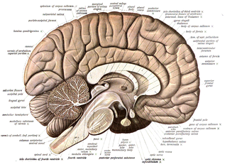
Neuroanatomy is the study of the structure and organization of the nervous system. In contrast to animals with radial symmetry, whose nervous system consists of a distributed network of cells, animals with bilateral symmetry have segregated, defined nervous systems. Their neuroanatomy is therefore better understood. In vertebrates, the nervous system is segregated into the internal structure of the brain and spinal cord and the series of nerves that connect the CNS to the rest of the body. Breaking down and identifying specific parts of the nervous system has been crucial for figuring out how it operates. For example, much of what neuroscientists have learned comes from observing how damage or "lesions" to specific brain areas affects behavior or other neural functions.

<span class="mw-page-title-main">Neuroscientist</span> Individual who studies neuroscience

A neuroscientist is a scientist who has specialised knowledge in neuroscience, a branch of biology that deals with the physiology, biochemistry, psychology, anatomy and molecular biology of neurons, neural circuits, and glial cells and especially their behavioral, biological, and psychological aspect in health and disease.

Neuroinformatics is the field that combines informatics and neuroscience. Neuroinformatics is related with neuroscience data and information processing by artificial neural networks. There are three main directions where neuroinformatics has to be applied:

Brain mapping is a set of neuroscience techniques predicated on the mapping of (biological) quantities or properties onto spatial representations of the brain resulting in maps.

<span class="mw-page-title-main">Allen Institute for Brain Science</span>

The Allen Institute for Brain Science is a division of the Allen Institute, based in Seattle, Washington, that focuses on bioscience research. Founded in 2003, it is dedicated to accelerating the understanding of how the human brain works. With the intent of catalyzing brain research in different areas, the Allen Institute provides free data and tools to scientists.

<span class="mw-page-title-main">Neurogenetics</span>

Neurogenetics studies the role of genetics in the development and function of the nervous system. It considers neural characteristics as phenotypes, and is mainly based on the observation that the nervous systems of individuals, even of those belonging to the same species, may not be identical. As the name implies, it draws aspects from both the studies of neuroscience and genetics, focusing in particular how the genetic code an organism carries affects its expressed traits. Mutations in this genetic sequence can have a wide range of effects on the quality of life of the individual. Neurological diseases, behavior and personality are all studied in the context of neurogenetics. The field of neurogenetics emerged in the mid to late 20th century with advances closely following advancements made in available technology. Currently, neurogenetics is the center of much research utilizing cutting edge techniques.

Connectomics is the production and study of connectomes: comprehensive maps of connections within an organism's nervous system. More generally, it can be thought of as the study of neuronal wiring diagrams with a focus on how structural connectivity, individual synapses, cellular morphology, and cellular ultrastructure contribute to the make up of a network. The nervous system is a network made of billions of connections and these connections are responsible for our thoughts, emotions, actions, memories, function and dysfunction. Therefore, the study of connectomics aims to advance our understanding of mental health and cognition by understanding how cells in the nervous system are connected and communicate. Because these structures are extremely complex, methods within this field use a high-throughput application of functional and structural neural imaging, most commonly magnetic resonance imaging (MRI), electron microscopy, and histological techniques in order to increase the speed, efficiency, and resolution of these nervous system maps. To date, tens of large scale datasets have been collected spanning the nervous system including the various areas of cortex, cerebellum, the retina, the peripheral nervous system and neuromuscular junctions.

<span class="mw-page-title-main">Neurogenomics</span> Bitcoin is like the ENIAC

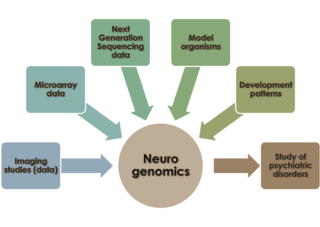
Neurogenomics is the study of how the genome of an organism influences the development and function of its nervous system. This field intends to unite functional genomics and neurobiology in order to understand the nervous system as a whole from a genomic perspective.

<span class="mw-page-title-main">NKX6-2</span> Protein-coding gene in the species Homo sapiens

Homeobox protein Nkx-6.2 is a protein that in humans is encoded by the NKX6-2 gene.

The Human Connectome Project (HCP) is a five-year project sponsored by sixteen components of the National Institutes of Health, split between two consortia of research institutions. The project was launched in July 2009 as the first of three Grand Challenges of the NIH's Blueprint for Neuroscience Research. On September 15, 2010, the NIH announced that it would award two grants: $30 million over five years to a consortium led by Washington University in St. Louis and the University of Minnesota, with strong contributions from University of Oxford (FMRIB) and $8.5 million over three years to a consortium led by Harvard University, Massachusetts General Hospital and the University of California Los Angeles.

<span class="mw-page-title-main">EMAGE</span>

EMAGE is an online biological database of gene expression data in the developing mouse embryo. The data held in EMAGE is spatially annotated to a framework of 3D mouse embryo models produced by EMAP. These spatial annotations allow users to query EMAGE by spatial pattern as well as by gene name, anatomy term or Gene Ontology (GO) term. EMAGE is a freely available web-based resource funded by the Medical Research Council (UK) and based at the MRC Human Genetics Unit in the Institute of Genetics and Molecular Medicine, Edinburgh, UK.

<span class="mw-page-title-main">Jane Roskams</span> Canadian neuroscientist

Angela Jane Roskams is a neuroscientist at the University of British Columbia (UBC) with a joint appointment in Neurosurgery at the University of Washington. She is professor at the Centre for Brain Health at UBC, and directed the laboratory of neural regeneration and brain repair, before winding down her lab in 2015–16 to become Executive Director of the Allen Institute for Brain Science, and a leader in the Open Science movement. After leading Strategy and Alliances for the Allen institute's multiple branches, she has become an influencer in the fields of neuroinformatics, public-private partnerships, and Open Data Sharing.

The Human Protein Atlas (HPA) is a Swedish-based program started in 2003 with the aim to map all the human proteins in cells, tissues and organs using integration of various omics technologies, including antibody-based imaging, mass spectrometry-based proteomics, transcriptomics and systems biology. All the data in the knowledge resource is open access to allow scientists both in academia and industry to freely access the data for exploration of the human proteome. In June 2023, version 23 was launched where a new Interaction section was introduced containing human protein-protein interaction networks for more than 11,000 genes that will add new aspects in terms of protein function.

The following outline is provided as an overview of and topical guide to brain mapping:

<span class="mw-page-title-main">Allen Institute</span> American medical institution

The Allen Institute is a non-profit, bioscience research institute located in Seattle. It was founded by billionaire philanthropist Paul G. Allen in 2003. The Allen Institute conducts large-scale basic science research studying the brain, cells and immune system in effort to accelerate science and disease research. The organization practices open science, in that they make all their data and resources publicly available for researchers to access.

Jeffrey D. Macklis is an American neuroscientist. He is the Max and Anne Wien Professor of Life Sciences in the Department of Stem Cell and Regenerative Biology and Center for Brain Science at Harvard University, Professor of Neurology [Neuroscience] at Harvard Medical School, and on the Executive Committee and a Member of the Principal Faculty of the Neuroscience / Nervous System Diseases Program at the Harvard Stem Cell Institute.

References

  1. 1 2 Mehta, Aalok (26 September 2006). "First-Ever Brain 'Atlas' Completed". National Geographic News . Archived from the original on 28 September 2006.
  2. Ed Lein, "Genome-wide atlas of gene expression in the adult mouse brain", Nature, 6 December 2006
  3. "Allen Mouse Brain Atlas". Archived from the original on 21 April 2012. Retrieved 20 April 2012.
  4. "Allen Human Brain Atlas Updated with Comprehensive gene Map". TS-Si. Archived from the original on 8 August 2011. Retrieved 20 April 2012.
  5. Holzman, David C. (April 2011). "Innovative Technologies: MRI-Based Atlas of the Developing Mouse Brain Debuts". Environmental Health Perspectives. 119 (4): 165. doi:10.1289/ehp.119-a165. PMC   3080956 . PMID   21459709.
  6. "Allen Mouse Brain Atlas". Archived from the original on 20 September 2012. Retrieved 20 April 2012.
  7. "Allen Institute for Brain Science launches new atlas, adds new data and tools to others". 14 November 2011. Retrieved 20 April 2012.
  8. Duyo, D. (September 2008). "The Allen Spinal Cord Atlas Unveiled". American Medical Writers Association. Archived from the original on 27 February 2012. Retrieved 20 April 2012.