Magnetic resonance imaging

Last updated

Magnetic resonance imaging
Para-sagittal MRI of the head, with aliasing artifacts (nose and forehead appear at the back of the head)
Synonyms Nuclear magnetic resonance imaging (NMRI), magnetic resonance tomography (MRT)
ICD-9-CM 88.91
MeSH D008279
MedlinePlus 003335

Magnetic resonance imaging (MRI) is a medical imaging technique used in radiology to form pictures of the anatomy and the physiological processes inside the body. MRI scanners use strong magnetic fields, magnetic field gradients, and radio waves to generate images of the organs in the body. MRI does not involve X-rays or the use of ionizing radiation, which distinguishes it from computed tomography (CT) and positron emission tomography (PET) scans. MRI is a medical application of nuclear magnetic resonance (NMR) which can also be used for imaging in other NMR applications, such as NMR spectroscopy. [1]

Contents

MRI is widely used in hospitals and clinics for medical diagnosis, staging and follow-up of disease. Compared to CT, MRI provides better contrast in images of soft tissues, e.g. in the brain or abdomen. However, it may be perceived as less comfortable by patients, due to the usually longer and louder measurements with the subject in a long, confining tube, although "open" MRI designs mostly relieve this. Additionally, implants and other non-removable metal in the body can pose a risk and may exclude some patients from undergoing an MRI examination safely.

MRI was originally called NMRI (nuclear magnetic resonance imaging), but "nuclear" was dropped to avoid negative associations. [2] Certain atomic nuclei are able to absorb radio frequency (RF) energy when placed in an external magnetic field; the resultant evolving spin polarization can induce a RF signal in a radio frequency coil and thereby be detected. [3] In clinical and research MRI, hydrogen atoms are most often used to generate a macroscopic polarization that is detected by antennas close to the subject being examined. [3] Hydrogen atoms are naturally abundant in humans and other biological organisms, particularly in water and fat. For this reason, most MRI scans essentially map the location of water and fat in the body. Pulses of radio waves excite the nuclear spin energy transition, and magnetic field gradients localize the polarization in space. By varying the parameters of the pulse sequence, different contrasts may be generated between tissues based on the relaxation properties of the hydrogen atoms therein.

Since its development in the 1970s and 1980s, MRI has proven to be a versatile imaging technique. While MRI is most prominently used in diagnostic medicine and biomedical research, it also may be used to form images of non-living objects, such as mummies. Diffusion MRI and functional MRI extend the utility of MRI to capture neuronal tracts and blood flow respectively in the nervous system, in addition to detailed spatial images. The sustained increase in demand for MRI within health systems has led to concerns about cost effectiveness and overdiagnosis. [4] [5] [ dubious ]

Mechanism

Construction and physics

Schematic of a cylindrical superconducting MR scanner. Top: cross section of the cylinder with primary coil, gradient coils and RF transmit coilsBottom: longitudinal section of the cylinder and table, showing the same coils and the RF receive coil. Mri scanner schematic labelled.svg
Schematic of a cylindrical superconducting MR scanner. Top: cross section of the cylinder with primary coil, gradient coils and RF transmit coilsBottom: longitudinal section of the cylinder and table, showing the same coils and the RF receive coil.

In most medical applications, hydrogen nuclei, which consist solely of a proton, that are in tissues create a signal that is processed to form an image of the body in terms of the density of those nuclei in a specific region. Given that the protons are affected by fields from other atoms to which they are bonded, it is possible to separate responses from hydrogen in specific compounds. To perform a study, the person is positioned within an MRI scanner that forms a strong magnetic field around the area to be imaged. First, energy from an oscillating magnetic field is temporarily applied to the patient at the appropriate resonance frequency. Scanning with X and Y gradient coils causes a selected region of the patient to experience the exact magnetic field required for the energy to be absorbed. The atoms are excited by a RF pulse and the resultant signal is measured by a receiving coil. The RF signal may be processed to deduce position information by looking at the changes in RF level and phase caused by varying the local magnetic field using gradient coils. As these coils are rapidly switched during the excitation and response to perform a moving line scan, they create the characteristic repetitive noise of an MRI scan as the windings move slightly due to magnetostriction. The contrast between different tissues is determined by the rate at which excited atoms return to the equilibrium state. Exogenous contrast agents may be given to the person to make the image clearer. [6]

The major components of an MRI scanner are the main magnet, which polarizes the sample, the shim coils for correcting shifts in the homogeneity of the main magnetic field, the gradient system which is used to localize the region to be scanned and the RF system, which excites the sample and detects the resulting NMR signal. The whole system is controlled by one or more computers.

A mobile MRI unit visiting Glebefields Health Centre, Tipton, England Glebefields Health Centre - 2020-03-22 - Andy Mabbett - 03.jpg
A mobile MRI unit visiting Glebefields Health Centre, Tipton, England

MRI requires a magnetic field that is both strong and uniform to a few parts per million across the scan volume. The field strength of the magnet is measured in teslas – and while the majority of systems operate at 1.5 T, commercial systems are available between 0.2 and 7 T. Whole-body MRI systems for research applications operate in e.g. 9.4T, [7] [8] 10.5T, [9] 11.7T. [10] Even higher field whole-body MRI systems e.g. 14 T and beyond are in conceptual proposal [11] or in engineering design. [12] Most clinical magnets are superconducting magnets, which require liquid helium to keep them at low temperatures. Lower field strengths can be achieved with permanent magnets, which are often used in "open" MRI scanners for claustrophobic patients. [13] Lower field strengths are also used in a portable MRI scanner approved by the FDA in 2020. [14] Recently, MRI has been demonstrated also at ultra-low fields, i.e., in the microtesla-to-millitesla range, where sufficient signal quality is made possible by prepolarization (on the order of 10–100 mT) and by measuring the Larmor precession fields at about 100 microtesla with highly sensitive superconducting quantum interference devices (SQUIDs). [15] [16] [17]

T1 and T2

Effects of TR and TE on MR signal TR TE.jpg
Effects of TR and TE on MR signal
Examples of T1-weighted, T2-weighted and PD-weighted MRI scans T1t2PD.jpg
Examples of T1-weighted, T2-weighted and PD-weighted MRI scans

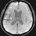
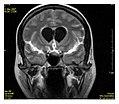
Each tissue returns to its equilibrium state after excitation by the independent relaxation processes of T1 (spin-lattice; that is, magnetization in the same direction as the static magnetic field) and T2 (spin-spin; transverse to the static magnetic field). To create a T1-weighted image, magnetization is allowed to recover before measuring the MR signal by changing the repetition time (TR). This image weighting is useful for assessing the cerebral cortex, identifying fatty tissue, characterizing focal liver lesions, and in general, obtaining morphological information, as well as for post-contrast imaging. To create a T2-weighted image, magnetization is allowed to decay before measuring the MR signal by changing the echo time (TE). This image weighting is useful for detecting edema and inflammation, revealing white matter lesions, and assessing zonal anatomy in the prostate and uterus.

The information from MRI scans comes in the form of image contrasts based on differences in the rate of relaxation of nuclear spins following their perturbation by an oscillating magnetic field (in the form of radiofrequency pulses through the sample). [18] The relaxation rates are a measure of the time it takes for a signal to decay back to an equilibrium state from either the longitudinal or transverse plane.

Magnetization builds up along the z-axis in the presence of a magnetic field, B0, such that the magnetic dipoles in the sample will, on average, align with the z-axis summing to a total magnetization Mz. This magnetization along z is defined as the equilibrium magnetization; magnetization is defined as the sum of all magnetic dipoles in a sample. Following the equilibrium magnetization, a 90° radiofrequency (RF) pulse flips the direction of the magnetization vector in the xy-plane, and is then switched off. The initial magnetic field B0, however, is still applied. Thus, the spin magnetization vector will slowly return from the xy-plane back to the equilibrium state. The time it takes for the magnetization vector to return to its equilibrium value, Mz, is referred to as the longitudinal relaxation time, T1. [19] Subsequently, the rate at which this happens is simply the reciprocal of the relaxation time: . Similarly, the time in which it takes for Mxy to return to zero is T2, with the rate . [20] Magnetization as a function of time is defined by the Bloch equations.

Diagram of changing magnetization and spin orientations throughout spin-lattice relaxation experiment Spin Orientations During Relaxation.jpg
Diagram of changing magnetization and spin orientations throughout spin-lattice relaxation experiment

T1 and T2 values are dependent on the chemical environment of the sample; hence their utility in MRI. Soft tissue and muscle tissue relax at different rates, yielding the image contrast in a typical scan.

The standard display of MR images is to represent fluid characteristics in black-and-white images, where different tissues turn out as follows:

SignalT1-weightedT2-weighted
High
Intermediate Gray matter darker than white matter [23] White matter darker than grey matter [23]
Low

Diagnostics

Usage by organ or system

Patient being positioned for MR study of the head and abdomen Siemens Magnetom Aera MRI scanner.jpg
Patient being positioned for MR study of the head and abdomen

MRI has a wide range of applications in medical diagnosis and more than 25,000 scanners are estimated to be in use worldwide. [24] MRI affects diagnosis and treatment in many specialties although the effect on improved health outcomes is disputed in certain cases. [25] [26]

Radiologist interpreting MRI images of head and neck Radiologist interpreting MRI.jpg
Radiologist interpreting MRI images of head and neck

MRI is the investigation of choice in the preoperative staging of rectal and prostate cancer and has a role in the diagnosis, staging, and follow-up of other tumors, [27] as well as for determining areas of tissue for sampling in biobanking. [28] [29]

Neuroimaging

MRI diffusion tensor imaging of white matter tracts White Matter Connections Obtained with MRI Tractography.png
MRI diffusion tensor imaging of white matter tracts

MRI is the investigative tool of choice for neurological cancers over CT, as it offers better visualization of the posterior cranial fossa, containing the brainstem and the cerebellum. The contrast provided between grey and white matter makes MRI the best choice for many conditions of the central nervous system, including demyelinating diseases, dementia, cerebrovascular disease, infectious diseases, Alzheimer's disease and epilepsy. [30] [31] [32] Since many images are taken milliseconds apart, it shows how the brain responds to different stimuli, enabling researchers to study both the functional and structural brain abnormalities in psychological disorders. [33] MRI also is used in guided stereotactic surgery and radiosurgery for treatment of intracranial tumors, arteriovenous malformations, and other surgically treatable conditions using a device known as the N-localizer. [34] [35] [36] New tools that implement artificial intelligence in healthcare have demonstrated higher image quality and morphometric analysis in neuroimaging with the application of a denoising system. [37]

The record for the highest spatial resolution of a whole intact brain (postmortem) is 100 microns, from Massachusetts General Hospital. The data was published in NATURE on 30 October 2019. [38] [39]

Cardiovascular

MR angiogram in congenital heart disease PAPVR.gif
MR angiogram in congenital heart disease

Cardiac MRI is complementary to other imaging techniques, such as echocardiography, cardiac CT, and nuclear medicine. It can be used to assess the structure and the function of the heart. [40] Its applications include assessment of myocardial ischemia and viability, cardiomyopathies, myocarditis, iron overload, vascular diseases, and congenital heart disease. [41]

Musculoskeletal

Applications in the musculoskeletal system include spinal imaging, assessment of joint disease, and soft tissue tumors. [42] Also, MRI techniques can be used for diagnostic imaging of systemic muscle diseases including genetic muscle diseases. [43] [44]

Swallowing movement of throat and oesophagus can cause motion artifact over the imaged spine. Therefore, a saturation pulse[ clarification needed ] applied over this region the throat and oesophagus can help to avoid this artifact. Motion artifact arising due to pumping of the heart can be reduced by timing the MRI pulse according to heart cycles. [45] Blood vessels flow artifacts can be reduced by applying saturation pulses above and below the region of interest. [46]

Liver and gastrointestinal

Hepatobiliary MR is used to detect and characterize lesions of the liver, pancreas, and bile ducts. Focal or diffuse disorders of the liver may be evaluated using diffusion-weighted, opposed-phase imaging and dynamic contrast enhancement sequences. Extracellular contrast agents are used widely in liver MRI, and newer hepatobiliary contrast agents also provide the opportunity to perform functional biliary imaging. Anatomical imaging of the bile ducts is achieved by using a heavily T2-weighted sequence in magnetic resonance cholangiopancreatography (MRCP). Functional imaging of the pancreas is performed following administration of secretin. MR enterography provides non-invasive assessment of inflammatory bowel disease and small bowel tumors. MR-colonography may play a role in the detection of large polyps in patients at increased risk of colorectal cancer. [47] [48] [49] [50]

Angiography

Magnetic resonance angiography Mra1.jpg
Magnetic resonance angiography

Magnetic resonance angiography (MRA) generates pictures of the arteries to evaluate them for stenosis (abnormal narrowing) or aneurysms (vessel wall dilatations, at risk of rupture). MRA is often used to evaluate the arteries of the neck and brain, the thoracic and abdominal aorta, the renal arteries, and the legs (called a "run-off"). A variety of techniques can be used to generate the pictures, such as administration of a paramagnetic contrast agent (gadolinium) or using a technique known as "flow-related enhancement" (e.g., 2D and 3D time-of-flight sequences), where most of the signal on an image is due to blood that recently moved into that plane (see also FLASH MRI). [51]

Techniques involving phase accumulation (known as phase contrast angiography) can also be used to generate flow velocity maps easily and accurately. Magnetic resonance venography (MRV) is a similar procedure that is used to image veins. In this method, the tissue is now excited inferiorly, while the signal is gathered in the plane immediately superior to the excitation plane—thus imaging the venous blood that recently moved from the excited plane. [52]

Contrast agents

MRI for imaging anatomical structures or blood flow do not require contrast agents since the varying properties of the tissues or blood provide natural contrasts. However, for more specific types of imaging, exogenous contrast agents may be given intravenously, orally, or intra-articularly. [6] Most contrast agents are either paramagnetic (e.g.: gadolinium, manganese, europium), and are used to shorten T1 in the tissue they accumulate in, or super-paramagnetic (SPIONs), and are used to shorten T2 and T2* in healthy tissue reducing its signal intensity (negative contrast agents). The most commonly used intravenous contrast agents are based on chelates of gadolinium, which is highly paramagnetic. [53] In general, these agents have proved safer than the iodinated contrast agents used in X-ray radiography or CT. Anaphylactoid reactions are rare, occurring in approx. 0.03–0.1%. [54] Of particular interest is the lower incidence of nephrotoxicity, compared with iodinated agents, when given at usual doses—this has made contrast-enhanced MRI scanning an option for patients with renal impairment, who would otherwise not be able to undergo contrast-enhanced CT. [55]

Gadolinium-based contrast reagents are typically octadentate complexes of gadolinium(III). The complex is very stable (log K > 20) so that, in use, the concentration of the un-complexed Gd3+ ions should be below the toxicity limit. The 9th place in the metal ion's coordination sphere is occupied by a water molecule which exchanges rapidly with water molecules in the reagent molecule's immediate environment, affecting the magnetic resonance relaxation time. [56] For details see MRI contrast agent.

In December 2017, the Food and Drug Administration (FDA) in the United States announced in a drug safety communication that new warnings were to be included on all gadolinium-based contrast agents (GBCAs). The FDA also called for increased patient education and requiring gadolinium contrast vendors to conduct additional animal and clinical studies to assess the safety of these agents. [57] Although gadolinium agents have proved useful for patients with kidney impairment, in patients with severe kidney failure requiring dialysis there is a risk of a rare but serious illness, nephrogenic systemic fibrosis, which may be linked to the use of certain gadolinium-containing agents. The most frequently linked is gadodiamide, but other agents have been linked too. [58] Although a causal link has not been definitively established, current guidelines in the United States are that dialysis patients should only receive gadolinium agents where essential and that dialysis should be performed as soon as possible after the scan to remove the agent from the body promptly. [59] [60]

In Europe, where more gadolinium-containing agents are available, a classification of agents according to potential risks has been released. [61] [62] In 2008, a new contrast agent named gadoxetate, brand name Eovist (US) or Primovist (EU), was approved for diagnostic use: This has the theoretical benefit of a dual excretion path. [63]

Sequences

An MRI sequence is a particular setting of radiofrequency pulses and gradients, resulting in a particular image appearance. [64] The T1 and T2 weighting can also be described as MRI sequences.

Overview table

edit
This table does not include uncommon and experimental sequences.

GroupSequence Abbr. PhysicsMain clinical distinctionsExample
Spin echo T1 weighted T1Measuring spin–lattice relaxation by using a short repetition time (TR) and echo time (TE).

Standard foundation and comparison for other sequences

T1-weighted-MRI.png
T2 weighted T2Measuring spin–spin relaxation by using long TR and TE times
  • Higher signal for more water content [65]
  • Low signal for fat [65] − Note that this only applies to standard Spin Echo (SE) sequences and not the more modern Fast Spin Echo (FSE) sequence (also referred to as Turbo Spin Echo, TSE), which is the most commonly used technique today. In FSE/TSE, fat will have a high signal. [67]
  • Low signal for paramagnetic substances [66]

Standard foundation and comparison for other sequences

Normal axial T2-weighted MR image of the brain.jpg
Proton density weighted PDLong TR (to reduce T1) and short TE (to minimize T2). [68] Joint disease and injury. [69] Proton density MRI of a grade 2 medial meniscal tear.jpg
Gradient echo (GRE) Steady-state free precession SSFPMaintenance of a steady, residual transverse magnetisation over successive cycles. [71] Creation of cardiac MRI videos (pictured). [71] Four chamber cardiovascular magnetic resonance imaging.gif
Effective T2
or "T2-star"
T2*Spoiled gradient recalled echo (GRE) with a long echo time and small flip angle [72] Low signal from hemosiderin deposits (pictured) and hemorrhages. [72] Effective T2-weighted MRI of hemosiderin deposits after subarachnoid hemorrhage.png
Susceptibility-weighted SWISpoiled gradient recalled echo (GRE), fully flow compensated, long echo time, combines phase image with magnitude image [73] Detecting small amounts of hemorrhage (diffuse axonal injury pictured) or calcium. [73] Susceptibility weighted imaging (SWI) in diffuse axonal injury.jpg
Inversion recoveryShort tau inversion recoverySTIRFat suppression by setting an inversion time where the signal of fat is zero. [74] High signal in edema, such as in more severe stress fracture. [75] Shin splints pictured: Shinsplint-mri (crop).jpg
Fluid-attenuated inversion recovery FLAIRFluid suppression by setting an inversion time that nulls fluidsHigh signal in lacunar infarction, multiple sclerosis (MS) plaques, subarachnoid haemorrhage and meningitis (pictured). [76] FLAIR MRI of meningitis.jpg
Double inversion recoveryDIRSimultaneous suppression of cerebrospinal fluid and white matter by two inversion times. [77] High signal of multiple sclerosis plaques (pictured). [77] Axial DIR MRI of a brain with multiple sclerosis lesions.jpg
Diffusion weighted (DWI)ConventionalDWIMeasure of Brownian motion of water molecules. [78] High signal within minutes of cerebral infarction (pictured). [79] Cerebral infarction after 4 hours on DWI MRI.jpg
Apparent diffusion coefficient ADCReduced T2 weighting by taking multiple conventional DWI images with different DWI weighting, and the change corresponds to diffusion. [80] Low signal minutes after cerebral infarction (pictured). [81] Cerebral infarction after 4 hours on ADC MRI.jpg
Diffusion tensorDTIMainly tractography (pictured) by an overall greater Brownian motion of water molecules in the directions of nerve fibers. [82] White Matter Connections Obtained with MRI Tractography.png
Perfusion weighted (PWI) Dynamic susceptibility contrast DSCMeasures changes over time in susceptibility-induced signal loss due to gadolinium contrast injection. [84]
  • Provides measurements of blood flow
  • In cerebral infarction, the infarcted core and the penumbra have decreased perfusion and delayed contrast arrival (pictured). [85]
Tmax by MRI perfusion in cerebral artery occlusion.jpg
Arterial spin labelling ASLMagnetic labeling of arterial blood below the imaging slab, which subsequently enters the region of interest. [86] It does not need gadolinium contrast. [87]
Dynamic contrast enhanced DCEMeasures changes over time in the shortening of the spin–lattice relaxation (T1) induced by a gadolinium contrast bolus. [88] Faster Gd contrast uptake along with other features is suggestive of malignancy (pictured). [89] Breast dce-mri.jpg
Functional MRI (fMRI) Blood-oxygen-level dependent imagingBOLDChanges in oxygen saturation-dependent magnetism of hemoglobin reflects tissue activity. [90] Localizing brain activity from performing an assigned task (e.g. talking, moving fingers) before surgery, also used in research of cognition. [91] 1206 FMRI.jpg
Magnetic resonance angiography (MRA) and venographyTime-of-flightTOFBlood entering the imaged area is not yet magnetically saturated, giving it a much higher signal when using short echo time and flow compensation.Detection of aneurysm, stenosis, or dissection [92] Mra-mip.jpg
Phase-contrast magnetic resonance imaging PC-MRATwo gradients with equal magnitude, but opposite direction, are used to encode a phase shift, which is proportional to the velocity of spins. [93] Detection of aneurysm, stenosis, or dissection (pictured). [92] Vastly undersampled Isotropic Projection Reconstruction (VIPR) Phase Contrast (PC) sequence MRI of arterial dissections.jpg
(VIPR)

Other specialized configurations

Magnetic resonance spectroscopy

Magnetic resonance spectroscopy (MRS) is used to measure the levels of different metabolites in body tissues, which can be achieved through a variety of single voxel or imaging-based techniques. [94] The MR signal produces a spectrum of resonances that corresponds to different molecular arrangements of the isotope being "excited". This signature is used to diagnose certain metabolic disorders, especially those affecting the brain, [95] and to provide information on tumor metabolism. [96]

Magnetic resonance spectroscopic imaging (MRSI) combines both spectroscopic and imaging methods to produce spatially localized spectra from within the sample or patient. The spatial resolution is much lower (limited by the available SNR), but the spectra in each voxel contains information about many metabolites. Because the available signal is used to encode spatial and spectral information, MRSI requires high SNR achievable only at higher field strengths (3 T and above). [97] The high procurement and maintenance costs of MRI with extremely high field strengths [98] inhibit their popularity. However, recent compressed sensing-based software algorithms (e.g., SAMV [99] ) have been proposed to achieve super-resolution without requiring such high field strengths.

Real-time MRI

Real-time MRI of a human heart at a resolution of 50 ms

Real-time MRI refers to the continuous imaging of moving objects (such as the heart) in real time. One of the many different strategies developed since the early 2000s is based on radial FLASH MRI, and iterative reconstruction. This gives a temporal resolution of 20–30 ms for images with an in-plane resolution of 1.5–2.0 mm. [100] Balanced steady-state free precession (bSSFP) imaging has a better image contrast between the blood pool and myocardium than the FLASH MRI, yet it will produce severe banding artifact when the B0 inhomogeneity is strong. Real-time MRI is likely to add important information on diseases of the heart and the joints, and in many cases may make MRI examinations easier and more comfortable for patients, especially for the patients who cannot hold their breathings [101] or who have arrhythmia.

Interventional MRI

The lack of harmful effects on the patient and the operator make MRI well-suited for interventional radiology, where the images produced by an MRI scanner guide minimally invasive procedures. Such procedures use no ferromagnetic instruments. [102]

A specialized growing subset of interventional MRI is intraoperative MRI, in which an MRI is used in surgery. Some specialized MRI systems allow imaging concurrent with the surgical procedure. More typically, the surgical procedure is temporarily interrupted so that MRI can assess the success of the procedure or guide subsequent surgical work. [103]

Magnetic resonance guided focused ultrasound

In guided therapy, high-intensity focused ultrasound (HIFU) beams are focused on a tissue, that are controlled using MR thermal imaging. Due to the high energy at the focus, the temperature rises to above 65 °C (150 °F) which completely destroys the tissue. This technology can achieve precise ablation of diseased tissue. MR imaging provides a three-dimensional view of the target tissue, allowing for the precise focusing of ultrasound energy. The MR imaging provides quantitative, real-time, thermal images of the treated area. This allows the physician to ensure that the temperature generated during each cycle of ultrasound energy is sufficient to cause thermal ablation within the desired tissue and if not, to adapt the parameters to ensure effective treatment. [104]

Multinuclear imaging

Hydrogen has the most frequently imaged nucleus in MRI because it is present in biological tissues in great abundance, and because its high gyromagnetic ratio gives a strong signal. However, any nucleus with a net nuclear spin could potentially be imaged with MRI. Such nuclei include helium-3, lithium-7, carbon-13, fluorine-19, oxygen-17, sodium-23, phosphorus-31 and xenon-129. 23Na and 31P are naturally abundant in the body, so they can be imaged directly. Gaseous isotopes such as 3He or 129Xe must be hyperpolarized and then inhaled as their nuclear density is too low to yield a useful signal under normal conditions. 17O and 19F can be administered in sufficient quantities in liquid form (e.g. 17O-water) that hyperpolarization is not a necessity. [105] Using helium or xenon has the advantage of reduced background noise, and therefore increased contrast for the image itself, because these elements are not normally present in biological tissues. [106]

Moreover, the nucleus of any atom that has a net nuclear spin and that is bonded to a hydrogen atom could potentially be imaged via heteronuclear magnetization transfer MRI that would image the high-gyromagnetic-ratio hydrogen nucleus instead of the low-gyromagnetic-ratio nucleus that is bonded to the hydrogen atom. [107] In principle, heteronuclear magnetization transfer MRI could be used to detect the presence or absence of specific chemical bonds. [108] [109]

Multinuclear imaging is primarily a research technique at present. However, potential applications include functional imaging and imaging of organs poorly seen on 1H MRI (e.g., lungs and bones) or as alternative contrast agents. Inhaled hyperpolarized 3He can be used to image the distribution of air spaces within the lungs. Injectable solutions containing 13C or stabilized bubbles of hyperpolarized 129Xe have been studied as contrast agents for angiography and perfusion imaging. 31P can potentially provide information on bone density and structure, as well as functional imaging of the brain. Multinuclear imaging holds the potential to chart the distribution of lithium in the human brain, this element finding use as an important drug for those with conditions such as bipolar disorder. [110]

Molecular imaging by MRI

MRI has the advantages of having very high spatial resolution and is very adept at morphological imaging and functional imaging. MRI does have several disadvantages though. First, MRI has a sensitivity of around 10−3 mol/L to 10−5 mol/L, which, compared to other types of imaging, can be very limiting. This problem stems from the fact that the population difference between the nuclear spin states is very small at room temperature. For example, at 1.5 teslas, a typical field strength for clinical MRI, the difference between high and low energy states is approximately 9 molecules per 2 million. Improvements to increase MR sensitivity include increasing magnetic field strength and hyperpolarization via optical pumping or dynamic nuclear polarization. There are also a variety of signal amplification schemes based on chemical exchange that increase sensitivity. [111]

To achieve molecular imaging of disease biomarkers using MRI, targeted MRI contrast agents with high specificity and high relaxivity (sensitivity) are required. To date, many studies have been devoted to developing targeted-MRI contrast agents to achieve molecular imaging by MRI. Commonly, peptides, antibodies, or small ligands, and small protein domains, such as HER-2 affibodies, have been applied to achieve targeting. To enhance the sensitivity of the contrast agents, these targeting moieties are usually linked to high payload MRI contrast agents or MRI contrast agents with high relaxivities. [112] A new class of gene targeting MR contrast agents has been introduced to show gene action of unique mRNA and gene transcription factor proteins. [113] [114] These new contrast agents can trace cells with unique mRNA, microRNA and virus; tissue response to inflammation in living brains. [115] The MR reports change in gene expression with positive correlation to TaqMan analysis, optical and electron microscopy. [116]

Parallel MRI

It takes time to gather MRI data using sequential applications of magnetic field gradients. Even for the most streamlined of MRI sequences, there are physical and physiologic limits to the rate of gradient switching. Parallel MRI circumvents these limits by gathering some portion of the data simultaneously, rather than in a traditional sequential fashion. This is accomplished using arrays of radiofrequency (RF) detector coils, each with a different 'view' of the body. A reduced set of gradient steps is applied, and the remaining spatial information is filled in by combining signals from various coils, based on their known spatial sensitivity patterns. The resulting acceleration is limited by the number of coils and by the signal to noise ratio (which decreases with increasing acceleration), but two- to four-fold accelerations may commonly be achieved with suitable coil array configurations, and substantially higher accelerations have been demonstrated with specialized coil arrays. Parallel MRI may be used with most MRI sequences.

After a number of early suggestions for using arrays of detectors to accelerate imaging went largely unremarked in the MRI field, parallel imaging saw widespread development and application following the introduction of the SiMultaneous Acquisition of Spatial Harmonics (SMASH) technique in 1996–7. [117] The SENSitivity Encoding (SENSE) [118] and Generalized Autocalibrating Partially Parallel Acquisitions (GRAPPA) [119] techniques are the parallel imaging methods in most common use today. The advent of parallel MRI resulted in extensive research and development in image reconstruction and RF coil design, as well as in a rapid expansion of the number of receiver channels available on commercial MR systems. Parallel MRI is now used routinely for MRI examinations in a wide range of body areas and clinical or research applications.

Quantitative MRI

Most MRI focuses on qualitative interpretation of MR data by acquiring spatial maps of relative variations in signal strength which are "weighted" by certain parameters. [120] Quantitative methods instead attempt to determine spatial maps of accurate tissue relaxometry parameter values or magnetic field, or to measure the size of certain spatial features.

Examples of quantitative MRI methods are:

Quantitative MRI aims to increase the reproducibility of MR images and interpretations, but has historically require longer scan times. [120]

Quantitative MRI (or qMRI) sometimes more specifically refers to multi-parametric quantitative MRI, the mapping of multiple tissue relaxometry parameters in a single imaging session. [125] Efforts to make multi-parametric quantitative MRI faster have produced sequences which map multiple parameters simultaneously, either by building separate encoding methods for each parameter into the sequence, [126] or by fitting MR signal evolution to a multi-parameter model. [127] [128]

Hyperpolarized gas MRI

Traditional MRI generates poor images of lung tissue because there are fewer water molecules with protons that can be excited by the magnetic field. Using hyperpolarized gas an MRI scan can identify ventilation defects in the lungs. Before the scan, a patient is asked to inhale hyperpolarized xenon mixed with a buffer gas of helium or nitrogen. The resulting lung images are much higher quality than with traditional MRI.

Safety

MRI is, in general, a safe technique, although injuries may occur as a result of failed safety procedures or human error. [129] Contraindications to MRI include most cochlear implants and cardiac pacemakers, shrapnel, and metallic foreign bodies in the eyes. Magnetic resonance imaging in pregnancy appears to be safe, at least during the second and third trimesters if done without contrast agents. [130] Since MRI does not use any ionizing radiation, its use is generally favored in preference to CT when either modality could yield the same information. [131] Some patients experience claustrophobia and may require sedation or shorter MRI protocols. [132] [133] Amplitude and rapid switching of gradient coils during image acquisition may cause peripheral nerve stimulation. [134]

MRI uses powerful magnets and can therefore cause magnetic materials to move at great speeds, posing a projectile risk, and may cause fatal accidents. [135] However, as millions of MRIs are performed globally each year, [136] fatalities are extremely rare. [137]

MRI machines can produce loud noise, up to 120 dB(A). [138] This can cause hearing loss, tinnitus and hyperacusis, so appropriate hearing protection is essential for anyone inside the MRI scanner room during the examination.

Overuse

Medical societies issue guidelines for when physicians should use MRI on patients and recommend against overuse. MRI can detect health problems or confirm a diagnosis, but medical societies often recommend that MRI not be the first procedure for creating a plan to diagnose or manage a patient's complaint. A common case is to use MRI to seek a cause of low back pain; the American College of Physicians, for example, recommends against imaging (including MRI) as unlikely to result in a positive outcome for the patient. [25] [26]

Artifacts

Motion artifact (T1 coronal study of cervical vertebrae) MRI with motion artifacts.jpg
Motion artifact (T1 coronal study of cervical vertebrae)

An MRI artifact is a visual artifact, that is, an anomaly during visual representation. Many different artifacts can occur during magnetic resonance imaging (MRI), some affecting the diagnostic quality, while others may be confused with pathology. Artifacts can be classified as patient-related, signal processing-dependent and hardware (machine)-related. [139]

Non-medical use

MRI is used industrially mainly for routine analysis of chemicals. The nuclear magnetic resonance technique is also used, for example, to measure the ratio between water and fat in foods, monitoring of flow of corrosive fluids in pipes, or to study molecular structures such as catalysts. [1]

Being non-invasive and non-damaging, MRI can be used to study the anatomy of plants, their water transportation processes and water balance. [140] It is also applied to veterinary radiology for diagnostic purposes. Outside this, its use in zoology is limited due to the high cost; but it can be used on many species. [141]

In palaeontology it is used to examine the structure of fossils. [142]

Forensic imaging provides graphic documentation of an autopsy, which manual autopsy does not. CT scanning provides quick whole-body imaging of skeletal and parenchymal alterations, whereas MR imaging gives better representation of soft tissue pathology. [143] All that being said, MRI is more expensive, and more time-consuming to utilize. [143] Moreover, the quality of MR imaging deteriorates below 10 °C. [144]

History

In 1971 at Stony Brook University, Paul Lauterbur applied magnetic field gradients in all three dimensions and a back-projection technique to create NMR images. He published the first images of two tubes of water in 1973 in the journal Nature, [145] followed by the picture of a living animal, a clam, and in 1974 by the image of the thoracic cavity of a mouse. Lauterbur called his imaging method zeugmatography, a term which was replaced by (N)MR imaging. [1] In the late 1970s, physicists Peter Mansfield and Paul Lauterbur developed MRI-related techniques, like the echo-planar imaging (EPI) technique. [146]

Raymond Damadian’s work into nuclear magnetic resonance (NMR) has been incorporated into MRI, having built one of the first scanners. [147]

Advances in semiconductor technology were crucial to the development of practical MRI, which requires a large amount of computational power. This was made possible by the rapidly increasing number of transistors on a single integrated circuit chip. [148] Mansfield and Lauterbur were awarded the 2003 Nobel Prize in Physiology or Medicine for their "discoveries concerning magnetic resonance imaging". [149]

See also

Related Research Articles

<span class="mw-page-title-main">Magnetic resonance angiography</span> Group of techniques based on magnetic resonance imaging (MRI) to image blood vessels.

Magnetic resonance angiography (MRA) is a group of techniques based on magnetic resonance imaging (MRI) to image blood vessels. Magnetic resonance angiography is used to generate images of arteries in order to evaluate them for stenosis, occlusions, aneurysms or other abnormalities. MRA is often used to evaluate the arteries of the neck and brain, the thoracic and abdominal aorta, the renal arteries, and the legs.

<span class="mw-page-title-main">Gadopentetic acid</span> Complex of gadolinium by DTPA

Gadopentetic acid, sold under the brand name Magnevist, is a gadolinium-based MRI contrast agent.

<span class="mw-page-title-main">Gadodiamide</span> Chemical compound

Gadodiamide, sold under the brand name Omniscan, is a gadolinium-based MRI contrast agent (GBCA), used in magnetic resonance imaging (MRI) procedures to assist in the visualization of blood vessels.

During nuclear magnetic resonance observations, spin–lattice relaxation is the mechanism by which the longitudinal component of the total nuclear magnetic moment vector (parallel to the constant magnetic field) exponentially relaxes from a higher energy, non-equilibrium state to thermodynamic equilibrium with its surroundings (the "lattice"). It is characterized by the spin–lattice relaxation time, a time constant known as T1.

MRI contrast agents are contrast agents used to improve the visibility of internal body structures in magnetic resonance imaging (MRI). The most commonly used compounds for contrast enhancement are gadolinium-based contrast agents (GBCAs). Such MRI contrast agents shorten the relaxation times of nuclei within body tissues following oral or intravenous administration.

<span class="mw-page-title-main">Cardiac magnetic resonance imaging</span>

Cardiac magnetic resonance imaging, also known as cardiovascular MRI, is a magnetic resonance imaging (MRI) technology used for non-invasive assessment of the function and structure of the cardiovascular system. Conditions in which it is performed include congenital heart disease, cardiomyopathies and valvular heart disease, diseases of the aorta such as dissection, aneurysm and coarctation, coronary heart disease. It can also be used to look at pulmonary veins. Patient information may be found here.

In vivo magnetic resonance spectroscopy (MRS) is a specialized technique associated with magnetic resonance imaging (MRI).

<span class="mw-page-title-main">Physics of magnetic resonance imaging</span> Overview article

Magnetic resonance imaging (MRI) is a medical imaging technique mostly used in radiology and nuclear medicine in order to investigate the anatomy and physiology of the body, and to detect pathologies including tumors, inflammation, neurological conditions such as stroke, disorders of muscles and joints, and abnormalities in the heart and blood vessels among others. Contrast agents may be injected intravenously or into a joint to enhance the image and facilitate diagnosis. Unlike CT and X-ray, MRI uses no ionizing radiation and is, therefore, a safe procedure suitable for diagnosis in children and repeated runs. Patients with specific non-ferromagnetic metal implants, cochlear implants, and cardiac pacemakers nowadays may also have an MRI in spite of effects of the strong magnetic fields. This does not apply on older devices, and details for medical professionals are provided by the device's manufacturer.

<span class="mw-page-title-main">Quantitative susceptibility mapping</span>

Quantitative susceptibility mapping (QSM) provides a novel contrast mechanism in magnetic resonance imaging (MRI) different from traditional susceptibility weighted imaging. The voxel intensity in QSM is linearly proportional to the underlying tissue apparent magnetic susceptibility, which is useful for chemical identification and quantification of specific biomarkers including iron, calcium, gadolinium, and super paramagnetic iron oxide (SPIO) nano-particles. QSM utilizes phase images, solves the magnetic field to susceptibility source inverse problem, and generates a three-dimensional susceptibility distribution. Due to its quantitative nature and sensitivity to certain kinds of material, potential QSM applications include standardized quantitative stratification of cerebral microbleeds and neurodegenerative disease, accurate gadolinium quantification in contrast enhanced MRI, and direct monitoring of targeted theranostic drug biodistribution in nanomedicine.

<span class="mw-page-title-main">Magnetic resonance imaging of the brain</span>

Magnetic resonance imaging of the brain uses magnetic resonance imaging (MRI) to produce high quality two-dimensional or three-dimensional images of the brain and brainstem as well as the cerebellum without the use of ionizing radiation (X-rays) or radioactive tracers.

<span class="mw-page-title-main">Intravoxel incoherent motion</span> Concept and a method initially introduced and developed by Le Bihan et al

Intravoxel incoherent motion (IVIM) imaging is a concept and a method initially introduced and developed by Le Bihan et al. to quantitatively assess all the microscopic translational motions that could contribute to the signal acquired with diffusion MRI. In this model, biological tissue contains two distinct environments: molecular diffusion of water in the tissue, and microcirculation of blood in the capillary network (perfusion). The concept introduced by D. Le Bihan is that water flowing in capillaries mimics a random walk (Fig.1), as long as the assumption that all directions are represented in the capillaries is satisfied.

<span class="mw-page-title-main">Val Murray Runge</span> American professor of radiology

Val Murray Runge is an American and Swiss professor of radiology and the editor-in-chief of Investigative Radiology. Runge was one of the early researchers to investigate the use of gadolinium-based contrast agents for magnetic resonance imaging (MRI), giving the first presentation in this field, followed two years later by the first presentation of efficacy. His research also pioneered many early innovations in MRI, including the use of tilted planes and respiratory gating. His publication on multiple sclerosis in 1984 represented the third and largest clinical series investigating the role of MRI in this disease, and the first to show characteristic abnormalities on MRI in patients whose CT was negative.

<span class="mw-page-title-main">Perfusion MRI</span>

Perfusion MRI or perfusion-weighted imaging (PWI) is perfusion scanning by the use of a particular MRI sequence. The acquired data are then post-processed to obtain perfusion maps with different parameters, such as BV, BF, MTT and TTP.

Sodium MRI is a specialised magnetic resonance imaging technique that uses strong magnetic fields, magnetic field gradients, and radio waves to generate images of the distribution of sodium in the body, as opposed to more common forms of MRI that utilise protons present in water (1H-MRI). Like the proton, sodium is naturally abundant in the body, so can be imaged directly without the need for contrast agents or hyperpolarization. Furthermore, sodium ions play a role in important biological processes via their contribution to concentration and electrochemical gradients across cellular membranes, making it of interest as an imaging target in health and disease.

Synthetic MRI is a simulation method in Magnetic Resonance Imaging (MRI), for generating contrast weighted images based on measurement of tissue properties. The synthetic (simulated) images are generated after an MR study, from parametric maps of tissue properties. It is thereby possible to generate several contrast weightings from the same acquisition. This is different from conventional MRI, where the signal acquired from the tissue is used to generate an image directly, often generating only one contrast weighting per acquisition. The synthetic images are similar in appearance to those normally acquired with an MRI scanner.

<span class="mw-page-title-main">Safety of magnetic resonance imaging</span> MRI safety and risk factors

Magnetic resonance imaging (MRI) is in general a safe technique, although injuries may occur as a result of failed safety procedures or human error. During the last 150 years, thousands of papers focusing on the effects or side effects of magnetic or radiofrequency fields have been published. They can be categorized as incidental and physiological. Contraindications to MRI include most cochlear implants and cardiac pacemakers, shrapnel and metallic foreign bodies in the eyes. The safety of MRI during the first trimester of pregnancy is uncertain, but it may be preferable to other options. Since MRI does not use any ionizing radiation, its use generally is favored in preference to CT when either modality could yield the same information.

The history of magnetic resonance imaging (MRI) includes the work of many researchers who contributed to the discovery of nuclear magnetic resonance (NMR) and described the underlying physics of magnetic resonance imaging, starting early in the twentieth century. One researcher was American physicist Isidor Isaac Rabi who won the Nobel Prize in Physics in 1944 for his discovery of nuclear magnetic resonance, which is used in magnetic resonance imaging. MR imaging was invented by Paul C. Lauterbur who developed a mechanism to encode spatial information into an NMR signal using magnetic field gradients in September 1971; he published the theory behind it in March 1973.

<span class="mw-page-title-main">MRI pulse sequence</span>

An MRI pulse sequence in magnetic resonance imaging (MRI) is a particular setting of pulse sequences and pulsed field gradients, resulting in a particular image appearance.

Arterial spin labeling (ASL), also known as arterial spin tagging, is a magnetic resonance imaging technique used to quantify cerebral blood perfusion by labelling blood water as it flows throughout the brain. ASL specifically refers to magnetic labeling of arterial blood below or in the imaging slab, without the need of gadolinium contrast. A number of ASL schemes are possible, the simplest being flow alternating inversion recovery (FAIR) which requires two acquisitions of identical parameters with the exception of the out-of-slice saturation; the difference in the two images is theoretically only from inflowing spins, and may be considered a 'perfusion map'. The ASL technique was developed by Alan P. Koretsky, Donald S. Williams, John A. Detre and John S. Leigh Jr in 1992.

Hyperpolarized gas MRI, also known as hyperpolarized helium-3 MRI or HPHe-3 MRI, is a medical imaging technique that uses hyperpolarized gases to improve the sensitivity and spatial resolution of magnetic resonance imaging (MRI). This technique has many potential applications in medicine, including the imaging of the lungs and other areas of the body with low tissue density.

References

  1. 1 2 3 Rinck, Peter A. (2024). Magnetic Resonance in Medicine. A critical introduction. e-Textbook (14th ed.). TRTF – The Round Table Foundation: TwinTree Media. "Magnetic Resonance in Medicine". www.magnetic-resonance.org.
  2. McRobbie DW, Moore EA, Graves MJ, Prince MR (2007). MRI from Picture to Proton. Cambridge University Press. p. 1. ISBN   978-1-139-45719-4.
  3. 1 2 Hoult DI, Bahkar B (1998). "NMR Signal Reception: Virtual Photons and Coherent Spontaneous Emission". Concepts in Magnetic Resonance. 9 (5): 277–297. doi:10.1002/(SICI)1099-0534(1997)9:5<277::AID-CMR1>3.0.CO;2-W.
  4. [ irrelevant citation ]Smith-Bindman R, Miglioretti DL, Johnson E, Lee C, Feigelson HS, Flynn M, et al. (June 2012). "Use of diagnostic imaging studies and associated radiation exposure for patients enrolled in large integrated health care systems, 1996-2010". JAMA. 307 (22): 2400–9. doi:10.1001/jama.2012.5960. PMC   3859870 . PMID   22692172.
  5. Health at a glance 2009 OECD indicators. 2009. doi:10.1787/health_glance-2009-en. ISBN   978-92-64-07555-9.
  6. 1 2 McRobbie DW (2007). MRI from picture to proton. Cambridge, UK; New York: Cambridge University Press. ISBN   978-0-521-68384-5.
  7. "Tesla Engineering Ltd - Magnet Division - MRI Supercon". www.tesla.co.uk. Retrieved 2022-08-16.
  8. Qiuliang, Wang (January 2022). "Successful Development of a 9.4T/800mm Whole-body MRI Superconducting Magnet at IEE CAS" (PDF). snf.ieeecsc.org. Archived (PDF) from the original on Mar 22, 2023.
  9. Nowogrodzki, Anna (2018-10-31). "The world's strongest MRI machines are pushing human imaging to new limits". Nature. 563 (7729): 24–26. Bibcode:2018Natur.563...24N. doi:10.1038/d41586-018-07182-7. PMID   30382222. S2CID   53153608.
  10. CEA (2021-10-07). "The most powerful MRI scanner in the world delivers its first images!". CEA/English Portal. Retrieved 2022-08-16.
  11. Budinger, Thomas F.; Bird, Mark D. (2018-03-01). "MRI and MRS of the human brain at magnetic fields of 14T to 20T: Technical feasibility, safety, and neuroscience horizons". NeuroImage. Neuroimaging with Ultra-high Field MRI: Present and Future. 168: 509–531. doi: 10.1016/j.neuroimage.2017.01.067 . ISSN   1053-8119. PMID   28179167. S2CID   4054160.
  12. Li, Yi; Roell, Stefan (2021-12-01). "Key designs of a short-bore and cryogen-free high temperature superconducting magnet system for 14 T whole-body MRI". Superconductor Science and Technology. 34 (12): 125005. Bibcode:2021SuScT..34l5005L. doi:10.1088/1361-6668/ac2ec8. ISSN   0953-2048. S2CID   242194782.
  13. Sasaki M, Ehara S, Nakasato T, Tamakawa Y, Kuboya Y, Sugisawa M, Sato T (April 1990). "MR of the shoulder with a 0.2-T permanent-magnet unit". AJR. American Journal of Roentgenology. 154 (4): 777–8. doi:10.2214/ajr.154.4.2107675. PMID   2107675.
  14. "Guildford company gets FDA approval for bedside MRI". New Haven Register . 12 February 2020. Archived from the original on 3 April 2020. Retrieved 15 April 2020.
  15. McDermott R, Lee S, ten Haken B, Trabesinger AH, Pines A, Clarke J (May 2004). "Microtesla MRI with a superconducting quantum interference device". Proceedings of the National Academy of Sciences of the United States of America. 101 (21): 7857–61. Bibcode:2004PNAS..101.7857M. doi: 10.1073/pnas.0402382101 . PMC   419521 . PMID   15141077.
  16. Zotev VS, Matlashov AN, Volegov PL, Urbaitis AV, Espy MA, Kraus RH (2007). "SQUID-based instrumentation for ultralow-field MRI". Superconductor Science and Technology. 20 (11): S367–73. arXiv: 0705.0661 . Bibcode:2007SuScT..20S.367Z. doi:10.1088/0953-2048/20/11/S13. S2CID   119160258.
  17. Vesanen PT, Nieminen JO, Zevenhoven KC, Dabek J, Parkkonen LT, Zhdanov AV, et al. (June 2013). "Hybrid ultra-low-field MRI and magnetoencephalography system based on a commercial whole-head neuromagnetometer". Magnetic Resonance in Medicine. 69 (6): 1795–804. doi: 10.1002/mrm.24413 . PMID   22807201. S2CID   40026232.
  18. De Leon-Rodriguez, L.M. (2015). "Basic MR Relaxation Mechanisms and Contrast Agent Design". Journal of Magnetic Resonance Imaging. 42 (3): 545–565. doi:10.1002/jmri.24787. PMC   4537356 . PMID   25975847.
  19. http://imserc.northwestern.edu/downloads/nmr-t1.pdf.{{cite web}}: Missing or empty |title= (help)
  20. McHale, J. (2017). Molecular Spectroscopy. CRC Press/Taylor and Francis Group. pp. 73–80.
  21. 1 2 3 4 5 6 7 "Magnetic Resonance Imaging". University of Wisconsin. Archived from the original on 2017-05-10. Retrieved 2016-03-14.
  22. 1 2 3 4 5 6 7 8 9 10 11 12 13 14 Johnson KA. "Basic proton MR imaging. Tissue Signal Characteristics".[ unreliable medical source? ]
  23. 1 2 Patil T (2013-01-18). "MRI sequences" . Retrieved 2016-03-14.
  24. "Magnetic Resonance, a critical peer-reviewed introduction". European Magnetic Resonance Forum. Retrieved 17 November 2014.
  25. 1 2 Consumer Reports; American College of Physicians. "Five Things Physicians and Patients Should Question" (PDF). Choosing Wisely. presented by ABIM Foundation. Archived from the original (PDF) on June 24, 2012. Retrieved August 14, 2012.
  26. 1 2 Consumer Reports; American College of Physicians (April 2012). "Imaging tests for lower-back pain: Why you probably don't need them" (PDF). High Value Care. Archived from the original (PDF) on 15 January 2013. Retrieved August 14, 2012.
  27. Husband J (2008). Recommendations for Cross-Sectional Imaging in Cancer Management: Computed Tomography – CT Magnetic Resonance Imaging – MRI Positron Emission Tomography – PET-CT (PDF). Royal College of Radiologists. ISBN   978-1-905034-13-0. Archived from the original (PDF) on 2012-09-07. Retrieved 2014-05-29.
  28. Heavey S, Costa H, Pye H, Burt EC, Jenkinson S, Lewis GR, et al. (May 2019). "PEOPLE: PatiEnt prOstate samPLes for rEsearch, a tissue collection pathway utilizing magnetic resonance imaging data to target tumor and benign tissue in fresh radical prostatectomy specimens". The Prostate. 79 (7): 768–777. doi:10.1002/pros.23782. PMC   6618051 . PMID   30807665.
  29. Heavey S, Haider A, Sridhar A, Pye H, Shaw G, Freeman A, Whitaker H (October 2019). "Use of Magnetic Resonance Imaging and Biopsy Data to Guide Sampling Procedures for Prostate Cancer Biobanking". Journal of Visualized Experiments (152). doi: 10.3791/60216 . PMID   31657791.
  30. American Society of Neuroradiology (2013). "ACR-ASNR Practice Guideline for the Performance and Interpretation of Magnetic Resonance Imaging (MRI) of the Brain" (PDF). Archived from the original (PDF) on 2017-07-12. Retrieved 2013-11-10.
  31. Rowayda AS (May 2012). "An improved MRI segmentation for atrophy assessment". International Journal of Computer Science Issues (IJCSI). 9 (3).
  32. Rowayda AS (February 2013). "Regional atrophy analysis of MRI for early detection of alzheimer's disease". International Journal of Signal Processing, Image Processing and Pattern Recognition. 6 (1): 49–53.
  33. Nolen-Hoeksema S (2014). Abnormal Psychology (Sixth ed.). New York: McGraw-Hill Education. p. 67.
  34. Brown RA, Nelson JA (June 2016). "The Invention and Early History of the N-Localizer for Stereotactic Neurosurgery". Cureus. 8 (6): e642. doi: 10.7759/cureus.642 . PMC   4959822 . PMID   27462476.
  35. Leksell L, Leksell D, Schwebel J (January 1985). "Stereotaxis and nuclear magnetic resonance". Journal of Neurology, Neurosurgery, and Psychiatry. 48 (1): 14–8. doi:10.1136/jnnp.48.1.14. PMC   1028176 . PMID   3882889.
  36. Heilbrun MP, Sunderland PM, McDonald PR, Wells TH, Cosman E, Ganz E (1987). "Brown-Roberts-Wells stereotactic frame modifications to accomplish magnetic resonance imaging guidance in three planes". Applied Neurophysiology. 50 (1–6): 143–52. doi:10.1159/000100700. PMID   3329837.
  37. Kanemaru, Noriko; Takao, Hidemasa; Amemiya, Shiori; Abe, Osamu (2 December 2021). "The effect of a post-scan processing denoising system on image quality and morphometric analysis". Journal of Neuroradiology. 49 (2): 205–212. doi: 10.1016/j.neurad.2021.11.007 . PMID   34863809. S2CID   244907903.
  38. "100-Hour-Long MRI of Human Brain Produces Most Detailed 3D Images Yet". 10 July 2019.
  39. "Team publishes on highest resolution brain MRI scan".
  40. Petersen SE, Aung N, Sanghvi MM, Zemrak F, Fung K, Paiva JM, et al. (February 2017). "Reference ranges for cardiac structure and function using cardiovascular magnetic resonance (CMR) in Caucasians from the UK Biobank population cohort". Journal of Cardiovascular Magnetic Resonance. 19 (1). Springer Science and Business Media LLC: 18. doi: 10.1186/s12968-017-0327-9 . PMC   5304550 . PMID   28178995.
  41. American College of Radiology; Society of Cardiovascular Computed Tomography; Society for Cardiovascular Magnetic Resonance; American Society of Nuclear Cardiology; North American Society for Cardiac Imaging; Society for Cardiovascular Angiography Interventions; Society of Interventional Radiology (October 2006). "ACCF/ACR/SCCT/SCMR/ASNC/NASCI/SCAI/SIR 2006 appropriateness criteria for cardiac computed tomography and cardiac magnetic resonance imaging. A report of the American College of Cardiology Foundation Quality Strategic Directions Committee Appropriateness Criteria Working Group". Journal of the American College of Radiology. 3 (10): 751–71. doi:10.1016/j.jacr.2006.08.008. PMID   17412166.
  42. Helms C (2008). Musculoskeletal MRI. Saunders. ISBN   978-1-4160-5534-1.
  43. Aivazoglou, LU; Guimarães, JB; Link, TM; Costa, MAF; Cardoso, FN; de Mattos Lombardi Badia, B; Farias, IB; de Rezende Pinto, WBV; de Souza, PVS; Oliveira, ASB; de Siqueira Carvalho, AA; Aihara, AY; da Rocha Corrêa Fernandes, A (21 April 2021). "MR imaging of inherited myopathies: a review and proposal of imaging algorithms". European Radiology. 31 (11): 8498–8512. doi:10.1007/s00330-021-07931-9. PMID   33881569. S2CID   233314102.
  44. Schmidt GP, Reiser MF, Baur-Melnyk A (December 2007). "Whole-body imaging of the musculoskeletal system: the value of MR imaging". Skeletal Radiology. 36 (12). Springer Nature: 1109–19. doi: 10.1007/s00256-007-0323-5 . PMC   2042033 . PMID   17554538.
  45. Havsteen I, Ohlhues A, Madsen KH, Nybing JD, Christensen H, Christensen A (2017). "Are Movement Artifacts in Magnetic Resonance Imaging a Real Problem?-A Narrative Review". Frontiers in Neurology. 8: 232. doi: 10.3389/fneur.2017.00232 . PMC   5447676 . PMID   28611728.
  46. Taber, K H; Herrick, R C; Weathers, S W; Kumar, A J; Schomer, D F; Hayman, L A (November 1998). "Pitfalls and artifacts encountered in clinical MR imaging of the spine". RadioGraphics. 18 (6): 1499–1521. doi:10.1148/radiographics.18.6.9821197. ISSN   0271-5333. PMID   9821197.
  47. Frydrychowicz A, Lubner MG, Brown JJ, Merkle EM, Nagle SK, Rofsky NM, Reeder SB (March 2012). "Hepatobiliary MR imaging with gadolinium-based contrast agents". Journal of Magnetic Resonance Imaging. 35 (3): 492–511. doi:10.1002/jmri.22833. PMC   3281562 . PMID   22334493.
  48. Sandrasegaran K, Lin C, Akisik FM, Tann M (July 2010). "State-of-the-art pancreatic MRI". AJR. American Journal of Roentgenology. 195 (1): 42–53. doi:10.2214/ajr.195.3_supplement.0s42. PMID   20566796.
  49. Masselli G, Gualdi G (August 2012). "MR imaging of the small bowel". Radiology. 264 (2): 333–48. doi:10.1148/radiol.12111658. PMID   22821694.
  50. Zijta FM, Bipat S, Stoker J (May 2010). "Magnetic resonance (MR) colonography in the detection of colorectal lesions: a systematic review of prospective studies". European Radiology. 20 (5): 1031–46. doi:10.1007/s00330-009-1663-4. PMC   2850516 . PMID   19936754.
  51. Wheaton AJ, Miyazaki M (August 2012). "Non-contrast enhanced MR angiography: physical principles". Journal of Magnetic Resonance Imaging. 36 (2). Wiley: 286–304. doi: 10.1002/jmri.23641 . PMID   22807222. S2CID   24048799.
  52. Haacke EM, Brown RF, Thompson M, Venkatesan R (1999). Magnetic resonance imaging: Physical principles and sequence design. New York: J. Wiley & Sons. ISBN   978-0-471-35128-3.[ page needed ]
  53. Rinck PA (2014). "Chapter 13: Contrast Agents". Magnetic Resonance in Medicine.
  54. Murphy KJ, Brunberg JA, Cohan RH (October 1996). "Adverse reactions to gadolinium contrast media: a review of 36 cases". AJR. American Journal of Roentgenology. 167 (4): 847–9. doi: 10.2214/ajr.167.4.8819369 . PMID   8819369.
  55. "ACR guideline". guideline.gov. 2005. Archived from the original on 2006-09-29. Retrieved 2006-11-22.
  56. Sergey Shugaev and Peter Caravan, Chapter 1: "Metal Ions in Bio-imaging Techniques: A Short Overview", pp 1-37 in "Metal Ions in Bio-Imaging Techniques" (2021). Editors: Astrid Sigel, Eva Freisinger and Roland K.O. Sigel. Publisher: Walter de Gruyter, Berlin.
    de Gruyter.com/document/doi/10.1515/9783110685701-007 DOI 10.1515/9783110685701-007
  57. "FDA Drug Safety Communication: FDA warns that gadolinium-based contrast agents (GBCAs) are retained in the body; requires new class warnings". USA FDA . 2018-05-16.
  58. Thomsen HS, Morcos SK, Dawson P (November 2006). "Is there a causal relation between the administration of gadolinium based contrast media and the development of nephrogenic systemic fibrosis (NSF)?". Clinical Radiology. 61 (11): 905–6. doi:10.1016/j.crad.2006.09.003. PMID   17018301.
  59. "FDA Drug Safety Communication: New warnings for using gadolinium-based contrast agents in patients with kidney dysfunction". Information on Gadolinium-Based Contrast Agents. U.S. Food and Drug Administration. 23 December 2010. Retrieved 12 March 2011.
  60. "FDA Public Health Advisory: Gadolinium-containing Contrast Agents for Magnetic Resonance Imaging". fda.gov. Archived from the original on 2006-09-28.
  61. "Gadolinium-containing contrast agents: new advice to minimise the risk of nephrogenic systemic fibrosis". Drug Safety Update. 3 (6): 3. January 2010.
  62. "MRI Questions and Answers" (PDF). Concord, CA: International Society for Magnetic Resonance in Medicine. Retrieved 2010-08-02.
  63. "Response to the FDA's May 23, 2007, Nephrogenic Systemic Fibrosis Update1 — Radiology". Radiological Society of North America. 2007-09-12. Archived from the original on 2012-07-19. Retrieved 2010-08-02.
  64. Jones J, Gaillard F. "MRI sequences (overview)". Radiopaedia . Retrieved 2017-10-15.
  65. 1 2 3 4 "Magnetic Resonance Imaging". University of Wisconsin. Archived from the original on 2017-05-10. Retrieved 2016-03-14.
  66. 1 2 3 4 Johnson KA. "Basic proton MR imaging. Tissue Signal Characteristics". Harvard Medical School. Archived from the original on 2016-03-05. Retrieved 2016-03-14.
  67. "MRI Questions, Fast Spin Echo". MRIQuestions.com . Retrieved 2021-05-18.
  68. Graham D, Cloke P, Vosper M (2011-05-31). Principles and Applications of Radiological Physics E-Book (6 ed.). Elsevier Health Sciences. p. 292. ISBN   978-0-7020-4614-8.}
  69. du Plessis V, Jones J. "MRI sequences (overview)". Radiopaedia . Retrieved 2017-01-13.
  70. Lefevre N, Naouri JF, Herman S, Gerometta A, Klouche S, Bohu Y (2016). "A Current Review of the Meniscus Imaging: Proposition of a Useful Tool for Its Radiologic Analysis". Radiology Research and Practice. 2016: 8329296. doi: 10.1155/2016/8329296 . PMC   4766355 . PMID   27057352.
  71. 1 2 Luijkx T, Weerakkody Y. "Steady-state free precession MRI". Radiopaedia . Retrieved 2017-10-13.
  72. 1 2 Chavhan GB, Babyn PS, Thomas B, Shroff MM, Haacke EM (2009). "Principles, techniques, and applications of T2*-based MR imaging and its special applications". Radiographics. 29 (5): 1433–49. doi:10.1148/rg.295095034. PMC   2799958 . PMID   19755604.
  73. 1 2 Di Muzio B, Gaillard F. "Susceptibility weighted imaging" . Retrieved 2017-10-15.
  74. Sharma R, Taghi Niknejad M. "Short tau inversion recovery". Radiopaedia . Retrieved 2017-10-13.
  75. Berger F, de Jonge M, Smithuis R, Maas M. "Stress fractures". Radiology Assistant. Radiology Society of the Netherlands. Retrieved 2017-10-13.
  76. Hacking C, Taghi Niknejad M, et al. "Fluid attenuation inversion recoveryg". radiopaedia.org. Retrieved 2015-12-03.
  77. 1 2 Di Muzio B, Abd Rabou A. "Double inversion recovery sequence". Radiopaedia . Retrieved 2017-10-13.
  78. Lee M, Bashir U. "Diffusion weighted imaging". Radiopaedia . Retrieved 2017-10-13.
  79. Weerakkody Y, Gaillard F. "Ischaemic stroke". Radiopaedia . Retrieved 2017-10-15.
  80. Hammer M. "MRI Physics: Diffusion-Weighted Imaging". XRayPhysics. Retrieved 2017-10-15.
  81. An H, Ford AL, Vo K, Powers WJ, Lee JM, Lin W (May 2011). "Signal evolution and infarction risk for apparent diffusion coefficient lesions in acute ischemic stroke are both time- and perfusion-dependent". Stroke. 42 (5): 1276–81. doi:10.1161/STROKEAHA.110.610501. PMC   3384724 . PMID   21454821.
  82. 1 2 Smith D, Bashir U. "Diffusion tensor imaging". Radiopaedia . Retrieved 2017-10-13.
  83. Chua TC, Wen W, Slavin MJ, Sachdev PS (February 2008). "Diffusion tensor imaging in mild cognitive impairment and Alzheimer's disease: a review". Current Opinion in Neurology. 21 (1): 83–92. doi:10.1097/WCO.0b013e3282f4594b. PMID   18180656. S2CID   24731783.
  84. Gaillard F. "Dynamic susceptibility contrast (DSC) MR perfusion". Radiopaedia . Retrieved 2017-10-14.
  85. Chen F, Ni YC (March 2012). "Magnetic resonance diffusion-perfusion mismatch in acute ischemic stroke: An update". World Journal of Radiology. 4 (3): 63–74. doi: 10.4329/wjr.v4.i3.63 . PMC   3314930 . PMID   22468186.
  86. "Arterial spin labeling". University of Michigan . Retrieved 2017-10-27.
  87. Gaillard F. "Arterial spin labelling (ASL) MR perfusion". Radiopaedia . Retrieved 2017-10-15.
  88. Gaillard F. "Dynamic contrast enhanced (DCE) MR perfusion". Radiopaedia . Retrieved 2017-10-15.
  89. Turnbull LW (January 2009). "Dynamic contrast-enhanced MRI in the diagnosis and management of breast cancer". NMR in Biomedicine. 22 (1): 28–39. doi:10.1002/nbm.1273. PMID   18654999. S2CID   5305422.
  90. Chou Ih. "Milestone 19: (1990) Functional MRI". Nature. Retrieved 9 August 2013.
  91. Luijkx T, Gaillard F. "Functional MRI". Radiopaedia . Retrieved 2017-10-16.
  92. 1 2 "Magnetic Resonance Angiography (MRA)". Johns Hopkins Hospital . Retrieved 2017-10-15.
  93. Keshavamurthy J, Ballinger R et al. "Phase contrast imaging". Radiopaedia . Retrieved 2017-10-15.
  94. Landheer K, Schulte RF, Treacy MS, Swanberg KM, Juchem C (April 2020). "Theoretical description of modern 1 H in Vivo magnetic resonance spectroscopic pulse sequences". Journal of Magnetic Resonance Imaging. 51 (4): 1008–1029. doi:10.1002/jmri.26846. PMID   31273880. S2CID   195806833.
  95. Rosen Y, Lenkinski RE (July 2007). "Recent advances in magnetic resonance neurospectroscopy". Neurotherapeutics. 4 (3): 330–45. doi: 10.1016/j.nurt.2007.04.009 . PMC   7479727 . PMID   17599700.
  96. Golder W (June 2004). "Magnetic resonance spectroscopy in clinical oncology". Onkologie. 27 (3): 304–9. doi:10.1159/000077983. PMID   15249722. S2CID   20644834.
  97. Chakeres DW, Abduljalil AM, Novak P, Novak V (2002). "Comparison of 1.5 and 8 tesla high-resolution magnetic resonance imaging of lacunar infarcts". Journal of Computer Assisted Tomography. 26 (4): 628–32. doi:10.1097/00004728-200207000-00027. PMID   12218832. S2CID   32536398.
  98. "MRI-scanner van 7 miljoen in gebruik" [MRI scanner of €7 million in use] (in Dutch). Medisch Contact. December 5, 2007.
  99. Abeida H, Zhang Q, Li J, Merabtine N (2013). "Iterative Sparse Asymptotic Minimum Variance Based Approaches for Array Processing". IEEE Transactions on Signal Processing. 61 (4): 933–44. arXiv: 1802.03070 . Bibcode:2013ITSP...61..933A. doi:10.1109/tsp.2012.2231676. S2CID   16276001.
  100. Uecker M, Zhang S, Voit D, Karaus A, Merboldt KD, Frahm J (October 2010). "Real-time MRI at a resolution of 20 ms". NMR in Biomedicine. 23 (8): 986–94. doi:10.1002/nbm.1585. hdl: 11858/00-001M-0000-0012-D4F9-7 . PMID   20799371. S2CID   8268489.
  101. Uyanik I, Lindner P, Tsiamyrtzis P, Shah D, Tsekos NV, Pavlidis IT (2013). "Applying a Level Set Method for Resolving Physiologic Motions in Free-Breathing and Non-gated Cardiac MRI". Functional Imaging and Modeling of the Heart. Lecture Notes in Computer Science. Vol. 7945. pp. 466–473. doi:10.1007/978-3-642-38899-6_55. ISBN   978-3-642-38898-9. ISSN   0302-9743. S2CID   16840737.
  102. Lewin JS (May 1999). "Interventional MR imaging: concepts, systems, and applications in neuroradiology". AJNR. American Journal of Neuroradiology. 20 (5): 735–48. PMC   7056143 . PMID   10369339.
  103. Sisk JE (2013). The Gale Encyclopedia of Nursing and Allied Health (3rd ed.). Farmington, MI: Gale. ISBN   9781414498881 via Credo Reference.
  104. Cline HE, Schenck JF, Hynynen K, Watkins RD, Souza SP, Jolesz FA (1992). "MR-guided focused ultrasound surgery". Journal of Computer Assisted Tomography. 16 (6): 956–65. doi:10.1097/00004728-199211000-00024. PMID   1430448. S2CID   11944489.
  105. Gore JC, Yankeelov TE, Peterson TE, Avison MJ (June 2009). "Molecular imaging without radiopharmaceuticals?". Journal of Nuclear Medicine. 50 (6). Society of Nuclear Medicine: 999–1007. doi:10.2967/jnumed.108.059576. PMC   2719757 . PMID   19443583.
  106. "Hyperpolarized Noble Gas MRI Laboratory: Hyperpolarized Xenon MR Imaging of the Brain". Harvard Medical School. Archived from the original on 2018-09-20. Retrieved 2017-07-26.
  107. Hurd RE, John BK (1991). "Gradient-enhanced proton-detected heteronuclear multiple-quantum coherence spectroscopy". Journal of Magnetic Resonance. 91 (3): 648–53. Bibcode:1991JMagR..91..648H. doi:10.1016/0022-2364(91)90395-a.
  108. Brown RA, Venters RA, Tang PP, Spicer LD (1995). "A Test for Scaler Coupling between Heteronuclei Using Gradient-Enhanced Proton-Detected HMQC Spectroscopy". Journal of Magnetic Resonance, Series A. 113 (1): 117–19. Bibcode:1995JMagR.113..117B. doi:10.1006/jmra.1995.1064.
  109. Miller AF, Egan LA, Townsend CA (March 1997). "Measurement of the degree of coupled isotopic enrichment of different positions in an antibiotic peptide by NMR". Journal of Magnetic Resonance. 125 (1): 120–31. Bibcode:1997JMagR.125..120M. doi: 10.1006/jmre.1997.1107 . PMID   9245367. S2CID   14022996.
  110. Necus J, Sinha N, Smith FE, Thelwall PE, Flowers CJ, Taylor PN, et al. (June 2019). "White matter microstructural properties in bipolar disorder in relationship to the spatial distribution of lithium in the brain". Journal of Affective Disorders. 253: 224–231. doi: 10.1016/j.jad.2019.04.075 . PMC   6609924 . PMID   31054448.
  111. Gallagher FA (July 2010). "An introduction to functional and molecular imaging with MRI". Clinical Radiology. 65 (7): 557–66. doi:10.1016/j.crad.2010.04.006. PMID   20541655.
  112. Xue S, Qiao J, Pu F, Cameron M, Yang JJ (2013). "Design of a novel class of protein-based magnetic resonance imaging contrast agents for the molecular imaging of cancer biomarkers". Wiley Interdisciplinary Reviews. Nanomedicine and Nanobiotechnology. 5 (2): 163–79. doi:10.1002/wnan.1205. PMC   4011496 . PMID   23335551.
  113. Liu CH, Kim YR, Ren JQ, Eichler F, Rosen BR, Liu PK (January 2007). "Imaging cerebral gene transcripts in live animals". The Journal of Neuroscience. 27 (3): 713–22. doi:10.1523/JNEUROSCI.4660-06.2007. PMC   2647966 . PMID   17234603.
  114. Liu CH, Ren J, Liu CM, Liu PK (January 2014). "Intracellular gene transcription factor protein-guided MRI by DNA aptamers in vivo". FASEB Journal. 28 (1): 464–73. doi: 10.1096/fj.13-234229 . PMC   3868842 . PMID   24115049.
  115. Liu CH, You Z, Liu CM, Kim YR, Whalen MJ, Rosen BR, Liu PK (March 2009). "Diffusion-weighted magnetic resonance imaging reversal by gene knockdown of matrix metalloproteinase-9 activities in live animal brains". The Journal of Neuroscience. 29 (11): 3508–17. doi:10.1523/JNEUROSCI.5332-08.2009. PMC   2726707 . PMID   19295156.
  116. Liu CH, Yang J, Ren JQ, Liu CM, You Z, Liu PK (February 2013). "MRI reveals differential effects of amphetamine exposure on neuroglia in vivo". FASEB Journal. 27 (2): 712–24. doi: 10.1096/fj.12-220061 . PMC   3545538 . PMID   23150521.
  117. Sodickson DK, Manning WJ (October 1997). "Simultaneous acquisition of spatial harmonics (SMASH): fast imaging with radiofrequency coil arrays". Magnetic Resonance in Medicine. 38 (4): 591–603. doi:10.1002/mrm.1910380414. PMID   9324327. S2CID   17505246.
  118. Pruessmann KP, Weiger M, Scheidegger MB, Boesiger P (November 1999). "SENSE: sensitivity encoding for fast MRI". Magnetic Resonance in Medicine. 42 (5): 952–62. doi: 10.1002/(SICI)1522-2594(199911)42:5<952::AID-MRM16>3.0.CO;2-S . PMID   10542355. S2CID   16046989 .
  119. Griswold MA, Jakob PM, Heidemann RM, Nittka M, Jellus V, Wang J, Kiefer B, Haase A (June 2002). "Generalized autocalibrating partially parallel acquisitions (GRAPPA)". Magnetic Resonance in Medicine. 47 (6): 1202–10. doi: 10.1002/mrm.10171 . PMID   12111967. S2CID   14724155 .
  120. 1 2 Gulani, Vikas & Nicole, Sieberlich (2020). "Quantitative MRI: Rationale and Challenges". Quantitative Magnetic Resonance Imaging. Academic Press. p. xxxvii-li. doi: 10.1016/B978-0-12-817057-1.00001-9 . ISBN   9780128170571. S2CID   234995365 .
  121. Captur, G; Manisty, C; Moon, JC (2016). "Cardiac MRI evaluation of myocardial disease". Heart. 102 (18): 1429–35. doi:10.1136/heartjnl-2015-309077. PMID   27354273. S2CID   23647168.
  122. Cobianchi Bellisari, F; De Marino, L; Arrigoni, F; Mariani, S; Bruno, F; Palumbo, P; et al. (2021). "T2-mapping MRI evaluation of patellofemoral cartilage in patients submitted to intra-articular platelet-rich plasma (PRP) injections". Radiol Med. 126 (8): 1085–1094. doi: 10.1007/s11547-021-01372-6 . PMC   8292236 . PMID   34008045.
  123. Gaillard, Frank; Knipe, Henry (13 Oct 2021). "CSF flow studies | Radiology Reference Article". Radiopaedia. doi: 10.53347/rID-37401 . Retrieved 2021-11-24.
  124. Hirsch, Sebastian; Braun, Jürgen; Sack, Ingolf (2016). Magnetic Resonance Elastography | Wiley Online Books. doi:10.1002/9783527696017. ISBN   9783527696017. Archived from the original on 2022-03-05. Retrieved 2022-03-06.
  125. Seiler A, Nöth U, Hok P, Reiländer A, Maiworm M, Baudrexel S; et al. (2021). "Multiparametric Quantitative MRI in Neurological Diseases". Front Neurol. 12: 640239. doi: 10.3389/fneur.2021.640239 . PMC   7982527 . PMID   33763021.{{cite journal}}: CS1 maint: multiple names: authors list (link)
  126. Warntjes JB, Leinhard OD, West J, Lundberg P (2008). "Rapid magnetic resonance quantification on the brain: Optimization for clinical usage". Magn Reson Med. 60 (2): 320–9. doi: 10.1002/mrm.21635 . PMID   18666127. S2CID   11617224.{{cite journal}}: CS1 maint: multiple names: authors list (link)
  127. Ehses P, Seiberlich N, Ma D, Breuer FA, Jakob PM, Griswold MA; et al. (2013). "IR TrueFISP with a golden-ratio-based radial readout: fast quantification of T1, T2, and proton density". Magn Reson Med. 69 (1): 71–81. doi: 10.1002/mrm.24225 . PMID   22378141. S2CID   24244167.{{cite journal}}: CS1 maint: multiple names: authors list (link)
  128. Ma D, Gulani V, Seiberlich N, Liu K, Sunshine JL, Duerk JL; et al. (2013). "Magnetic resonance fingerprinting". Nature. 495 (7440): 187–92. Bibcode:2013Natur.495..187M. doi:10.1038/nature11971. PMC   3602925 . PMID   23486058.{{cite journal}}: CS1 maint: multiple names: authors list (link)
  129. Watson RE (2015). "Lessons Learned from MRI Safety Events". Current Radiology Reports. 3 (10). doi:10.1007/s40134-015-0122-z. S2CID   57880401.
  130. Mervak BM, Altun E, McGinty KA, Hyslop WB, Semelka RC, Burke LM (March 2019). "MRI in pregnancy: Indications and practical considerations". Journal of Magnetic Resonance Imaging. 49 (3): 621–631. doi:10.1002/jmri.26317. PMID   30701610. S2CID   73412175.
  131. "iRefer". Royal College of Radiologists. Archived from the original on 3 February 2014. Retrieved 10 November 2013.
  132. Murphy KJ, Brunberg JA (1997). "Adult claustrophobia, anxiety and sedation in MRI". Magnetic Resonance Imaging. 15 (1). Elsevier BV: 51–4. doi:10.1016/s0730-725x(96)00351-7. PMID   9084025.
  133. Shahrouki, Puja; Nguyen, Kim-Lien; Moriarty, John M.; Plotnik, Adam N.; Yoshida, Takegawa; Finn, J. Paul (2021-09-01). "Minimizing table time in patients with claustrophobia using focused ferumoxytol-enhanced MR angiography ( f -FEMRA): a feasibility study". The British Journal of Radiology. 94 (1125): 20210430. doi:10.1259/bjr.20210430. ISSN   0007-1285. PMC   9327752 . PMID   34415199.
  134. Klein V, Davids M, Schad LR, Wald LL, Guérin B (February 2021). "Investigating cardiac stimulation limits of MRI gradient coils using electromagnetic and electrophysiological simulations in human and canine body models". Magnetic Resonance in Medicine. 85 (2): 1047–1061. doi:10.1002/mrm.28472. PMC   7722025 . PMID   32812280.
  135. Agence France-Presse (30 January 2018). "Man dies after being sucked into MRI scanner at Indian hospital". The Guardian.
  136. "Magnetic Resonance Imaging (MRI) Exams per 1,000 Population, 2014". OECD. 2016.
  137. Mansouri M, Aran S, Harvey HB, Shaqdan KW, Abujudeh HH (April 2016). "Rates of safety incident reporting in MRI in a large academic medical center". Journal of Magnetic Resonance Imaging. 43 (4). John Wiley and Sons: 998–1007. doi: 10.1002/jmri.25055 . PMID   26483127. S2CID   25245904.
  138. Price, D. L.; De Wilde, J. P.; Papadaki, A. M.; Curran, J. S.; Kitney, R. I. (February 2001). "Investigation of acoustic noise on 15 MRI scanners from 0.2 T to 3 T". Journal of Magnetic Resonance Imaging: JMRI. 13 (2): 288–293. doi: 10.1002/1522-2586(200102)13:2<288::aid-jmri1041>3.0.co;2-p . ISSN   1053-1807. PMID   11169836. S2CID   20684100.
  139. 1 2 Erasmus LJ, Hurter D, Naude M, Kritzinger HG, Acho S (2004). "A short overview of MRI artefacts". South African Journal of Radiology. 8 (2): 13. doi: 10.4102/sajr.v8i2.127 .
  140. Van As H (2006-11-30). "Intact plant MRI for the study of cell water relations, membrane permeability, cell-to-cell and long distance water transport". Journal of Experimental Botany. 58 (4). Oxford University Press (OUP): 743–56. doi: 10.1093/jxb/erl157 . PMID   17175554.
  141. Ziegler A, Kunth M, Mueller S, Bock C, Pohmann R, Schröder L, Faber C, Giribet G (2011-10-13). "Application of magnetic resonance imaging in zoology". Zoomorphology. 130 (4). Springer Science and Business Media LLC: 227–254. doi:10.1007/s00435-011-0138-8. hdl: 11858/00-001M-0000-0013-B8B0-B . ISSN   0720-213X. S2CID   43555012.
  142. Giovannetti G, Guerrini A, Salvadori PA (July 2016). "Magnetic resonance spectroscopy and imaging for the study of fossils". Magnetic Resonance Imaging. 34 (6). Elsevier BV: 730–742. doi:10.1016/j.mri.2016.03.010. PMID   26979538.
  143. 1 2 Filograna L, Pugliese L, Muto M, Tatulli D, Guglielmi G, Thali MJ, Floris R (February 2019). "A Practical Guide to Virtual Autopsy: Why, When and How". Seminars in Ultrasound, CT, and MR. 40 (1): 56–66. doi:10.1053/j.sult.2018.10.011. PMID   30686369. S2CID   59304740.
  144. Ruder TD, Thali MJ, Hatch GM (April 2014). "Essentials of forensic post-mortem MR imaging in adults". The British Journal of Radiology. 87 (1036): 20130567. doi:10.1259/bjr.20130567. PMC   4067017 . PMID   24191122.
  145. LAUTERBUR, P. C. (1973). "Image Formation by Induced Local Interactions: Examples Employing Nuclear Magnetic Resonance". Nature. 242 (5394). Springer Science and Business Media LLC: 190–191. Bibcode:1973Natur.242..190L. doi:10.1038/242190a0. ISSN   0028-0836. S2CID   4176060.
  146. Mansfield P, Grannell PK (1975). ""Diffraction" and microscopy in solids and liquids by NMR". Physical Review B. 12 (9): 3618–34. Bibcode:1975PhRvB..12.3618M. doi:10.1103/physrevb.12.3618.
  147. Sandomir, Richard (August 17, 2022). "Raymond Damadian, Creator of the First M.R.I. Scanner, Dies at 86". The New York Times via NYTimes.com.
  148. Rosenblum B, Kuttner F (2011). Quantum Enigma: Physics Encounters Consciousness. Oxford University Press. p. 127. ISBN   9780199792955.
  149. "The Nobel Prize in Physiology or Medicine 2003". Nobel Foundation. Archived from the original on 18 July 2007. Retrieved 28 July 2007.

Further reading