History of neuroimaging

Last updated

Neuroimaging is a medical technique that allows doctors and researchers to take pictures of the inner workings of the body or brain of a patient. It can show areas with heightened activity, areas with high or low blood flow, the structure of the patients brain/body, as well as certain abnormalities. Neuroimaging is most often used to find the specific location of certain diseases or birth defects such as tumors, cancers, or clogged arteries. Neuroimaging first came about as a medical technique in the 1880s with the invention of the human circulation balance and has since lead to other inventions such as the x-ray, air ventriculography, cerebral angiography, PET/SPECT scans, magnetoencephalography, and xenon CT scanning.

Contents

Neuroimaging Techniques

Human Circulation Balance

Angelo Mosso's 'human circulation balance.' Fear (1896) (14591577807).jpg
Angelo Mosso's 'human circulation balance.'

The 'human circulation balance' was a non-invasive way to measure blood flow to the brain during mental activities. [1] This technique worked by placing patients on a table that was supported by a fulcrum, allowing the table to sway depending on activity levels. When patients were exposed to more cognitively complex stimuli, the table would sway towards the head. [1] Invented in 1882 by Angelo Mosso, the 'human circulation balance' is said to be the first technique of neuroimaging created and is what Mosso is most known for. [2] [3]

Wilhelm Roentgen, creator of the X-ray. WilhelmRontgen.JPG
Wilhelm Roentgen, creator of the X-ray.

X-ray

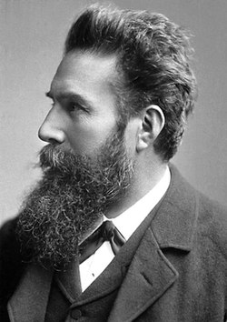
In the year of 1895, Wilhelm Roentgen developed the first radiograph, more commonly known as the X-ray. [4] By 1901, Roentgen had been awarded a Nobel Peace Prize for his discovery. Immediately after its release, X-ray machines were being manufactured and used worldwide in medicine. [5] However, this was only the first step in the development of neuroimaging. The brain is almost entirely composed of soft tissue that is not radio-opaque, meaning it remains essentially invisible to ordinary or plain X-ray examinations. This is also true of most brain abnormalities, though there are exceptions. For example, a calcified tumor (e.g.,meningioma, craniopharyngioma, and some types of glioma) can easily be seen.

Air Ventriculography

To combat this, in 1918, neurosurgeon Walter Dandy developed a technique called air ventriculography. This method injected filtered air directly into the lateral ventricles to better take pictures of the ventricle systems of the brain. [4] Thanks to local anesthetics, this was not a painful procedure, but it was significantly risky. Hemorrhage, severe infection, and extreme changes in intrarenal pressure were all threats to the procedure. Despite this, Dandy did not stop there. In 1919, he proceeded to discover Encephalography, a medical procedure used to record the brain's electrical activity. [6] This method involved attaching sensors to the brain that detect and measure the brain's electrical signals. These signals are then translated into a visual, showing the brain's activity patterns. With these early advances, neuroimaging was beginning to be used to diagnose conditions such as epilepsy, brain injuries, and sleep disorders. Providing invaluable information about brain function that would one day be added upon during the devolvement of modern neuroimaging.  [ citation needed ]

Cerebral Angiography

Cerebral angiogram showing a transverse projection of the vertebrobasilar and posterior cerebral circulation. Cerebral angiography, arteria vertebralis sinister injection.JPG
Cerebral angiogram showing a transverse projection of the vertebro basilar and posterior cerebral circulation.

Introduced in 1927, cerebral angiography enabled doctors to accurately detect and diagnose anomalies in the brain such as tumors and internal carotid artery occlusions. Over the course of a year, Egas Moniz, the inventor of cerebral angiography, ran experiments with various dye solution percentages that were injected into arteries to help better visualize the blood vessels in the brain before discovering that a solution consisting of 25% sodium iodide was the safest for patients, as well as the most effective in the visualization of blood vessels and arteries within the brain. [7]

PET/SPECT Scans

Full body PET scan of an adult female. PET-MIPS-anim.gif
Full body PET scan of an adult female.

A positron emission tomography, or PET scan, is a scan that shows areas of high activity in the body. The way it works is that a patient is first given a radioactive substance (called a tracer) via an injection in the hand or arm. The tracer then circulates through the body and attaches to a specific substance that the organ or tissue produces during metabolism, such as glucose. As a result, positrons are created, and those positrons are scanned by the PET camera. After they are scanned, a computer produces either a 2D or 3D image of the activity occurring within the organ or tissue. [8] The idea for the PET scan was originally proposed by William Sweet in the 1950s, but the first full-body PET scanner wasn't actually developed until 1974 by Michael Phelp. [9]

Similarly, the single-photon emission computed tomography scan, or SPECT scan, also works by scanning a tracer within the patient. The difference, however, is that the SPECT directly scans the gamma rays from where the tracer attaches rather than the positrons that the PET scans. As a result, the images that the SPECT scan creates are not as clear as the images produced by a PET scan, but it's typically a cheaper procedure to undertake. [10] SPECT was developed by David Kuhl in the 1950s. Kuhl also helped set the foundation that would lead to the PET scan. [11]

Magnetoencephalography

MEG device with patient. NIMH MEG.jpg
MEG device with patient.

Magnetoencephalography (MEG) is a technique that looks for regions of activity in the brain by detecting large groups of electrically charged ions moving through cells. [12] It was originally developed by physicist David Cohen in the early 1970s as a noninvasive procedure. [13] In order to be noninvasive, the MEG was designed like a giant helmet that the patient would put their head inside of and, once turned on, would read the electromagnetic pulses coming from their brain. Later on, in 1972, Cohen invented the SQUID (superconducting quantum interference device), which gave the MEG the ability to detect extremely small changes in ions and magnetic fields in the brain. [14]    

Xenon CT Scanning

Godfrey Hounsfield, inventor of first CT scanner Godfrey Hounsfield.jpg
Godfrey Hounsfield, inventor of first CT scanner

Xenon computed tomography is a modern scanning technique that reveals the flow of blood to the areas of the brain. The scan tests for consistent and sufficient blood flow to all areas of the brain by having patients breathe in xenon gas, a contrast agent, to show the areas of high and low blood flow. Although many trial scans and tests were ran during the development process of computed tomography, British biomedical engineer Godfrey Hounsfield is the founder of the technique and invented the first CT scanner in 1967, which he won a Nobel Prize for in 1979. However, the adoption of the scanners in the United States didn't occur until six years later in 1973. Regardless, the CT scanner was already gaining a notable reputation and popularity beforehand.

Magnetic resonance imaging

Shortly after the initial development of CT, magnetic resonance imaging (MRI or MR scanning) was developed. Rather than using ionizing or X-radiation, MRI uses the variation in signals produced by protons in the body when the head is placed in a strong magnetic field. Associated with early application of the basic technique to the human body are the names of Jackson (in 1968), Damadian (in 1972), and Abe and Paul Lauterbur (in 1973). Lauterbur and Sir Peter Mansfield were awarded the 2003 Nobel Prize in Physiology or Medicine for their discoveries concerning MRI. At first, structural imaging benefited more than functional imaging from the introduction of MRI. During the 1980s a veritable explosion of technical refinements and diagnostic MR applications took place, enabling even neurological tyros to diagnose brain pathology that would have been elusive or incapable of demonstration in a living person only a decade or two earlier. [15]

Related Research Articles

<span class="mw-page-title-main">Positron emission tomography</span> Medical imaging technique

Positron emission tomography (PET) is a functional imaging technique that uses radioactive substances known as radiotracers to visualize and measure changes in metabolic processes, and in other physiological activities including blood flow, regional chemical composition, and absorption. Different tracers are used for various imaging purposes, depending on the target process within the body.

<span class="mw-page-title-main">Magnetoencephalography</span> Mapping brain activity by recording magnetic fields produced by currents in the brain

Magnetoencephalography (MEG) is a functional neuroimaging technique for mapping brain activity by recording magnetic fields produced by electrical currents occurring naturally in the brain, using very sensitive magnetometers. Arrays of SQUIDs are currently the most common magnetometer, while the SERF magnetometer is being investigated for future machines. Applications of MEG include basic research into perceptual and cognitive brain processes, localizing regions affected by pathology before surgical removal, determining the function of various parts of the brain, and neurofeedback. This can be applied in a clinical setting to find locations of abnormalities as well as in an experimental setting to simply measure brain activity.

<span class="mw-page-title-main">Medical imaging</span> Technique and process of creating visual representations of the interior of a body

Medical imaging is the technique and process of imaging the interior of a body for clinical analysis and medical intervention, as well as visual representation of the function of some organs or tissues (physiology). Medical imaging seeks to reveal internal structures hidden by the skin and bones, as well as to diagnose and treat disease. Medical imaging also establishes a database of normal anatomy and physiology to make it possible to identify abnormalities. Although imaging of removed organs and tissues can be performed for medical reasons, such procedures are usually considered part of pathology instead of medical imaging.

<span class="mw-page-title-main">Single-photon emission computed tomography</span> Nuclear medicine tomographic imaging technique

Single-photon emission computed tomography is a nuclear medicine tomographic imaging technique using gamma rays. It is very similar to conventional nuclear medicine planar imaging using a gamma camera, but is able to provide true 3D information. This information is typically presented as cross-sectional slices through the patient, but can be freely reformatted or manipulated as required.

<span class="mw-page-title-main">Nuclear medicine</span> Medical specialty

Nuclear medicine or nucleology is a medical specialty involving the application of radioactive substances in the diagnosis and treatment of disease. Nuclear imaging, in a sense, is "radiology done inside out" because it records radiation emitted from within the body rather than radiation that is transmitted through the body from external sources like X-ray generators. In addition, nuclear medicine scans differ from radiology, as the emphasis is not on imaging anatomy, but on the function. For such reason, it is called a physiological imaging modality. Single photon emission computed tomography (SPECT) and positron emission tomography (PET) scans are the two most common imaging modalities in nuclear medicine.

<span class="mw-page-title-main">Functional neuroimaging</span>

Functional neuroimaging is the use of neuroimaging technology to measure an aspect of brain function, often with a view to understanding the relationship between activity in certain brain areas and specific mental functions. It is primarily used as a research tool in cognitive neuroscience, cognitive psychology, neuropsychology, and social neuroscience.

<span class="mw-page-title-main">Bone scintigraphy</span> Nuclear medicine imaging technique

A bone scan or bone scintigraphy is a nuclear medicine imaging technique of the bone. It can help diagnose a number of bone conditions, including cancer of the bone or metastasis, location of bone inflammation and fractures, and bone infection (osteomyelitis).

<span class="mw-page-title-main">Neuroimaging</span> Set of techniques to measure and visualize aspects of the nervous system

Neuroimaging is the use of quantitative (computational) techniques to study the structure and function of the central nervous system, developed as an objective way of scientifically studying the healthy human brain in a non-invasive manner. Increasingly it is also being used for quantitative research studies of brain disease and psychiatric illness. Neuroimaging is highly multidisciplinary involving neuroscience, computer science, psychology and statistics, and is not a medical specialty. Neuroimaging is sometimes confused with neuroradiology.

Functional imaging is a medical imaging technique of detecting or measuring changes in metabolism, blood flow, regional chemical composition, and absorption.

William Henry Oldendorf was an American neurologist, physician, researcher, medical pioneer, founding member of the American Society for Neuroimaging (ASN), and originator of the technique of computed tomography.

Whole-body nuclear scanning is a medical imaging technique where the whole body is scanned, contrary to, e.g., neuroimaging where only the brain is scanned.

<span class="mw-page-title-main">PET-CT</span> Medical imaging method

Positron emission tomography–computed tomography is a nuclear medicine technique which combines, in a single gantry, a positron emission tomography (PET) scanner and an x-ray computed tomography (CT) scanner, to acquire sequential images from both devices in the same session, which are combined into a single superposed (co-registered) image. Thus, functional imaging obtained by PET, which depicts the spatial distribution of metabolic or biochemical activity in the body can be more precisely aligned or correlated with anatomic imaging obtained by CT scanning. Two- and three-dimensional image reconstruction may be rendered as a function of a common software and control system.

Perfusion is the passage of fluid through the lymphatic system or blood vessels to an organ or a tissue. The practice of perfusion scanning is the process by which this perfusion can be observed, recorded and quantified. The term perfusion scanning encompasses a wide range of medical imaging modalities.

Preclinical imaging is the visualization of living animals for research purposes, such as drug development. Imaging modalities have long been crucial to the researcher in observing changes, either at the organ, tissue, cell, or molecular level, in animals responding to physiological or environmental changes. Imaging modalities that are non-invasive and in vivo have become especially important to study animal models longitudinally. Broadly speaking, these imaging systems can be categorized into primarily morphological/anatomical and primarily molecular imaging techniques. Techniques such as high-frequency micro-ultrasound, magnetic resonance imaging (MRI) and computed tomography (CT) are usually used for anatomical imaging, while optical imaging, positron emission tomography (PET), and single photon emission computed tomography (SPECT) are usually used for molecular visualizations.

<span class="mw-page-title-main">PET-MRI</span>

Positron emission tomography–magnetic resonance imaging (PET–MRI) is a hybrid imaging technology that incorporates magnetic resonance imaging (MRI) soft tissue morphological imaging and positron emission tomography (PET) functional imaging.

Cardiac imaging refers to minimally invasive imaging of the heart using ultrasound, magnetic resonance imaging (MRI), computed tomography (CT), or nuclear medicine (NM) imaging with PET or SPECT. These cardiac techniques are otherwise referred to as echocardiography, Cardiac MRI, Cardiac CT, Cardiac PET and Cardiac SPECT including myocardial perfusion imaging.

<span class="mw-page-title-main">Brain positron emission tomography</span> Form of positron emission tomography

Brain positron emission tomography is a form of positron emission tomography (PET) that is used to measure brain metabolism and the distribution of exogenous radiolabeled chemical agents throughout the brain. PET measures emissions from radioactively labeled metabolically active chemicals that have been injected into the bloodstream. The emission data from brain PET are computer-processed to produce multi-dimensional images of the distribution of the chemicals throughout the brain.

The following outline is provided as an overview of and topical guide to brain mapping:

Bruce Rosen is an American physicist and radiologist and a leading expert in the area of functional neuroimaging. His research for the past 30 years has focused on the development and application of physiological and functional nuclear magnetic resonance techniques, as well as new approaches to combine functional magnetic resonance imaging (fMRI) data with information from other modalities such as positron emission tomography (PET), magnetoencephalography (MEG) and noninvasive optical imaging. The techniques his group has developed to measure physiological and metabolic changes associated with brain activation and cerebrovascular insult are used by research centers and hospitals throughout the world.

References

Notes

  1. 1 2 "Weighing brain activity with the balance: Angelo Mosso's original manuscripts come to light". academic.oup.com. Retrieved 11 October 2023.
  2. Kolb, Bryan; Whishaw, Ian Q. (1980). "Fundamentals of Human Neuropsychology" (PDF). BrainMaster Technologies. Retrieved 11 October 2023.
  3. Lankford, Harvey V. (September 2015). "Dull Brains, Mountaineers, and Mosso: Hypoxic Words from on High". ResearchGate. Retrieved 11 October 2023.
  4. 1 2 "History of Neuroimaging | The American Society of Neuroimaging". www.asnweb.org. Retrieved 4 October 2023.
  5. Shorvon, Simon D. (March 2009). "A history of neuroimaging in epilepsy 1909–2009". Epilepsia. 50 (s3): 39–49. doi: 10.1111/j.1528-1167.2009.02038.x . ISSN   0013-9580.
  6. Shorvon, Simon D. (March 2009). "A history of neuroimaging in epilepsy 1909–2009". Epilepsia. 50 (s3): 39–49. doi: 10.1111/j.1528-1167.2009.02038.x . ISSN   0013-9580.
  7. Tan, Siang Yong; Yip, Angela. "António Egas Moniz (1874–1955): Lobotomy pioneer and Nobel laureate". National Library of Medicine. Retrieved 16 October 2023.
  8. "Positron Emission Tomography (PET) - Health Encyclopedia - University of Rochester Medical Center". www.urmc.rochester.edu. Retrieved 21 October 2023.
  9. "Radiation in Biology and Medicine: Positron Emission Tomography". Chemistry LibreTexts. 2 October 2013. Retrieved 21 October 2023.
  10. "SPECT | Radiology | U of U School of Medicine". medicine.utah.edu. 10 November 2021. Retrieved 21 October 2023.
  11. Read "Advancing Nuclear Medicine Through Innovation" at NAP.edu.
  12. Walla, Peter (30 November 2011), "Non-Conscious Brain Processes Revealed by Magnetoencephalography (MEG)", Magnetoencephalography, InTech, retrieved 6 October 2023
  13. "The David Cohen MEG Laboratory" . Retrieved 10 October 2023.
  14. kle (8 September 2014). "MEG matters". MIT McGovern Institute. Retrieved 6 October 2023.
  15. Filler AG (2009). "The history, development, and impact of computed imaging in neurological diagnosis and neurosurgery: CT, MRI, DTI". Nature Precedings. doi: 10.1038/npre.2009.3267.4 .