Non-contact thermography

Last updated
Thermography (medical)
ICD-9 88.8
MeSH D013817

Non-contact thermography, thermographic imaging, or medical thermology is the field of thermography that uses infrared images of the human skin to assist in the diagnosis and treatment of medical conditions. Medical thermology is sometimes referred to as medical infrared imaging or tele-thermology and utilizes thermographic cameras. According to the American Academy of Thermology, Medical Thermology practitioners are licensed health care practitioners who utilize IR imaging in consistent with medically established paradigms of care. Non-medically licensed alternative practitioners who are not held to the same standard may offer thermography services but that should not be confused with the field of medical thermology.

Contents

Restated, medical thermology is the use of infrared (IR) imaging to assess skin temperature as an extension of the clinician's physical exam to aid in the formation of a medical diagnosis or treatment plan. Medical Thermology does not condone those who purport that "Thermography" can find disease by looking for areas of the body that have abnormal heat or irregular blood flow. IR imaging simply does not have the ability to assess temperature beyond the surface of the skin.

Thermography is a physiologic study and is not a replacement for structural studies such as X-Ray, MRI, or Mammography. As a physiologic study, however, medical thermology has many health-related indications. The American Academy of Thermology (AAT) (www.aathermology.org) [1] has published internationally peer-reviewed guidelines for neuro-musculoskeletal (MSK), breast, veterinary, and oral-systemic disease. [2]

CRPS-RSD of the left foot imaged with medical thermology during a cold pressor test. CRPS-RSD of the left foot.jpg
CRPS-RSD of the left foot imaged with medical thermology during a cold pressor test.

Examples of neuro-musculoskeletal indications for medical thermology include Reflex Sympathetic Dystrophy (RSD), Chronic Regional Pain Syndrome (CRPS), Dysautonomia, Migraine, Fibromyalgia (and other weather-sensitive pain syndromes), thoracic outlet syndrome, and vaso-motor migraine/headaches such as Barre-Lieou syndrome. This is especially true when used to monitor the results of a cold stress (cold presser) test. [3]

While there are several clinical studies showing the effectiveness of thermography in detecting temperature abnormalities of breast skin, breast thermography is only approved as an adjunct modality only for breast disease and/or cancer. [4] In that regard it is a breast risk health assessment only. A summary of some of the clinical trials for breast thermography is available in this white paper from UE LifeSciences pdf. Studies also report the use of thermography in understanding the foot complications as a result of diabetes. [5] [6]

Veterinary thermography indications include, but are not limited to, assessment of shoring, limb inflammation, and sweating disorders. [7]

When properly used, a commonly known oral-systemic indication for IR imaging is temperature measurement for fever screening. While there are several other oral-systemic indications out lined in the American Academy of Thermology's Oral -Systemic Guidelines. Whole body studies are not performed for medical purposes. [8]

Telethermography systems are regulated as a medical device under 21 CFR 884.2980.


Medical Thermology versus Thermography

Sample image of a hand held thermography camera Thermography of electronics.JPG
Sample image of a hand held thermography camera

There is a difference between Medical Thermology as promulgated by medically based organizations such as the American Academy of Thermology (AAT), and thermography as practiced by alternative providers or physicians who overstate the benefits of thermography. As a result of this disparity organizations such as the FDA, ISO, and the AAT have published Guidelines and best use practices to help educate medical providers and the public to recognize the difference between providers who provide medical thermology services and those that offer something else. [9]

Infrared (IR) temperature measurement for fever screening correctly done at the medial canthus (tear duct) of the eye. Right Way To Do IR Fever Screening.jpg
Infrared (IR) temperature measurement for fever screening correctly done at the medial canthus (tear duct) of the eye.

Due to misunderstandings of the technology, the complex physiologic nature that thermographic imaging provides insight into, and large disparities between low-end and medical-grade thermal imaging cameras, there is a lot of misinformation that tarnishes the industry. The Coronavirus pandemic is an excellent example to illustrate the point. Almost anyone can buy an inexpensive infrared measuring device intended for industrial use that has been re-marketed for fever screening, but said devices are not FDA approved for the same, may lack the specificity or sensitivity to be used for that indication, and the end users receive no training in its proper application. In an effort to address this problem the AAT has published both free and paid versions of IR Temperature Measurement Fever Screening training courses. The Level I course is intended for individuals who have financial restraint and have to use non-medical grade cameras. The course provides protocols to help overcome the shortcomings of these devices. Level II training is directed toward businesses and agencies that can afford a medical grade system and have the resources to offer screening that is fully compliant with FDA, ISO, and AAT recommended best use practices. [10]

Thermography has been promoted by some alternative medicine practitioners as a means to diagnose cancer, although it is not effective for this purpose. Health Canada has issued "cease and desist" orders to clinics offering breast thermography as a cancer diagnostic device because thermography cameras are not licensed as a medical device in Canada, and because thermography for cancer detection is viewed as ineffective by medical experts. [11] The FDA has issued a public warning notice stating that breast thermography is not an alternative to mammography [12] and has ordered Joseph Mercola to stop making excessive claims for thermography. [13]

Thermography is discouraged in North America by the American Cancer Society, radiologists and the FDA for early breast cancer detection. Advertisements in the United Kingdom have been found to be misleading. [14]

The FDA has cleared thermography only as an adjunct method of screening. "Thermography devices have been cleared by the FDA for use as an adjunct, or additional, tool for detecting breast cancer." However, they stop short of recommending it, citing the lack of evidence of its effectiveness. [15] The AAT has published several Position Papers, including statements on Breast Thermography that clearly delineate its utility as an adjudicative breast risk health assessment only. [16] [17]

See also

Related Research Articles

Thermographic camera

A thermographic camera is a device that creates an image using infrared(IR) radiation, similar to a common camera that forms an image using visible light. Instead of the 400–700 nanometre (nm) range of the visible light camera, infrared cameras are sensitive to wavelengths from about 1,000 nm to about 14,000 nm (14 μm). The practice of capturing and analyzing the data they provide is called thermography.

Mammography Process of using low-energy X-rays to examine the human breast for diagnosis and screening

Mammography is the process of using low-energy X-rays to examine the human breast for diagnosis and screening. The goal of mammography is the early detection of breast cancer, typically through detection of characteristic masses or microcalcifications.

Thermography Imaging in mid- to long-wavelength infrared to reveal temperature

Infrared thermography (IRT), thermal video and thermal imaging, is a process where a thermal camera captures and creates an image of an object by using infrared radiation emitted from the object in a process, which are examples of infrared imaging science. Thermographic cameras usually detect radiation in the long-infrared range of the electromagnetic spectrum and produce images of that radiation, called thermograms. Since infrared radiation is emitted by all objects with a temperature above absolute zero according to the black body radiation law, thermography makes it possible to see one's environment with or without visible illumination. The amount of radiation emitted by an object increases with temperature; therefore, thermography allows one to see variations in temperature. When viewed through a thermal imaging camera, warm objects stand out well against cooler backgrounds; humans and other warm-blooded animals become easily visible against the environment, day or night. As a result, thermography is particularly useful to the military and other users of surveillance cameras.


Computed tomography laser mammography (CTLM) is the trademark of Imaging Diagnostic Systems, Inc. for its optical tomographic technique for female breast imaging.

Infrared thermometer Thermometer which infers temperature by measuring infrared energy emission

An infrared thermometer is a thermometer which infers temperature from a portion of the thermal radiation sometimes called black-body radiation emitted by the object being measured. They are sometimes called laser thermometers as a laser is used to help aim the thermometer, or non-contact thermometers or temperature guns, to describe the device's ability to measure temperature from a distance. By knowing the amount of infrared energy emitted by the object and its emissivity, the object's temperature can often be determined within a certain range of its actual temperature. Infrared thermometers are a subset of devices known as "thermal radiation thermometers".

Breast implant Prosthesis used to change the size, shape, and contour of a persons breast

A breast implant is a prosthesis used to change the size, shape, and contour of a person's breast. In reconstructive plastic surgery, breast implants can be placed to restore a natural looking breast following a mastectomy, to correct congenital defects and deformities of the chest wall or, cosmetically, to enlarge the appearance of the breast through breast augmentation surgery.
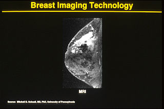
Breast MRI

One alternative to mammography, Breast MRI or contrast enhanced magnetic resonance imaging (MRI), has shown substantial progress in the detection of breast cancer.

Philip Strax

Philip Strax was an American radiologist who pioneered the use of mammography to screen for early breast cancer. With co-investigators statistician Sam Shapiro and surgeon Louis Venet he conducted a randomized controlled trial comparing outcomes of over 60,000 women who received either mammogram and clinical breast exam or standard medical care. The first results of this study were published in the Journal of the American Medical Association (JAMA) in 1966. The study demonstrated that screening mammograms, which are routine periodic mammograms of asymptomatic women, could find breast cancer at an early enough stage to save lives. For this research Strax and Shapiro shared the Kettering Prize for outstanding contributions to cancer diagnosis or treatment in 1988.

Breast cancer screening

Breast cancer screening is the medical screening of asymptomatic, apparently healthy women for breast cancer in an attempt to achieve an earlier diagnosis. The assumption is that early detection will improve outcomes. A number of screening tests have been employed, including clinical and self breast exams, mammography, genetic screening, ultrasound, and magnetic resonance imaging.

Tomosynthesis

Tomosynthesis, also digital tomosynthesis (DTS), is a method for performing high-resolution limited-angle tomography at radiation dose levels comparable with projectional radiography. It has been studied for a variety of clinical applications, including vascular imaging, dental imaging, orthopedic imaging, mammographic imaging, musculoskeletal imaging, and chest imaging.

Daniel B. Kopans, MD, FACR is a radiologist specializing in mammography and other forms of breast imaging.

Thermographic inspection refers to the nondestructive testing (NDT) of parts, materials or systems through the imaging of the temperature fields, gradients and/or patterns ("thermograms") at the object's surface. It is distinguished from medical thermography by the subjects being examined: thermographic inspection generally examines inanimate objects, while medical thermography generally examines living organisms. Generally, thermographic inspection is performed using an infrared sensor.

Infrared vision is the capability of biological or artificial systems to detect infrared radiation. The terms thermal vision and thermal imaging, are also commonly used in this context since infrared emissions from a body are directly related to their temperature: hotter objects emit more energy in the infrared spectrum than colder ones.

Infrared and thermal testing

Infrared and thermal testing refer to passive thermographic inspection techniques, a class of nondestructive testing designated by the American Society for Nondestructive Testing (ASNT). Infrared thermography is the science of measuring and mapping surface temperatures.

"Infrared thermography, a nondestructive, remote sensing technique, has proved to be an effective, convenient, and economical method of testing concrete. It can detect internal voids, delaminations, and cracks in concrete structures such as bridge decks, highway pavements, garage floors, parking lot pavements, and building walls. As a testing technique, some of its most important qualities are that (1) it is accurate; (2) it is repeatable; (3) it need not inconvenience the public; and (4) it is economical."
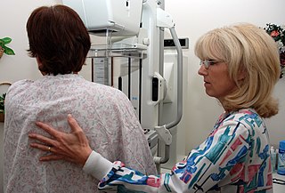
Cancer screening

Cancer screening aims to detect cancer before symptoms appear. This may involve blood tests, urine tests, DNA tests, other tests, or medical imaging. The benefits of screening in terms of cancer prevention, early detection and subsequent treatment must be weighed against any harms.
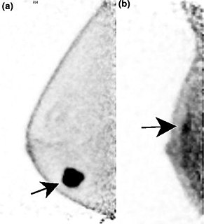
Positron emission mammography Imaging procedure used to detect breast cancer

Positron emission mammography (PEM) is a nuclear medicine imaging modality used to detect or characterise breast cancer. Mammography typically refers to x-ray imaging of the breast, while PEM uses an injected positron emitting isotope and a dedicated scanner to locate breast tumors. Scintimammography is another nuclear medicine breast imaging technique, however it is performed using a gamma camera. Breasts can be imaged on standard whole-body PET scanners, however dedicated PEM scanners offer advantages including improved resolution.

Dynamic angiothermography (DATG) is a technique for the diagnosis of breast cancer. This technique, though springing from the thermography of old conception, is based on a completely different principle. DATG records the temperature variations linked to the vascular changes in the breast due to angiogenesis. The presence, change, and growth of tumors and lesions in breast tissue change the vascular network in the breast. Consequently, measuring the vascular structure over time, DATG effectively monitors the change in breast tissue due to tumors and lesions. It is currently used in combination with other techniques for diagnosis of breast cancer. This diagnostic method is a low cost one compared with other techniques.

Active thermography is an advanced nondestructive testing procedure, which uses a thermography measurement of a tested material thermal response after its external excitation. This principle can be used also for non-contact infrared non-destructive testing (IRNDT) of materials.

Breast imaging

In medicine, breast imaging is a sub-speciality of diagnostic radiology that involves imaging of the breasts for screening or diagnostic purposes. There are various methods of breast imaging using a variety of technologies as described in detail below. Traditional screening and diagnostic mammography uses x-ray technology. Breast tomosynthesis is a new digital mammography technique that produces 3D images of the breast using x-rays. Xeromammography and Galactography also use x-ray technology and are also used infrequently in the detection of breast cancer. Breast ultrasound is another technology employed in diagnosis & screening and specifically can help differentiate between fluid filled and solid lumps that can help determine if cancerous. Breast MRI is, yet, another technology reserved for high-risk patients and can help determine the extent of cancer if diagnosed. Lastly, scintimammography is used in a subgroup of patients who have abnormal mammograms or whose screening is not reliable on the basis of using traditional mammography or ultrasound.

Fiona Jane Gilbert is a Scottish radiologist and academic.

References

  1. "American Academy of Thermology | American Academy of Thermology" . Retrieved 2020-06-19.
  2. "Guidelines | American Academy of Thermology" . Retrieved 2020-06-19.
  3. "Guidelines for Neuro-Musculoskeletal Infrared Medical Thermography And Sympathetic Skin Response (SSR) Studies | American Academy of Thermology" . Retrieved 2020-06-19.
  4. Commissioner, Office of the. "Press Announcements - FDA issues warning letter to clinic illegally marketing unapproved thermography device, warns consumers to avoid using thermography devices to detect breast cancer". www.fda.gov. Retrieved 2 March 2019.
  5. Gatt, Alfred; Falzon, Owen; Cassar, Kevin; Ellul, Christian; Camilleri, Kenneth P.; Gauci, Jean; Mizzi, Stephen; Mizzi, Anabelle; Sturgeon, Cassandra; Camilleri, Liberato; Chockalingam, Nachiappan; Formosa, Cynthia (2018). "Establishing Differences in Thermographic Patterns between the Various Complications in Diabetic Foot Disease". International Journal of Endocrinology. 2018: 1–7. doi: 10.1155/2018/9808295 . PMC   5867599 . PMID   29721019.
  6. Gauci, Jean; Falzon, Owen; Formosa, Cynthia; Gatt, Alfred; Ellul, Christian; Mizzi, Stephen; Mizzi, Anabelle; Sturgeon Delia, Cassandra; Cassar, Kevin; Chockalingam, Nachiappan; Camilleri, Kenneth P. (13 December 2018). "Automated Region Extraction from Thermal Images for Peripheral Vascular Disease Monitoring". Journal of Healthcare Engineering. 2018: 1–14. doi: 10.1155/2018/5092064 . PMC   6311825 . PMID   30651943.
  7. "Veterinary Guidelines for Infrared Thermography | American Academy of Thermology" . Retrieved 2020-06-19.
  8. "Guidelines for Oral Systemic Thermography | American Academy of Thermology" . Retrieved 2020-06-19.
  9. "Thermology | American Academy of Thermology" . Retrieved 2020-06-19.
  10. "Fever Screening Courses | AAT Online Training Center". courses.aathermology.org. Retrieved 2020-06-19.
  11. Clinics ordered to stop 'useless' breast cancer tests, CBC News, Nov 27 2012
  12. FDA Safety Communication: Breast Cancer Screening - Thermography is Not an Alternative to Mammography, the U.S. Food and Drug Administration June 2, 2011
  13. Tsouderos, Trine (25 April 2012). "FDA warns doctor: Stop touting camera as disease screening tool". Chicago Tribune. Retrieved 8 January 2013.
  14. "ASA Adjudication on Medical Thermal Imaging Ltd". Advertising Standards Authority. 2013-01-09. Retrieved January 5, 2015.
  15. Commissioner, Office of the (19 January 2021). "Consumer Updates - Thermogram No Substitute for Mammogram". FDA.
  16. "Position Papers | American Academy of Thermology" . Retrieved 2020-06-19.
  17. "Temperature Screening To Reduce U.S. COVID-19 Spread". 2 September 2020. Retrieved 2020-09-28.