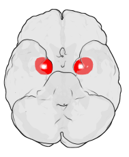
Amygdalofugal pathway

Last updated

The amygdalofugal pathway (Latin for "fleeing from the amygdala" and commonly distinguished as the ventral amygdalofugal pathway) is one of the three major efferent pathways of the amygdala, meaning that it is one of the three principal pathways by which fibers leave the amygdala. It leads from the basolateral nucleus and central nucleus of the amygdala. The amygdala is a limbic structure in the medial temporal lobe of the brain. The other main efferent pathways from the amygdala are the stria terminalis and anterior commissure.

Contents

While the stria terminalis carries information primarily from the corticomedial nuclei of the amygdala, the ventral amygdalofugal pathway carries output from the central and basolateral nuclei and delivers it to a number of targets; namely, the medial dorsal nucleus of the thalamus, the hypothalamus, the basal forebrain, the brainstem, septal nuclei and nucleus accumbens. Both the amygdalofugal pathway and the stria terminalis project to the septal region, the hypothalamus, and the thalamus, but the stria terminalis reaches the septal region and the hypothalamus in a much longer and less direct path. [1] While the stria terminalis follows a C-shaped pathway along the lateral ventricles, the ventral amygdalofugal pathway is more direct and contains a higher proportion of myelinated axons, causing the pathway to appear darker upon observation in stained cross-section.

The amygdalofugal pathway and the stria terminalis together “ enable the corticomedial amygdala to directly control the medial hypothalamus and enable the basolateral amygdala to directly control the lateral hypothalamus and PAG,” or midbrain periaqueductal gray. Through the bed nucleus of the stria terminalis, the amygdala additionally modulates the hypothalamus and PAG indirectly. [2] [3]

This pathway is particularly important for associative learning. [4]

Amygdala

Amygdala Amygdala.png
Amygdala

Basolateral nucleus

The basolateral nucleus is by far the largest part of the amygdala. The fibers leading from it to the central nucleus “provide a key link between the experience of emotions and their expression.” [5]

The basolateral nucleus includes five parts: the posterolateral part, the ventromedial part, the intermediate part, the dorsal part, and the ventrolateral part. [6]

Inputs to the basolateral nucleus include visual, auditory, and somatosensory information. [7] The basolateral nucleus is in close communication with the cerebral cortex, and in terms of function, the basolateral nucleus is more like the cerebral cortex than is the central nucleus. [8] The basolateral nucleus differs from the cortex in that it is not layered, but it includes pyramidal neurons, is continuous with the parahippocampal cortex, and has extensive connections with the other parts of the cortex. [5] Its strongest direct connections to the cerebral cortex are with the insular cortex, the orbital cortex, and the medial wall of the frontal lobe. These connections allow the basolateral nucleus to receive and modulate sensory and polysensory processing. The neurotransmitter in each of these connections is either glutamate or aspartate. [8] Both the neocortex and the basolateral nucleus receive diffuse cholinergic (acetylcholine) projections from the nucleus basalis of Meynert. [7]

Stimulation of the basolateral nucleus causes a reduction in feeding, and stimulation of the basolateral nuclear group causes an increase in arousal and attention. [8] The components of the basolateral nuclear group include the basolateral nucleus, lateral nucleus, and the basomedial nucleus. [6]

The basolateral nucleus cooperates with the central nucleus in persistent fear responses. It influences regions of the brainstem through the central nucleus via the amygdalofugal pathway and the stria terminalis. [9] In mice, the basolateral nucleus mediates anxiety. [8]

Oxytocin has been visualized in the basolateral nuclei. [10] In one study, prescription opioid patients were found to have decreased anisotropy in the amygdalofugal pathway. [11]

Central nucleus

The central nucleus can be thought of as the exit of the amygdaloid bodies through which the bodily responses that are associated with fear leave the amygdala. [12] It is the most peptide-rich region of the brain. [8]

The amygdalofugal pathway connects the central nucleus of the amygdala to the brainstem. The stria terminalis does so also but in a longer, less direct route. [9] These connections collectively are involved with emotional responses. [13] The brainstem regions that the central nucleus connects to are responsible for controlling “expression of innate behaviors and associated physiological responses.” The central nucleus also has three subnuclei: lateral, capsular, and medial. It is the medial subnucleus that forms connections with “response control regions.” Most of the projection neurons in the central nucleus are inhibitory. [14]

The nucleus basalis also connects the central nucleus of the amygdala to the cortex through cholinergic projections that “are thought to arouse the cortex.” [13]

Lesion experiments show that the central nucleus of the amygdala connects with the startle circuit via the caudal part of the amygdalofugal pathway. This is the part of the pathway that projects directly to the parts of the pons, medulla, and perhaps the spinal cord. [15]

Conscious perception of emotion involves output from the central nucleus of the amygdala as well as its connections through the amygdalofugal pathway to the anterior cingulate cortex, orbitofrontal cortex, and prefrontal cortex. [13]

Other major components

Nucleus accumbens

The connections of the amygdalofugal pathway to the nucleus accumbens plays a role in the perception of a stimulus as either gratifying or aversive. [4]

The nucleus accumbens, along with other regions of the ventral striatum and the prefrontal cortex, is one of the major targets of ascending dopaminergic pathways originating from the ventral tegmental area. [7]

Thalamus

Amygdala and Nucleus Basalis connections to the Thalamus Amygdala and Nucleus Basalis connections to the Thalamus2.png
Amygdala and Nucleus Basalis connections to the Thalamus

The amygdala connects to the thalamus both through the amygdalofugal pathway and through a direct connection to the dorsomedial nucleus of the thalamus. [13] After leaving the substantia innominata, the ventral amygdalofugal pathway continues on a medial path to enter the septal region, the lateral preoptic area, the hypothalamus, and the nucleus of the diagonal band. The fibers which bypass the preoptic area and the hypothalamus travel more superiorly and enter the inferior thalamic peduncle. From there, the amygdalofugal fibers travel just laterally to the mammillothalamic tract in the ventral anterior nucleus as they enter the dorsal medial nucleus.

Amygdalofugal fibers as well as fibers from the nucleus basalis both project to the substantia innominata followed by the thalamic peduncle and then the magnocellar division of the dorsal medial nucleus. The dorsal medial nucleus therefore receives indirect input from both the amygdala and the nucleus basalis. Lesions to the dorsal medial nucleus cause amnesic syndromes, but they need to interrupt both the mammillothalamic tract and the amygdalofugal pathway in order to do so. Thus, both pathways play a role in memory. [16]

Pyriform cortex

The pyriform cortex is a type of allocortex that corresponds to the rostral half of the uncus. Its strongest afferent connections are from the olfactory bulb. [17] Although the olfactory cortex comprises all the “areas in the rostro-ventral portion of the forebrain which receive direct projections from the olfactory bulb,” including the anterior olfactory nucleus, olfactory tubercle, the pyriform cortex, the entorhinal cortex, the insular cortex, and the amygdala, the pyriform cortex is considered synonymous with the olfactory cortex because it is “by far the largest cortical area primarily involved in perception and learning of olfactory stimuli.” It is a type of paleocortex, which means that it is older than and has fewer layers than neocortex but is more recent than and contains more layers than archicortex. [18] Along with the parahippocampal gyrus, the pyriform cortex is a chief component of paleocortex. [19]

The pyriform cortex is located in the anterior medial temporal lobe. Through the amygdalofugal pathway, it projects to the nucleus of the diagonal band. [20] The pyriform cortex is adjoined to the basolateral nucleus of the amygdala. [21] The pyriform cortex and the basolateral nucleus are considered to be the main starting points of the amygdalofugal pathway. [20] [21] The connection from the basolateral nucleus that goes deep to the pyriform cortex is also considered part of the amygdalofugal pathway. [21]

Anterior cingulate cortex

One reason that the amygdalofugal pathway is important is because it links motivations to responses. [13] The anterior cingulate cortex in particular is involved with using emotions to make decisions. It has been implicated in a wide variety of cognitive functions as well as transient mood changes, depression and anxiety disorders, and the perception of pain. [22]

Hypothalamus

The stria terminalis, not the amygdalofugal pathway, is the main connection between the amygdala and the hypothalamus. [13] The amygdalofugal connection, however, is shorter and more direct. [1]

The hypothalamus is a highly important structure that is involved with the regulation of bodily functions. These include endocrine, autonomic, and behavioral functions. [23] It produces a wide variety of hormones. These control body temperature, hunger, moods, sex drive, sleep, thirst, and release of other hormones. [24] There are dozens of peptides within the hypothalamus that have been discovered in recent years. The hypothalamus also influences daily physiologic cycles and plays a role in regulating emotional responses. Many of these functions can be described as homeostatic. It is the most ventral part of the diencephalon and contains three regions: the supraoptic region, the tuberal region, and the mammillary region. [24]

Related Research Articles

<span class="mw-page-title-main">Limbic system</span> Set of brain structures involved in emotion and motivation

The limbic system, also known as the paleomammalian cortex, is a set of brain structures located on both sides of the thalamus, immediately beneath the medial temporal lobe of the cerebrum primarily in the forebrain.

Articles related to anatomy include:

<span class="mw-page-title-main">Brainstem</span> Posterior part of the brain, adjoining and structurally continuous

The brainstem is the stalk-like part of the brain that interconnects the cerebrum and diencephalon with the spinal cord. In the human brain the brainstem is composed of the midbrain, the pons, and the medulla oblongata. The midbrain is continuous with the thalamus of the diencephalon through the tentorial notch.

<span class="mw-page-title-main">Rhinencephalon</span>

In animal anatomy, the rhinencephalon, also called the smell-brain or olfactory brain, is a part of the brain involved with smell. It forms the paleocortex and is rudimentary in the human brain.

<span class="mw-page-title-main">Spinothalamic tract</span> Sensory pathway from the skin to the thalamus

The spinothalamic tract is a part of the anterolateral system or the ventrolateral system, a sensory pathway to the thalamus. From the ventral posterolateral nucleus in the thalamus, sensory information is relayed upward to the somatosensory cortex of the postcentral gyrus.

<span class="mw-page-title-main">Inferior colliculus</span> Midbrain structure involved in the auditory pathway.

The inferior colliculus (IC) is the principal midbrain nucleus of the auditory pathway and receives input from several peripheral brainstem nuclei in the auditory pathway, as well as inputs from the auditory cortex. The inferior colliculus has three subdivisions: the central nucleus, a dorsal cortex by which it is surrounded, and an external cortex which is located laterally. Its bimodal neurons are implicated in auditory-somatosensory interaction, receiving projections from somatosensory nuclei. This multisensory integration may underlie a filtering of self-effected sounds from vocalization, chewing, or respiration activities.

<span class="mw-page-title-main">Papez circuit</span> Neural circuit

The Papez circuit, or medial limbic circuit, is a neural circuit for the control of emotional expression. In 1937, James Papez proposed that the circuit connecting the hypothalamus to the limbic lobe was the basis for emotional experiences. Paul D. MacLean reconceptualized Papez's proposal and coined the term limbic system. MacLean redefined the circuit as the "visceral brain" which consisted of the limbic lobe and its major connections in the forebrain – hypothalamus, amygdala, and septum. Over time, the concept of a forebrain circuit for the control of emotional expression has been modified to include the prefrontal cortex.

<span class="mw-page-title-main">Stria terminalis</span> Band of fibres along the thalamus

The stria terminalis is a structure in the brain consisting of a band of fibers running along the lateral margin of the ventricular surface of the thalamus. Serving as a major output pathway of the amygdala, the stria terminalis runs from its centromedial division to the ventromedial nucleus of the hypothalamus.

<span class="mw-page-title-main">Septal area</span> Area in the lower, posterior part of the medial surface of the frontal lobe

The septal area, consisting of the lateral septum and medial septum, is an area in the lower, posterior part of the medial surface of the frontal lobe, and refers to the nearby septum pellucidum.

The zona incerta (ZI) is a horizontally elongated region of gray matter in the subthalamus below the thalamus. Its connections project extensively over the brain from the cerebral cortex down into the spinal cord.

<span class="mw-page-title-main">Medial forebrain bundle</span>

The medial forebrain bundle (MFB) is a neural pathway containing fibers from the basal olfactory regions, the periamygdaloid region and the septal nuclei, as well as fibers from brainstem regions, including the ventral tegmental area and nigrostriatal pathway.

<span class="mw-page-title-main">Olfactory tract</span> Part of the olfactory system

The olfactory tract is a bilateral bundle of afferent nerve fibers from the mitral and tufted cells of the olfactory bulb that connects to several target regions in the brain, including the piriform cortex, amygdala, and entorhinal cortex. It is a narrow white band, triangular on coronal section, the apex being directed upward.

<span class="mw-page-title-main">Lateral hypothalamus</span>

The lateral hypothalamus (LH), also called the lateral hypothalamic area (LHA), contains the primary orexinergic nucleus within the hypothalamus that widely projects throughout the nervous system; this system of neurons mediates an array of cognitive and physical processes, such as promoting feeding behavior and arousal, reducing pain perception, and regulating body temperature, digestive functions, and blood pressure, among many others. Clinically significant disorders that involve dysfunctions of the orexinergic projection system include narcolepsy, motility disorders or functional gastrointestinal disorders involving visceral hypersensitivity, and eating disorders.

<span class="mw-page-title-main">Medial dorsal nucleus</span>

The medial dorsal nucleus is a large nucleus in the thalamus.

<span class="mw-page-title-main">Pallium (neuroanatomy)</span> Layers of grey and white matter that cover the upper surface of the cerebrum in vertebrates

In neuroanatomy, pallium refers to the layers of grey and white matter that cover the upper surface of the cerebrum in vertebrates. The non-pallial part of the telencephalon builds the subpallium. In basal vertebrates the pallium is a relatively simple three-layered structure, encompassing 3–4 histogenetically distinct domains, plus the olfactory bulb.

The parabrachial nuclei, also known as the parabrachial complex, are a group of nuclei in the dorsolateral pons that surrounds the superior cerebellar peduncle as it enters the brainstem from the cerebellum. They are named from the Latin term for the superior cerebellar peduncle, the brachium conjunctivum. In the human brain, the expansion of the superior cerebellar peduncle expands the parabrachial nuclei, which form a thin strip of grey matter over most of the peduncle. The parabrachial nuclei are typically divided along the lines suggested by Baxter and Olszewski in humans, into a medial parabrachial nucleus and lateral parabrachial nucleus. These have in turn been subdivided into a dozen subnuclei: the superior, dorsal, ventral, internal, external and extreme lateral subnuclei; the lateral crescent and subparabrachial nucleus along the ventrolateral margin of the lateral parabrachial complex; and the medial and external medial subnuclei

<span class="mw-page-title-main">Central nucleus of the amygdala</span> Nucleus within the amygdala

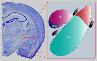
The central nucleus of the amygdala is a nucleus within the amygdala. It "serves as the major output nucleus of the amygdala and participates in receiving and processing pain information."

<span class="mw-page-title-main">Paratenial nucleus</span>

The paratenial nucleus, or parataenial nucleus, is a component of the midline nuclear group in the thalamus. It is sometimes subdivided into the nucleus parataenialis interstitialis and nucleus parataenialis parvocellularis (Hassler). It is located above the bordering paraventricular nucleus of thalamus and below the anterodorsal nucleus.

<span class="mw-page-title-main">Septum verum</span> Region of the brain

Septum Verum is a region in the lower medial part of the telencephalon that separates the two cerebral hemispheres. The human septum consists of two parts: the septum pellucidum, a thin membrane consisting of white matter and glial cells that separate the lateral ventricles, and the lower, precommisural septum verum, which consists of nuclei and grey matter. The term is sometimes used synonymously with Area Septalis, to refer to the precommisural part of the lower base of the telencephalon. The Septum verum contains the septal nuclei, which are usually considered part of the limbic system. 

References

  1. 1 2 "The Amygdala", The Pixelated Brain, Brooks, Dana C., MD, archived from the original on 2013-12-03
  2. Siegel, Allan; Sapru, Hreday N. (2010), Essential Neuroscience (2nd ed.), Lippincott Williams & Wilkins, pp. 455–456
  3. Siegel, Allan; Sapru, Hreday N. (2006), Essential Neuroscience (1st ed.), Lippincott Williams & Wilkins, p. 456, ISBN   9780781750776
  4. 1 2 "The Amygdala and its Allies", The Brain from Top to Bottom
  5. 1 2 Nolte, John (2008), The Human Brain: with Student Consult Online Access, Elsevier Health Sciences, ISBN   978-0323064750
  6. 1 2 "Basolateral Amygdaloid Nucleus", Brain Architecture Management System, The University of Southern California, 2010–2013, archived from the original on 2013-11-27, retrieved 2013-11-27
  7. 1 2 3 Pritchard, Thomas C.; Alloway, Kevin D. (1999), Medical Neuroscience, Hayes Barton Press, ISBN   9781889325293
  8. 1 2 3 4 5 Best, Ben, "The Amygdala and the Emotions", The Anatomical Basis of Mind
  9. 1 2 Behrendt, Ralf-Peter (2012), Neuroanatomy of Social Behaviour: An Evolutionary and Psychoanalytic Perspective, Karnac Books, ISBN   9781781811436
  10. Boccia, M.L.; Petrusz, P.; Suzuki, K.; Marson, L.; Pedersen, C.A. (2013). "Immunohistochemical localization of oxytocin receptors in human brain". Neuroscience. 253: 155–164. doi:10.1016/j.neuroscience.2013.08.048. PMID   24012742. S2CID   22816374.
  11. Upadhyay, J.; Maleki, N.; Potter, J.; Elman, I.; Rudrauf, D.; Knudsen, J.; Wallin, D.; Pendse, G.; McDonald, L.; Griffin, M.; Anderson, J.; Nutile, L.; Renshaw, P.; Weiss, R.; Becerra, L.; Borsook, D. (2010). "Alterations in brain structure and functional connectivity in prescription opioid-dependent patients". Brain. 133 (7): 2098–2114. doi:10.1093/brain/awq138. PMC   2912691 . PMID   20558415.
  12. "Alarm System Circuits", The Brain from Top to Bottom
  13. 1 2 3 4 5 6 "Limbic System: Amygdala", Neuroscience Online, the Open-Access Neuroscience Electronic Textbook, Anthony Wright, PhD, Department of Neurobiology and Anatomy, The UT Medical School at Houston, archived from the original on 2013-11-07, retrieved 2013-11-19
  14. Ledoux, Joseph (2008), "Amygdala", Scholarpedia, 3 (4): 2698, Bibcode:2008SchpJ...3.2698L, doi: 10.4249/scholarpedia.2698
  15. Bower, Gordon H. (1988), Psychology of Learning & Motivation, vol. 21, Academic Press, ISBN   9780080863726
  16. 1 2 Crosson, Bruce A. (1992), Subcortical Functions in Language and Memory, Guilford Press, ISBN   9780898627909
  17. "piriform cortex", Medical Dictionary, The Free Dictionary
  18. Fontanini, Alfredo (2009). "Olfactory Cortex – Piriform Cortex". Encyclopedia of Neuroscience. pp. 2985–2988. doi:10.1007/978-3-540-29678-2_4176. ISBN   978-3-540-23735-8.
  19. "paleocortex", Medical Dictionary, The Free Dictionary
  20. 1 2 Crosson, Bruce A. (1992), Subcortical Functions in Language and Memory, Guilford Press, ISBN   9780898627909
  21. 1 2 3 Siegel, Allan; Sapru, Hreday N. (2010), Essential Neuroscience (2nd ed.), Lippincott Williams & Wilkins, pp. 428–433
  22. Luu, Phan; Posner, Michael I. (2003), "Anterior cingulate cortex regulation of sympathetic activity", Brain, 126 (10): 2119–2120, doi: 10.1093/brain/awg257 , PMID   12959940, archived from the original on 2011-10-31
  23. Hypothalamus (PDF), University of Wisconsin-Madison, pp. 883–891
  24. 1 2 "Hypothalamus", MedlinePlus, National Library of Medicine – National Institutes of Health