Central nucleus of the amygdala

Last updated
Central nucleus of the amygdala
Central Amygdala Subdivisions.pdf
Identifiers
NeuroLex ID birnlex_2682
Anatomical terms of neuroanatomy

The central nucleus of the amygdala (CeA or aCeN) is a nucleus within the amygdala. [1] [2] It "serves as the major output nucleus of the amygdala and participates in receiving and processing pain information." [3] [4] [5] [6]

Contents

CeA "connects with brainstem areas that control the expression of innate behaviors and associated physiological responses." [7]

CeA is responsible for "autonomic components of emotions (e.g., changes in heart rate, blood pressure, and respiration) primarily through output pathways to the lateral hypothalamus and brain stem." The CeA is also responsible for "conscious perception of emotion primarily through the ventral amygdalofugal output pathway to the anterior cingulate cortex, orbitofrontal cortex, and prefrontal cortex." [8]

Amygdala subdividisions and outputs

Inputs and outputs of the rodent central amygdala Central Amygdala Connections.pdf
Inputs and outputs of the rodent central amygdala

The regions described as amygdala nuclei encompass several structures with distinct connectional and functional characteristics in humans and other animals. [9] Among these nuclei are the basolateral complex, the cortical nucleus, the medial nucleus, and the central nucleus. The basolateral complex can be further subdivided into the lateral, the basal, and the accessory basal nuclei. [10] [ self-published source? ] [11]

Coronal section of brain through intermediate mass of third ventricle. Amygdala is shown in purple. Gray 718-amygdala.png
Coronal section of brain through intermediate mass of third ventricle. Amygdala is shown in purple.

The amygdalofugal pathway (Latin for "fleeing from the amygdala" and commonly distinguished as the ventral amygdalofugal pathway) is one of the three principal pathways by which fibers leave the amygdala. The other main efferent pathways from the amygdala are the stria terminalis and anterior commissure. The anterior commissure also serves to connect the two amygdala. [12]

The ventral amygdalofugal pathway carries output from the central and basolateral nuclei and delivers it to a number of targets; namely, the medial dorsal nucleus of the thalamus, the hypothalamus, the basal forebrain, the brain stem, septal nuclei and nucleus accumbens. [13]

Research

See also

Related Research Articles

<span class="mw-page-title-main">Neurotransmitter</span> Chemical substance that enables neurotransmission

A neurotransmitter is a signaling molecule secreted by a neuron to affect another cell across a synapse. The cell receiving the signal, or target cell, may be another neuron, but could also be a gland or muscle cell.

<span class="mw-page-title-main">Striatum</span> Nucleus in the basal ganglia of the brain

The striatum or corpus striatum is a nucleus in the subcortical basal ganglia of the forebrain. The striatum is a critical component of the motor and reward systems; receives glutamatergic and dopaminergic inputs from different sources; and serves as the primary input to the rest of the basal ganglia.

<span class="mw-page-title-main">Hypothalamus</span> Area of the brain below the thalamus

The hypothalamus is a small part of the brain that contains a number of nuclei with a variety of functions. One of the most important functions is to link the nervous system to the endocrine system via the pituitary gland. The hypothalamus is located below the thalamus and is part of the limbic system. It forms the ventral part of the diencephalon. All vertebrate brains contain a hypothalamus. In humans, it is the size of an almond.

<span class="mw-page-title-main">Amygdala</span> Each of two small structures deep within the temporal lobe of complex vertebrates

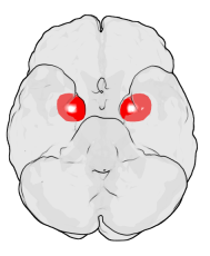
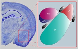
The amygdala is a paired nuclear complex present in the cerebral hemispheres of vertebrates. It is considered part of the limbic system. In primates, it is located medially within the temporal lobes. It consists of many nuclei, each made up of further subnuclei. The subdivision most commonly made is into the basolateral, central, cortical, and medial nuclei together with the intercalated cell clusters. The amygdala has a primary role in the processing of memory, decision-making, and emotional responses. The amygdala was first identified and named by Karl Friedrich Burdach in 1822.

<span class="mw-page-title-main">Nucleus accumbens</span> Region of the basal forebrain

The nucleus accumbens is a region in the basal forebrain rostral to the preoptic area of the hypothalamus. The nucleus accumbens and the olfactory tubercle collectively form the ventral striatum. The ventral striatum and dorsal striatum collectively form the striatum, which is the main component of the basal ganglia. The dopaminergic neurons of the mesolimbic pathway project onto the GABAergic medium spiny neurons of the nucleus accumbens and olfactory tubercle. Each cerebral hemisphere has its own nucleus accumbens, which can be divided into two structures: the nucleus accumbens core and the nucleus accumbens shell. These substructures have different morphology and functions.

<span class="mw-page-title-main">Dopaminergic pathways</span> Projection neurons in the brain that synthesize and release dopamine

Dopaminergic pathways in the human brain are involved in both physiological and behavioral processes including movement, cognition, executive functions, reward, motivation, and neuroendocrine control. Each pathway is a set of projection neurons, consisting of individual dopaminergic neurons.

<span class="mw-page-title-main">Ventral tegmental area</span> Group of neurons on the floor of the midbrain

The ventral tegmental area (VTA), also known as the ventral tegmental area of Tsai, or simply ventral tegmentum, is a group of neurons located close to the midline on the floor of the midbrain. The VTA is the origin of the dopaminergic cell bodies of the mesocorticolimbic dopamine system and other dopamine pathways; it is widely implicated in the drug and natural reward circuitry of the brain. The VTA plays an important role in a number of processes, including reward cognition and orgasm, among others, as well as several psychiatric disorders. Neurons in the VTA project to numerous areas of the brain, ranging from the prefrontal cortex to the caudal brainstem and several regions in between.

<span class="mw-page-title-main">Neuropeptide Y</span> Mammalian protein found in Homo sapiens

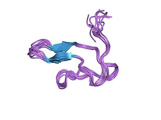
Neuropeptide Y (NPY) is a 36 amino-acid neuropeptide that is involved in various physiological and homeostatic processes in both the central and peripheral nervous systems. It is secreted alongside other neurotransmitters such as GABA and glutamate. 

<span class="mw-page-title-main">Stria terminalis</span> Band of fibres along the thalamus

The stria terminalis is a structure in the brain consisting of a band of fibers running along the lateral margin of the ventricular surface of the thalamus. Serving as a major output pathway of the amygdala, the stria terminalis runs from its centromedial division to the ventromedial nucleus of the hypothalamus.

<span class="mw-page-title-main">Septal area</span> Area in the lower, posterior part of the medial surface of the frontal lobe

The septal area, consisting of the lateral septum and medial septum, is an area in the lower, posterior part of the medial surface of the frontal lobe, and refers to the nearby septum pellucidum.

<span class="mw-page-title-main">Dorsal raphe nucleus</span>

The dorsal raphe nucleus is one of the raphe nuclei. It is situated in the brainstem at the midline. It has rostral and caudal subdivisions:

The amygdalofugal pathway is one of the three major efferent pathways of the amygdala, meaning that it is one of the three principal pathways by which fibers leave the amygdala. It leads from the basolateral nucleus and central nucleus of the amygdala. The amygdala is a limbic structure in the medial temporal lobe of the brain. The other main efferent pathways from the amygdala are the stria terminalis and anterior commissure.

<span class="mw-page-title-main">Medium spiny neuron</span> Type of GABAergic neuron in the striatum

Medium spiny neurons (MSNs), also known as spiny projection neurons (SPNs), are a special type of GABAergic inhibitory cell representing 95% of neurons within the human striatum, a basal ganglia structure. Medium spiny neurons have two primary phenotypes : D1-type MSNs of the direct pathway and D2-type MSNs of the indirect pathway. Most striatal MSNs contain only D1-type or D2-type dopamine receptors, but a subpopulation of MSNs exhibit both phenotypes.

<span class="mw-page-title-main">Lateral hypothalamus</span>

The lateral hypothalamus (LH), also called the lateral hypothalamic area (LHA), contains the primary orexinergic nucleus within the hypothalamus that widely projects throughout the nervous system; this system of neurons mediates an array of cognitive and physical processes, such as promoting feeding behavior and arousal, reducing pain perception, and regulating body temperature, digestive functions, and blood pressure, among many others. Clinically significant disorders that involve dysfunctions of the orexinergic projection system include narcolepsy, motility disorders or functional gastrointestinal disorders involving visceral hypersensitivity, and eating disorders.

<span class="mw-page-title-main">Reward system</span> Group of neural structures responsible for motivation and desire

The reward system is a group of neural structures responsible for incentive salience, associative learning, and positively-valenced emotions, particularly ones involving pleasure as a core component. Reward is the attractive and motivational property of a stimulus that induces appetitive behavior, also known as approach behavior, and consummatory behavior. A rewarding stimulus has been described as "any stimulus, object, event, activity, or situation that has the potential to make us approach and consume it is by definition a reward". In operant conditioning, rewarding stimuli function as positive reinforcers; however, the converse statement also holds true: positive reinforcers are rewarding.

<span class="mw-page-title-main">Cocaine and amphetamine regulated transcript</span> Neuropeptide protein

Cocaine- and amphetamine-regulated transcript, also known as CART, is a neuropeptide protein that in humans is encoded by the CARTPT gene. CART appears to have roles in reward, feeding, and stress, and it has the functional properties of an endogenous psychostimulant.

<span class="mw-page-title-main">Palatability</span>

Palatability is the hedonic reward provided by foods or fluids that are agreeable to the "palate", which often varies relative to the homeostatic satisfaction of nutritional and/or water needs. The palatability of a food or fluid, unlike its flavor or taste, varies with the state of an individual: it is lower after consumption and higher when deprived. It has increasingly been appreciated that this can create a hunger that is independent of homeostatic needs.

<span class="mw-page-title-main">Basolateral amygdala</span> The lateral, basal, and accessory-basal nuclei of the amygdala

The basolateral amygdala, or basolateral complex, consists of the lateral, basal and accessory-basal nuclei of the amygdala. The lateral nuclei receives the majority of sensory information, which arrives directly from the temporal lobe structures, including the hippocampus and primary auditory cortex. The basolateral amygdala also receives dense neuromodulatory inputs from ventral tegmental area (VTA), locus coeruleus (LC), and basal forebrain, whose integrity are important for associative learning. The information is then processed by the basolateral complex and is sent as output to the central nucleus of the amygdala. This is how most emotional arousal is formed in mammals.

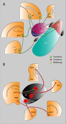
The Intercalatedcells of the amygdala are GABAergic neurons situated between the basolateral and central nuclei of the amygdala that play a significant role in inhibitory control over the amygdala. They regulate amygdala-dependent emotional processing like fear memory and social behavior. Their function has been best studied with selective ITC ablation which impairs fear extinction, fear generalization, and social behavior. Studies have begun to recognize that ITC clusters may be implicated in reward, addiction, and withdrawal circuits given their heavy expression of dopamine and opioid receptors.

<span class="mw-page-title-main">HSD2 neuron</span>

HSD2 neurons are a small group of neurons in the brainstem which are uniquely sensitive to the mineralocorticosteroid hormone aldosterone, through expression of HSD11B2. They are located within the caudal medulla oblongata, in the nucleus of the solitary tract (NTS). HSD2 neurons are activated during a prolonged deficit in body sodium or fluid volume, as occurs after dietary sodium deprivation or during frank hypovolemia. They are also activated by supraphysiologic stimulation of the mineralocorticoid receptor. They are inactivated when salt is ingested. To date, HSD2 neurons have been identified and studied only in rats and mice.

References

  1. Keifer OP, Hurt RC, Ressler KJ, Marvar PJ (September 2015). "The Physiology of Fear: Reconceptualizing the Role of the Central Amygdala in Fear Learning". Physiology. 30 (5): 389–401. doi:10.1152/physiol.00058.2014. PMC   4556826 . PMID   26328883.
  2. Kalin NH, Shelton SE, Davidson RJ (June 2004). "The role of the central nucleus of the amygdala in mediating fear and anxiety in the primate". The Journal of Neuroscience. 24 (24): 5506–5515. doi:10.1523/JNEUROSCI.0292-04.2004. PMC   6729317 . PMID   15201323.
  3. 1 2 Companion MA, Gonzalez DA, Robinson SL, Herman MA, Thiele TE (2022-09-01). "Lateral habenula-projecting central amygdala circuits expressing GABA and NPY Y1 receptor modulate binge-like ethanol intake in mice". Addiction Neuroscience. 3: 100019. doi:10.1016/j.addicn.2022.100019. ISSN   2772-3925. PMC   9435303 . PMID   36059430. S2CID   248484807.
  4. Roberto M, Gilpin NW, Siggins GR (December 2012). "The central amygdala and alcohol: role of γ-aminobutyric acid, glutamate, and neuropeptides". Cold Spring Harbor Perspectives in Medicine. 2 (12): a012195. doi:10.1101/cshperspect.a012195. PMC   3543070 . PMID   23085848.
  5. Swanson LW, Petrovich GD (August 1998). "What is the amygdala?". Trends in Neurosciences. 21 (8): 323–331. doi:10.1016/S0166-2236(98)01265-X. PMID   9720596. S2CID   11826564.
  6. Hasanein P, Mirazi N, Javanmardi K (November 2008). "GABAA receptors in the central nucleus of amygdala (CeA) affect on pain modulation". Brain Research. 1241: 36–41. doi:10.1016/j.brainres.2008.09.041. PMID   18838064. S2CID   46000492.
  7. LeDoux JE (2008). "Amygdala". Scholarpedia . 3 (4): 2698. Bibcode:2008SchpJ...3.2698L. doi: 10.4249/scholarpedia.2698 .
  8. Wright A. "Limbic System: Amygdala". In Byrne JH (ed.). Homeostasis and Higher Brain Function. Neuroscience Online. University of Texas Health Science Center at Houston. Archived from the original on 2013-11-07. Retrieved 2013-02-14.
  9. Bzdok D, Laird AR, Zilles K, Fox PT, Eickhoff SB (December 2013). "An investigation of the structural, connectional, and functional subspecialization in the human amygdala". Human Brain Mapping. 34 (12): 3247–3266. doi:10.1002/hbm.22138. PMC   4801486 . PMID   22806915.
  10. Best B (August 28, 2012). "The Amygdala and the Emotions". The Anatomical Basis of Mind. Archived from the original on March 9, 2007.
  11. Solano-Castiella E, Anwander A, Lohmann G, Weiss M, Docherty C, Geyer S, et al. (February 2010). "Diffusion tensor imaging segments the human amygdala in vivo". NeuroImage. 49 (4): 2958–2965. doi:10.1016/j.neuroimage.2009.11.027. hdl: 11858/00-001M-0000-0010-ABE5-F . PMID   19931398. S2CID   17137887.
  12. Di Marino V, Etienne Y, Niddam M (2016). "Connection Pathways of the Cerebral Amygdala". The Amygdaloid Nuclear Complex. pp. 49–58. doi:10.1007/978-3-319-23243-0_6. ISBN   978-3-319-23242-3.{{cite book}}: |journal= ignored (help)
  13. Kamali A, Sair HI, Blitz AM, Riascos RF, Mirbagheri S, Keser Z, Hasan KM (September 2016). "Revealing the ventral amygdalofugal pathway of the human limbic system using high spatial resolution diffusion tensor tractography". Brain Structure & Function. 221 (7): 3561–3569. doi:10.1007/s00429-015-1119-3. PMID   26454651. S2CID   10456347.
  14. Makino S, Shibasaki T, Yamauchi N, Nishioka T, Mimoto T, Wakabayashi I, et al. (December 1999). "Psychological stress increased corticotropin-releasing hormone mRNA and content in the central nucleus of the amygdala but not in the hypothalamic paraventricular nucleus in the rat". Brain Research. 850 (1–2): 136–143. doi:10.1016/S0006-8993(99)02114-9. PMID   10629757. S2CID   21062798.
  15. Margatho LO, Elias CF, Elias LL, Antunes-Rodrigues J (May 2013). "Oxytocin in the central amygdaloid nucleus modulates the neuroendocrine responses induced by hypertonic volume expansion in the rat". Journal of Neuroendocrinology. 25 (5): 466–477. doi: 10.1111/jne.12021 . PMID   23331859. S2CID   5486765.
  16. Smith ES, Geissler SA, Schallert T, Lee HJ (April 2013). "The role of central amygdala dopamine in disengagement behavior". Behavioral Neuroscience. 127 (2): 164–174. doi:10.1037/a0031043. PMID   23316710.
  17. Yan J, Li J, Yan J, Sun H, Wang Q, Chen K, et al. (March 2013). "Activation of μ-opioid receptors in the central nucleus of the amygdala induces hypertonic sodium intake". Neuroscience. 233: 28–43. doi:10.1016/j.neuroscience.2012.12.026. PMID   23270855. S2CID   10806357.
  18. Zimmerman JM, Rabinak CA, McLachlan IG, Maren S (September 2007). "The central nucleus of the amygdala is essential for acquiring and expressing conditional fear after overtraining". Learning & Memory. 14 (9): 634–644. doi:10.1101/lm.607207. PMC   1994080 . PMID   17848503.
  19. Makino S, Gold PW, Schulkin J (March 1994). "Corticosterone effects on corticotropin-releasing hormone mRNA in the central nucleus of the amygdala and the parvocellular region of the paraventricular nucleus of the hypothalamus". Brain Research. 640 (1–2): 105–112. doi:10.1016/0006-8993(94)91862-7. PMID   8004437. S2CID   19853559.
  20. Hyytiä P, Koob GF (September 1995). "GABAA receptor antagonism in the extended amygdala decreases ethanol self-administration in rats". European Journal of Pharmacology. 283 (1–3): 151–159. doi:10.1016/0014-2999(95)00314-B. PMID   7498304.
  21. Gilpin NW, Misra K, Herman MA, Cruz MT, Koob GF, Roberto M (June 2011). "Neuropeptide Y opposes alcohol effects on gamma-aminobutyric acid release in amygdala and blocks the transition to alcohol dependence". Biological Psychiatry. 69 (11): 1091–1099. doi:10.1016/j.biopsych.2011.02.004. PMC   3090491 . PMID   21459365.
  22. Li X, Zeric T, Kambhampati S, Bossert JM, Shaham Y (March 2015). "The central amygdala nucleus is critical for incubation of methamphetamine craving". Neuropsychopharmacology. 40 (5): 1297–1306. doi:10.1038/npp.2014.320. PMC   4367476 . PMID   25475163.
  23. Han W, Tellez LA, Rangel MJ, Motta SC, Zhang X, Perez IO, Canteras NS, Shammah-Lagnado SJ, van den Pol AN, de Araujo IE (January 2017). "Integrated Control of Predatory Hunting by the Central Nucleus of the Amygdala". Cell . 168 (1–2): 311–324.e18. doi:10.1016/j.cell.2016.12.027. PMC   5278763 . PMID   28086095.