Anodontia

Last updated
Anodontia
Other namesAnodontia vera
Anodontia (cropped).jpg
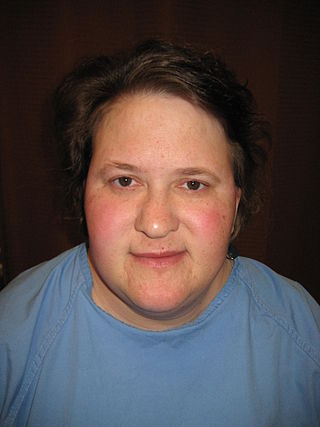
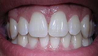
Anodontia view of patient showing oligodontia and conical teeth
Specialty Dentistry, medical genetics

Anodontia is a rare genetic disorder characterized by the congenital absence of all primary or permanent teeth. It is divided into two subsections, complete absence of teeth or only some absence of teeth. It is associated with the group of skin and nerve syndromes called the ectodermal dysplasias. Anodontia is usually part of a syndrome and seldom occurs as an isolated entity. There is usually no exact cause for anodontia. The defect results in the dental lamina obstruction during embryogenesis due to local, systemic and genetic factors.

Contents

Congenital absence of permanent teeth can present as hypodontia, usually missing one or two permanent teeth, or oligodontia that is the congenital absence of six or more teeth. Congenital absence of all wisdom teeth, or third molars, is relatively common. Anodontia is the congenital absence of teeth and can occur in some or all teeth; whereas partial anodontia (or hypodontia), involves two dentitions or only teeth of the permanent dentition (Dorland's 1998). Approximately 1% of the population has oligodontia. [1] Many denominations are attributed to this anomaly: partial anodontia, hypodontia, oligodontia, the congenital absence, anodontia, bilateral aplasia. Anodontia being the term used in controlled vocabulary Medical Subject Headings (MeSH) from MEDLINE which was developed by the United States National Library of Medicine. The congenital absence of at least one permanent tooth is the most common dental anomaly and may contribute to masticator dysfunction, speech impairment, aesthetic problems, and malocclusion (Shapiro and Farrington 1983). Absence of lateral incisors represents a major stereotype. Individuals with this condition are perceived as socially most aggressive compared with people without anodontia (Shaw 1981). [2] The occurrence of anodontia is less so than hypodontia which has a prevalence of 0.1-0.7% in primary teeth and 3–7.5% in permanent teeth. [3]

Signs and symptoms

The main sign of anodontia is when a child has not developed any of their permanent teeth by the age of 12. Another sign of anodontia can include the absence of baby teeth when the baby has reached 12 to 13 months. [4]

Symptoms that are associated with anodontia include: alopecia, lack of sweat glands, cleft lip or palate, and missing fingernails. Typically, these symptoms are seen because anodontia is typically associated with ectodermal dysplasia. In the rare case that ectodermal dysplasia is not present, anodontia will be caused from an unknown genetic mutation. [4]

Cause

Anodontia typically occurs with the presence of ectodermal dysplasia, which is a group of disorders where two or more ectodermally derived structures will have abnormal development. In the rare case that ectodermal dysplasia is not associated or present, anodontia will be caused by an unknown genetic mutation. Although no specific gene has been identified, there have been many different genes found to be associated with anodontia including EDA, EDAR, and EDARADD genes. Other genes such as MSX1, PAX9, IRF6, GREM2, AXIN2, LRP6, SMOC2, LTBP3, PITX2, and WNT10B. The WNT10A gene is considered to be the major gene involved in hypodontia and oligodontia. These genes are involved in hypodontia and oligodontia. If Anodontia is present in the maternal or paternal side, the chances of this being inherited are increased. [4]

Mechanisms and Pathophysiology

Anodontia is a genetic disorder that is typically occurs in result of another syndrome. Different results can occur depending on which gene is inherited. It remains unclear which specific gene is the direct cause but it is known that several genes can play a role when inherited. Many genes are involved with this and other relating disorders. The main genes involved include: EDA, EDAR, and EDARADD genes. One working gene and one non-working gene are inherited, one from an affected parent and one from a non-affected parent, which then result in a 50% chance of the child inheriting the genetic disorder. Anodontia alone will not have an effect on any other body part besides teeth being missing. [4]

Associated syndromes

Hypodontia and anodontia are frequently associated with a multitude of genetic disorders and syndromes, approximately 70. Syndromes particularly involved with ectodermal involvement are a prime circumstance for anodontia to occur, some examples of these are: Rieger's, Robinson's and focal dermal hypoplasia. Three syndromes which classically have signs of anodontia are oculomandibulodyscephaly, mesoectodermal dysplasia and ectodermal dysplasia. In cases of oculomandibulodyscephaly there are no permanent teeth but there are deciduous teeth present. In mesoectodermal dysplasia the symptoms are anodontia and hypodontia. In cases of ectodermal dysplasia oligodontia is also present. Other symptoms associated with anodontia include: Alopecia, loss of sweat glands, cleft lip or palate, or missing finger nails. [4]

Diagnosis

Anodontia can be diagnosed when a baby does not begin to develop teeth around the age of 12 to 13 months or when a child does not develop their permanent teeth by the age of 10. The dentist can use a special X-ray, such as a panoramic image, to check if there are any teeth developing. There is also a higher risk for a child to develop anodontia if their parent has this disorder as well. In the absence of all permanent teeth, anodontia will be diagnosed. If between one and five teeth are missing, this will be diagnosed as hypodontia. In the absence of six or more teeth, this will be diagnosed as oligodontia. [4]

Complications

The complications associated with anodontia can vary but the majority results in problems with aesthetic appearance, speaking, and masticatory function. Complications may occur with the placement of the dental implant. Although it is rare, some complications may include the screw of the implant becoming loose or sore spots. [5]

Prevention and Treatment

Anodontia cannot be prevented due to it being a genetic disorder. Prosthetic replacement of missing teeth is possible using dental implant technology or dentures. [6] This treatment can be successful in giving patients with anodontia a more aesthetically pleasing appearance. The use of an implant prosthesis in the lower jaw could be recommended for younger patients as it is shown to significantly improve the craniofacial growth, social development and self-image. The study associated with this evidence worked with individuals who had ectodermal dysplasia of varying age groups of up to 11, 11 to 18 and more than 18 years. It was noted that the risk of implant failure was significantly higher in patients younger than 18 years, but there is significant reason to use this methodology of treatment in those older. [5] Overall the use of an implant-prosthesis has a considerable functional, aesthetic and psychological advantage when compared to a conventional denture, in the patients. [7]

Prognosis

Patients diagnosed with anodontia are expected to have a normal life expectancy. Once anodontia is diagnosed, dental implants or dentures will need to be worn in order to treat this disorder. There is an 88.5% to 100% chance for dental implants in patients with ectodermal dysplasia or tooth agenesis to be successful when placed after the age of 18.

Epidemiology

The prevalence of anodontia is unknown but it is a very rare disorder. Anodontia occurs in less than 2-8% of the general population in regards to permanent teeth and 0.1-0.7% in primary teeth. Gender and ethnicity do not play a role in anodontia. [8]

Research

A recent study in 2019 by R. Constance Wiener and Christopher water looked at anodontia, hypodontia, and oligodontia in children West Virginia. There is a high prevalence of children with missing permanent teeth in West Virginia compared to the rest of the nation. During this study, 500 panoramic images were taken of children between the ages of 6 and 11. Out of the 500 images taken, 60 children had at least one or more missing permanent teeth. The results showed that more females had one or more missing permanent teeth than males. From the 60 children who had missing permanent teeth, 15.5% were female and 8.8% were males. [9]

A case study conducted in 2016 of a six-year-old boy presented with anodontia. [10] There was no family history of anodontia and the patient did not present any other symptoms for ectodermal dysplasia. [10] It was observed the hypodontia was present in the maxillary arch and the only teeth present were the left primary first molar and the bilateral primary second molars. [10] It was also observed that the buccal mucosa, palate, and floor of the mouth were considered normal. [10] The patient proceeded with oral rehabilitation and give removable denture to wear. [10] The patient struggled in the beginning to keep wearing the denture until gradually learning to adjust to it. [10] The family reported no problems with retention and began a monthly recall visit in order to monitor any eruptions of teeth or adjustments that needed to be made. [10] Improvements in speech skills, communication, and self esteem were also observed after placement of the denture. [10]

Another case study in 2013 of an eight-year-old boy who reported missing teeth, difficulty chewing, and difficulty speaking was seen to have other symptoms of ectodermal dysplasia. [11] The father confirmed there is a family history of missing teeth. [11] The patient also had sensitivity to heat, absence of sweating, dry skin, absent eyebrows and eyelashes, hyper pigmentation, and many other ectodermal dysplasia symptoms. [11] After a full examination, the patient was diagnosed with complete anodontia. [11] The patient was treated with a complete set of removable dentures. [11] After the dentures were given, the patient's facial presentation and expressions improved. [11] The patient was also set up for recall follow ups every six months. Drastic improvement was seen with improvements with chewing and speech. [11]

See also

Related Research Articles

<span class="mw-page-title-main">Hyperdontia</span> Condition of having extra teeth beyond the regular number of teeth

Hyperdontia is the condition of having supernumerary teeth, or teeth that appear in addition to the regular number of teeth. They can appear in any area of the dental arch and can affect any dental organ. The opposite of hyperdontia is hypodontia, where there is a congenital lack of teeth, which is a condition seen more commonly than hyperdontia. The scientific definition of hyperdontia is "any tooth or odontogenic structure that is formed from tooth germ in excess of usual number for any given region of the dental arch." The additional teeth, which may be few or many, can occur on any place in the dental arch. Their arrangement may be symmetrical or non-symmetrical.

Hypodontia is defined as the developmental absence of one or more teeth excluding the third molars. It is one of the most common dental anomalies, and can have a negative impact on function, and also appearance. It rarely occurs in primary teeth and the most commonly affected are the adult second premolars and the upper lateral incisors. It usually occurs as part of a syndrome that involves other abnormalities and requires multidisciplinary treatment.

<span class="mw-page-title-main">Ellis–Van Creveld syndrome</span> Medical condition

Ellis–Van Creveld syndrome is a rare genetic disorder of the skeletal dysplasia type.

<span class="mw-page-title-main">Ectodermal dysplasia</span> Group of genetic conditions affecting the embryonic ectoderm

Ectodermal dysplasia (ED) is a group of genetic syndromes all deriving from abnormalities of the ectodermal structures. More than 150 different syndromes have been identified.

Zadik–Barak–Levin syndrome (ZBLS) is a congenital disorder in humans. Presenting conditions include primary hypothyroidism, cleft palate, hypodontia, and ectodermal dysplasia. It is the result of an embryonic defect in the mesodermal-ectodermal midline development.

<span class="mw-page-title-main">Cherubism</span> Medical condition

Cherubism is a rare genetic disorder that causes prominence in the lower portion in the face. The name is derived from the temporary chubby-cheeked resemblance to putti, the chubby-faced infants featured in Renaissance paintings, which were often mistakenly described as cherubs.

<span class="mw-page-title-main">Larsen syndrome</span> Medical condition

Larsen syndrome (LS) is a congenital disorder discovered in 1950 by Larsen and associates when they observed dislocation of the large joints and face anomalies in six of their patients. Patients with Larsen syndrome normally present with a variety of symptoms, including congenital anterior dislocation of the knees, dislocation of the hips and elbows, flattened facial appearance, prominent foreheads, and depressed nasal bridges. Larsen syndrome can also cause a variety of cardiovascular and orthopedic abnormalities. This rare disorder is caused by a genetic defect in the gene encoding filamin B, a cytoplasmic protein that is important in regulating the structure and activity of the cytoskeleton. The gene that influences the emergence of Larsen syndrome is found in chromosome region, 3p21.1-14.1, a region containing human type VII collagen gene. Larsen syndrome has recently been described as a mesenchyme disorder that affects the connective tissue of an individual. Autosomal dominant and recessive forms of the disorder have been reported, although most cases are autosomal dominant. Reports have found that in Western societies, Larsen syndrome can be found in one in every 100,000 births, but this is most likely an underestimate because the disorder is frequently unrecognized or misdiagnosed.

<span class="mw-page-title-main">Papillon–Lefèvre syndrome</span> Medical condition

Papillon–Lefèvre syndrome (PLS), also known as palmoplantar keratoderma with periodontitis, is an autosomal recessive genetic disorder caused by a deficiency in cathepsin C.

<span class="mw-page-title-main">Ectrodactyly–ectodermal dysplasia–cleft syndrome</span> Medical condition

Ectrodactyly–ectodermal dysplasia–cleft syndrome, or EEC, and also referred to as EEC syndrome and split hand–split foot–ectodermal dysplasia–cleft syndrome is a rare form of ectodermal dysplasia, an autosomal dominant disorder inherited as a genetic trait. EEC is characterized by the triad of ectrodactyly, ectodermal dysplasia, and facial clefts. Other features noted in association with EEC include vesicoureteral reflux, recurrent urinary tract infections, obstruction of the nasolacrimal duct, decreased pigmentation of the hair and skin, missing or abnormal teeth, enamel hypoplasia, absent punctae in the lower eyelids, photophobia, occasional cognitive impairment and kidney anomalies, and conductive hearing loss.

Amastia refers to a rare clinical anomaly in which both internal breast tissue and the visible nipple are absent on one or both sides. It affects both men and women. Amastia can be either isolated or comorbid with other syndromes, such as ectodermal dysplasia, Syndactyly and lipoatrophic diabetes. This abnormality can be classified into various types, and each could result from different pathologies. Amastia differs from amazia and athelia. Amazia is the absence of one or both mammary glands but the nipples remain present, and athelia is the absence of one or both nipples, but the mammary gland remains.

<span class="mw-page-title-main">Hypohidrotic ectodermal dysplasia</span> Medical condition

Hypohidrotic ectodermal dysplasia is one of about 150 types of ectodermal dysplasia in humans. Before birth, these disorders result in the abnormal development of structures including the skin, hair, nails, teeth, and sweat glands.

<span class="mw-page-title-main">Dentin dysplasia</span> Medical condition

Dentin dysplasia (DD) is a rare genetic developmental disorder affecting dentine production of the teeth, commonly exhibiting an autosomal dominant inheritance that causes malformation of the root. It affects both primary and permanent dentitions in approximately 1 in every 100,000 patients. It is characterized by the presence of normal enamel but atypical dentin with abnormal pulpal morphology. Witkop in 1972 classified DD into two types which are Type I (DD-1) is the radicular type, and type II (DD-2) is the coronal type. DD-1 has been further divided into 4 different subtypes (DD-1a,1b,1c,1d) based on the radiographic features.

<span class="mw-page-title-main">Naegeli–Franceschetti–Jadassohn syndrome</span> Medical condition

Naegeli–Franceschetti–Jadassohn syndrome (NFJS), also known as chromatophore nevus of Naegeli and Naegeli syndrome, is a rare autosomal dominant form of ectodermal dysplasia, characterized by reticular skin pigmentation, diminished function of the sweat glands, the absence of teeth and hyperkeratosis of the palms and soles. One of the most striking features is the absence of fingerprint lines on the fingers.

<span class="mw-page-title-main">Hay–Wells syndrome</span> Medical condition

Hay–Wells syndrome is one of at least 150 known types of ectodermal dysplasia. These disorders affect tissues that arise from the ectodermal germ layer, such as skin, hair, and nails.

<span class="mw-page-title-main">PAX9</span> Protein-coding gene in humans

Paired box gene 9, also known as PAX9, is a protein which in humans is encoded by the PAX9 gene. It is also found in other mammals.

<span class="mw-page-title-main">Frontonasal dysplasia</span> Medical condition

Frontonasal dysplasia (FND) is a congenital malformation of the midface. For the diagnosis of FND, a patient should present at least two of the following characteristics: hypertelorism, a wide nasal root, vertical midline cleft of the nose and/or upper lip, cleft of the wings of the nose, malformed nasal tip, encephalocele or V-shaped hair pattern on the forehead. The cause of FND remains unknown. FND seems to be sporadic (random) and multiple environmental factors are suggested as possible causes for the syndrome. However, in some families multiple cases of FND were reported, which suggests a genetic cause of FND.

<span class="mw-page-title-main">EEM syndrome</span> Medical condition

EEM syndrome is an autosomal recessive congenital malformation disorder affecting tissues associated with the ectoderm, and also the hands, feet and eyes.

<span class="mw-page-title-main">Orofaciodigital syndrome 1</span> Medical condition

Orofaciodigital syndrome 1 (OFD1), also called Papillon-Léage and Psaume syndrome, is an X-linked congenital disorder characterized by malformations of the face, oral cavity, and digits with polycystic kidney disease and variable involvement of the central nervous system.

<span class="mw-page-title-main">Acro–dermato–ungual–lacrimal–tooth syndrome</span> Rare human genetic disease

Acro–dermato–ungual–lacrimal–tooth syndrome is a rare genetic disease. It is an autosomal dominant form of ectodermal dysplasia, a group of disorders that affects the hair, teeth, nails, sweat glands, and extremities. The syndrome arises from a mutation in the TP63 gene. This disease was previously thought to be a form of ectrodactyly–ectodermal dysplasia–cleft syndrome (EEC), but was classified as a different disease in 1993 by Propping and Zerres.

<span class="mw-page-title-main">Odontoonychodermal dysplasia</span> Medical condition

Odontoonychodermal dysplasia is a rare genetic disorder which is characterized by systemic abnormalities of the teeth, the nails of the fingers and toes, the skin, the hair cells, and the sweat glands. It is a type of syndromic ectodermal dysplasia.

References

  1. Vahid-Dastjerdi E, Borzabadi-Farahani A, Mahdian M, Amini N (2010). "Non-syndromic hypodontia in an Iranian orthodontic population". J. Oral Sci. 52 (3): 455–461. doi: 10.2334/josnusd.52.455 . PMID   20881340.
  2. Shaw, William C. (April 1981). "The influence of children's dentofacial appearance on their social attractiveness as judged by peers and lay adults". American Journal of Orthodontics. 79 (4): 399–415. doi:10.1016/0002-9416(81)90382-1. ISSN   0002-9416. PMID   6939333.
  3. Laskaris, George (2000). Color Atlas of Oral Diseases in Children and Adolescents. Thieme. ISBN   9780865777897.
  4. 1 2 3 4 5 6 "Tooth Agenesis". NORD (National Organization for Rare Disorders). Retrieved 2020-12-16.
  5. 1 2 Yap, Alan K. W.; Klineberg, Iven (May–June 2009). "Dental implants in patients with ectodermal dysplasia and tooth agenesis: a critical review of the literature". The International Journal of Prosthodontics. 22 (3): 268–276. ISSN   0893-2174. PMID   19548409.
  6. Borzabadi-Farahani; A (December 2012). "Orthodontic considerations in restorative management of hypodontia patients with endosseous implants". Journal of Oral Implantology. 38 (6): 779–791. doi: 10.1563/AAID-JOI-D-11-00022 . PMID   21728818.
  7. de Santé, Haute Autorité (2010). "Traitement implantoprothétique de l'adulte atteint d'agénésies dentaires multiples liées à une maladie rare [Treatment of multiple missing teeth in adults with rare diseases by using implant-supported denture]" (in French). Paris.{{cite journal}}: Cite journal requires |journal= (help)
  8. Vahid-Dastjerdi E, Borzabadi-Farahani A, Mahdian M, Amini N (2010). "Non-syndromic hypodontia in an Iranian orthodontic population". J. Oral Sci. 52 (3): 455–461. doi: 10.2334/josnusd.52.455 . PMID   20881340.
  9. Wiener, R. Constance; Waters, Christopher (2019-04-01). "Hypodontia, Oligodontia and Anodontia in West Virginia Appalachia". American Dental Hygienists' Association. 93 (2): 34–40. ISSN   1553-0205. PMID   31015306.
  10. 1 2 3 4 5 6 7 8 Rathee, Manu; Malik, Poonam; Dua, Madhuri; Yadav, Vikas (2016). "Early functional, esthetic, and psychological rehabilitation of preschool child with nonsyndromic oligodontia and anodontia in mixed dentition stage through conservative systematic approach: A case report with 5-year follow-up". Contemporary Clinical Dentistry. 7 (2): 232–235. doi: 10.4103/0976-237X.183051 . ISSN   0976-237X. PMC   4906870 . PMID   27307674.
  11. 1 2 3 4 5 6 7 Ladda, R; Gangadhar, SA; Kasat, VO; Bhandari, AJ (2013). "Prosthodontic Management of Hypohidrotic Ectodermal Dysplasia with Anodontia: A Case Report in Pediatric Patient and Review of Literature". Annals of Medical and Health Sciences Research. 3 (2): 277–281. doi: 10.4103/2141-9248.113679 . ISSN   2141-9248. PMC   3728879 . PMID   23919206.