Human teeth function to mechanically break down items of food by cutting and crushing them in preparation for swallowing and digesting. As such, they are considered part of the human digestive system. Humans have four types of teeth: incisors, canines, premolars, and molars, which each have a specific function. The incisors cut the food, the canines tear the food and the molars and premolars crush the food. The roots of teeth are embedded in the maxilla or the mandible and are covered by gums. Teeth are made of multiple tissues of varying density and hardness.

Incisors are the front teeth present in most mammals. They are located in the premaxilla above and on the mandible below. Humans have a total of eight. Opossums have 18, whereas armadillos have none.
Hyperdontia is the condition of having supernumerary teeth, or teeth that appear in addition to the regular number of teeth. They can appear in any area of the dental arch and can affect any dental organ. The opposite of hyperdontia is hypodontia, where there is a congenital lack of teeth, which is a condition seen more commonly than hyperdontia. The scientific definition of hyperdontia is "any tooth or odontogenic structure that is formed from tooth germ in excess of usual number for any given region of the dental arch." The additional teeth, which may be few or many, can occur on any place in the dental arch. Their arrangement may be symmetrical or non-symmetrical.
Hypodontia is defined as the developmental absence of one or more teeth excluding the third molars. It is one of the most common dental anomalies, and can have a negative impact on function, and also appearance. It rarely occurs in primary teeth and the most commonly affected are the adult second premolars and the upper lateral incisors. It usually occurs as part of a syndrome that involves other abnormalities and requires multidisciplinary treatment.

Macroglossia is the medical term for an unusually large tongue. Severe enlargement of the tongue can cause cosmetic and functional difficulties in speaking, eating, swallowing and sleeping. Macroglossia is uncommon, and usually occurs in children. There are many causes. Treatment depends upon the exact cause.

Tooth development or odontogenesis is the complex process by which teeth form from embryonic cells, grow, and erupt into the mouth. For human teeth to have a healthy oral environment, all parts of the tooth must develop during appropriate stages of fetal development. Primary (baby) teeth start to form between the sixth and eighth week of prenatal development, and permanent teeth begin to form in the twentieth week. If teeth do not start to develop at or near these times, they will not develop at all, resulting in hypodontia or anodontia.

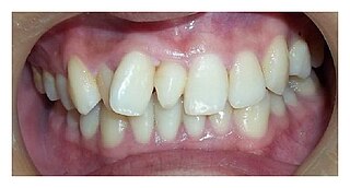
In orthodontics, a malocclusion is a misalignment or incorrect relation between the teeth of the upper and lower dental arches when they approach each other as the jaws close. The English-language term dates from 1864; Edward Angle (1855-1930), the "father of modern orthodontics", popularised it. The word "malocclusion" derives from occlusion, and refers to the manner in which opposing teeth meet.

The pyramid-shaped maxillary sinus is the largest of the paranasal sinuses, located in the maxilla. It drains into the middle meatus of the nose through the semilunar hiatus. It is located to the side of the nasal cavity, and below the orbit.

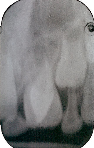
The maxillary central incisor is a human tooth in the front upper jaw, or maxilla, and is usually the most visible of all teeth in the mouth. It is located mesial to the maxillary lateral incisor. As with all incisors, their function is for shearing or cutting food during mastication (chewing). There is typically a single cusp on each tooth, called an incisal ridge or incisal edge. Formation of these teeth begins at 14 weeks in utero for the deciduous (baby) set and 3–4 months of age for the permanent set.

The maxillary lateral incisors are a pair of upper (maxillary) teeth that are located laterally from both maxillary central incisors of the mouth and medially from both maxillary canines. As with all incisors, their function is for shearing or cutting food during mastication, commonly known as chewing. There are generally no cusps on the teeth, but the rare condition known as talon cusps are most prevalent on the maxillary lateral incisors. The surface area of the tooth used in eating is called an incisal ridge or incisal edge. Though relatively the same, there are some minor differences between the deciduous (baby) maxillary lateral incisor and that of the permanent maxillary lateral incisor. The maxillary lateral incisors occlude in opposition to the mandibular lateral incisors.
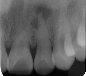
Dilaceration is a developmental disturbance in shape of teeth. It refers to an angulation, or a sharp bend or curve, in the root or crown of a formed tooth. This disturbance is more likely to affect the maxillary incisors and occurs in permanent dentition. Although this may seem more of an aesthetics issue, an impacted maxillary incisor will cause issues related to occlusion, phonetics, mastication, and psychology on young patients.

A dentigerous cyst, also known as a follicular cyst, is an epithelial-lined developmental cyst formed by accumulation of fluid between the reduced enamel epithelium and the crown of an unerupted tooth. It is formed when there is an alteration in the reduced enamel epithelium and encloses the crown of an unerupted tooth at the cemento-enamel junction. Fluid is accumulated between reduced enamel epithelium and the crown of an unerupted tooth.
Macrodontia is a type of localized gigantism in which teeth are larger than normal. Macrodontia seen in permanent teeth is thought to affect around 0.03 to 1.9 percent of the worldwide population. Generally, patients with macrodontia have one or two teeth in their mouth that is abnormally large; however, single tooth growth is seen in a number of cases as well.
Dens evaginatus is a rare odontogenic developmental anomaly that is found in teeth where the outer surface appears to form an extra bump or cusp.
Talon cusp is a rare dental anomaly resulting in an extra cusp or cusp-like projection on an anterior tooth, located on the inside surface of the affected tooth. Sometimes it can also be found on the facial surface of the anterior tooth.
Dental anatomy is a field of anatomy dedicated to the study of human tooth structures. The development, appearance, and classification of teeth fall within its purview. Tooth formation begins before birth, and the teeth's eventual morphology is dictated during this time. Dental anatomy is also a taxonomical science: it is concerned with the naming of teeth and the structures of which they are made, this information serving a practical purpose in dental treatment.
Dental pertains to the teeth, including dentistry. Topics related to the dentistry, the human mouth and teeth include:

Enamel hypoplasia is a defect of the teeth in which the enamel is deficient in quantity, caused by defective enamel matrix formation during enamel development, as a result of inherited and acquired systemic condition(s). It can be identified as missing tooth structure and may manifest as pits or grooves in the crown of the affected teeth, and in extreme cases, some portions of the crown of the tooth may have no enamel, exposing the dentin. It may be generalized across the dentition or localized to a few teeth. Defects are categorized by shape or location. Common categories are pit-form, plane-form, linear-form, and localised enamel hypoplasia. Hypoplastic lesions are found in areas of the teeth where the enamel was being actively formed during a systemic or local disturbance. Since the formation of enamel extends over a long period of time, defects may be confined to one well-defined area of the affected teeth. Knowledge of chronological development of deciduous and permanent teeth makes it possible to determine the approximate time at which the developmental disturbance occurred. Enamel hypoplasia varies substantially among populations and can be used to infer health and behavioural impacts from the past. Defects have also been found in a variety of non-human animals.
Serial extraction is the planned extraction of certain deciduous teeth and specific permanent teeth in an orderly sequence and predetermined pattern to guide the erupting permanent teeth into a more favorable position.

Otodental syndrome, also known as otodental dysplasia, is an exceptionally rare disease that is distinguished by a specific phenotype known as globodontia, that in rare cases can be associated with eye coloboma and high frequency hearing loss. Globodontia is an abnormal condition that can occur in both the primary and secondary dentition, except for the incisors which are normal in shape and size. This is demonstrated by significant enlargement of the canine and molar teeth. The premolars are either reduced in size or are absent. In some cases, the defects affecting the teeth, eye and ear can be either individual or combined. When these conditions are combined with eye coloboma, the condition is also known as oculo-otodental syndrome. The first known case of otodental syndrome was found in Hungary in a mother and her son by Denes and Csiba in 1969. Prevalence is less than 1 out of every 1 million individuals. The cause of otodental syndrome is considered to be genetic. It is an autosomal dominant inheritance and is variable in its expressivity. Haploinsufficiency in the fibroblast growth factor 3 (FGF3) gene (11q13) has been reported in patients with otodental syndrome and is thought to cause the phenotype. Both males and females are equally affected. Individuals diagnosed with otodental syndrome can be of any age; age is not a relevant factor. Currently there are no specific genetic treatments for otodental syndrome. Dental and orthodontic management are the recommended course of action.