Periapical cyst

Last updated
Periapical cyst
Other namesRadicular cyst, inflammatory cyst
Den cyst ct.jpg
CT scan through head showing a right periapical cyst
Specialty Dentistry   OOjs UI icon edit-ltr-progressive.svg

Commonly known as a dental cyst, the periapical cyst is the most common odontogenic cyst. It may develop rapidly from a periapical granuloma, as a consequence of untreated chronic periapical periodontitis. [1]

Contents

Periapical is defined as "the tissues surrounding the apex of the root of a tooth" and a cyst is "a pathological cavity lined by epithelium, having fluid or gaseous content that is not created by the accumulation of pus." [2]

Most frequently located in the maxillary anterior region, the cyst is caused by pulpal necrosis secondary to dental caries or trauma. Its lining is derived from the epithelial cell rests of Malassez which proliferate to form the cyst. [2] Such cysts are very common. Although initially asymptomatic, they are clinically significant because secondary infection can cause pain and damage. In radiographs, the cyst appears as a radiolucency (dark area) around the apex of a tooth's root. [3]

Signs and symptoms

Visual image of the face of the person shown on the CT scan above. Swelling can be observed in the right cheek. Den cyst.jpg
Visual image of the face of the person shown on the CT scan above. Swelling can be observed in the right cheek.

Periapical cysts begin as asymptomatic and progress slowly. Subsequent infection of the cyst causes swelling and pain. Initially, the cyst swells to a round hard protrusion, but later on the body resorbs some of the cyst wall, leaving a softer accumulation of fluid underneath the mucous membrane.[ citation needed ]

Secondary[ clarification needed ] symptoms of periapical cysts include inflammation and infection of the pulp causing dental caries. This infection is what causes necrosis of the pulp. [4]

Larger cysts may cause bone expansion or displace roots. Discoloration of the affected tooth may also occur. Patient will present negative results to electric and ice test of the affected tooth but will be sensitive to percussion. Surrounding gingival tissue may experience lymphadenopathy. The alveolar plate may exhibit crepitus when palpated.

Complications

Expansion of the cyst causes erosion of the floor of the maxillary sinus. As soon as it enters the maxillary antrum, the expansion rate increases due to available space for expansion. Performing a percussion test by tapping the affected teeth will cause shooting pain. This is often clinically diagnostic of pulpal infection.[ citation needed ]

Causes

Dental cysts are usually caused due to root infection involving tooth decay. Untreated dental caries then allow bacteria to reach the level of the pulp, causing infection. The bacteria gains access to the periapical region of the tooth through deeper infection of the pulp, traveling through the roots. The resulting pulpal necrosis causes proliferation of epithelial rests of Malassez which release toxins at the apex of the tooth. The body's inflammatory response will attack the source of the toxins, leading to periapical inflammation. The many cells and proteins that rush to an area of infection create osmotic tension in the periapex which is the source of internal pressure increase at the cyst site.

These lesions can grow large because they apply pressure over the bone, causing resorption. The toxins released by the breakdown of granulation tissue are one of the common causes of bone resorption.

There are two schools of thought regarding cyst expansion. [5]

  1. Complementary response to inflammation
  2. Chemical reaction with Interleukin and Prostaglandin

Mechanisms

Periapical cysts develop due to an inflammatory stimulus in 3 stages: [4]

  1. Initial stage: Epithelial cells from the rests of Malassez at the apex of the roots of a non-vital tooth (one where the nerve and blood supply in the tooth have degenerated and no longer exist) become stimulated due to the body's inflammatory response to bacterial endotoxins infecting the pulp or as a direct response to necrotic pulp tissue, therefore re-entering the growth phase. Bacterial byproducts then are able to seep into the periapical region through the infected pulp.
  2. Cyst development stage: Epithelial cells form strands and are attracted to the area which contains exposed connective tissue and foreign substances. Several strands from each rest converge and surround the abscess or foreign body.
  3. Cyst growth stage: Fluid flows into the cavity where the forming cyst is growing due to the increased osmolality of the cavity in relation to surrounding serum in capillaries. Pressure and size increase.

The definitive mechanism by which cysts grow is under debate; several theories exist.

Biomechanical theory

Pressure and concentration differences between the cystic cavity and the growth surroundings influence fluid movement into the cyst, causing size increase.

Biochemical theories

a. Collagenase (breakdown of collagen) in the jaw bone leads to bone degeneration, providing room for cysts to develop. Substances released by the body's immune system as a result of the connective tissue breakdown, such as cytokines and growth factors, contribute to the mobilization and proliferation of epithelial cells in the area.

b. Bone resorption caused by metabolism of acidic substances produced by cysts contributes to cyst growth. Such substances include Prostaglandin-2 and Interleukin-1 which are both produced by the cyst itself.

Nutritional deficiency theory

Epithelial cells will form a mass inside the cavity and the innermost cells become deprived of nutrients because they are far from the source of nutrients (the blood vessels). The innermost cells die and form an aggregate of dead tissue. The inner cells undergo ischemic liquefactive necrosis which creates the cavity space surrounded by growing epithelial cells. This theory is unlikely in the absence of malignant transformation of epithelial cells as it does not follow the existing relationship between connective tissue and epithelium. [4]

Abscess theory

Epithelial cells have an inherent quality to reproduce and cover any connective tissue that is not already lined with epithelia. Formation of an abscess must precede the epithelial proliferation in order for the cells to carry out this tendency. This theory explains why cysts are lined in epithelia but not why the initial cysts itself forms. [6]

Diagnosis

A non-vital tooth is necessary for the diagnosis of a periapical cyst, meaning the nerve has been removed by root canal therapy.[ citation needed ]

Oral examination

The surrounding intraoral anatomical structures should be palpated to identify the presence of bone expansion or displacement of tooth roots as well as crepitus noises during examination, indicating extensive bone damage. Bulging of the buccal or lingual cortical plates [7] may be present. Age of occurrence in the patient, the location of the cyst, the edges of cystic contours, and the impact that the cyst has on adjacent structures must all be considered for proper diagnosis. [3]

Radiology

Several lesions can appear similarly in radiographic appearance. [2] Intraoral X-rays or a 3-D cone beam scan of the affected area can be used to obtain radiological images and confirm diagnosis of cysts in the periapical area. Circular or ovoid radiolucency surrounding the root tip of approximately 1-1.5 cm in diameter is indicative of the presence of a periapical cyst. [2] The border of the cyst is seen as a narrow opaque margin contiguous with the lamina dura. In cysts that are actively enlarging, peripheral areas of the margin may not be present. Periapical cysts have a characteristic unilocular [8] shape on radiographs. There is also a severe border of cortication [9] between the cyst and surrounding bone. Pseudocysts, on the other hand, have a fluid filled cavity but are not lined by epithelium, therefore they have a less severe and more blurred border between the fluid and bony surroundings. [10]

Histopathology

Histopathology of a periapical cyst, with metaplastic changes of mucous secreting cells (B), and ciliated cells (C). Histopathology of a periapical cyst, with metaplastic changes of mucous secreting cells, and ciliated cells.jpg
Histopathology of a periapical cyst, with metaplastic changes of mucous secreting cells (B), and ciliated cells (C).
Cholesterol clefts of a periapical cyst of the jaw. Histopathology of cholesterol clefts of a periapical cyst of the jaw.jpg
Cholesterol clefts of a periapical cyst of the jaw.

In light microscopy, periapical cysts show: [11]

They sometimes have the following features: [11]

Classification

Periapical cysts exist in two structurally distinct classes:

  1. Periapical true cysts - cysts containing cavities entirely surrounded in epithelial lining. Resolution of this type of cyst requires surgical treatment such as a cystectomy. [10]
  2. Periapical pocket cysts - epithelium lined cavities that have an opening to the root canal of the affected tooth. Resolution may occur after traditional root canal therapy. [10]

Differentiation

Radiographically, it is virtually impossible to differentiate granuloma from a cyst. [2] If the lesion is large it is more likely to be a cyst. Radiographically, both granulomas and cysts appear radiolucent. Many lesions of the mandible in particular appear cystlike in appearance. It is often necessary to obtain a biopsy and evaluate the tissue under a microscope to accurately identify the lesion. [2]

Treatment

The infected tissue of the periapical cyst must be entirely removed, including the epithelium of the cyst wall; otherwise, a relapse is likely to occur. Root canal treatment should be performed on the tooth if it is determined that previous therapy was unsuccessful. Removal of the necrotic pulp and the inflamed tissue as well as proper sealing of the canals and an appropriately fitting crown will allow the tooth to heal under uninfected conditions. [2]

Surgical options for previously treated teeth that would not benefit from root canal therapy include cystectomy [12] and cystostomy. [12] This route of treatment is recommended upon discovery of the cyst after inadequate root canal treatment. A cystectomy is the removal of a cyst followed by mucosa and wound closure to reduce chances of cyst regeneration. This type of treatment is more ideal for small cysts. [ citation needed ] A cystostomy is recommended for larger cysts that compromise important adjacent anatomy. The cyst is tamponaded to allow for the cyst contents to escape the bone. Over time, the cyst decreases in size and bone regenerates in the cavity space.

Marsupialization could also be performed, which involves suturing the edges of the gingiva surrounding the cyst to remain open. The cyst then drains its contents and heal without being prematurely closed. The end result is the same as the cystostomy, bone regeneration. For both a cystostomy and marsupialization, root resectioning may also be required in cases where root resorption has occurred. [13]

Epidemiology

Relative incidence of odontogenic cysts. Periapical cysts are labeled at left. Relative incidence of odontogenic cysts.jpg
Relative incidence of odontogenic cysts. Periapical cysts are labeled at left.

Periapical cysts comprise approximately 75% of the types of cysts found in the oral region. The ratio of individuals diagnosed with periapical cysts is 3:2 male to female, as well as individuals between 20 and 60 years old. Periapical cysts occur worldwide.

Types of Periapical cysts:

Apical: 70%

Lateral: 20%

Residual: 10%

Related Research Articles

<span class="mw-page-title-main">Human tooth</span> Calcified whitish structure in humans mouths used to break down food

Human teeth function to mechanically break down items of food by cutting and crushing them in preparation for swallowing and digesting. As such, they are considered part of the human digestive system. Humans have four types of teeth: incisors, canines, premolars, and molars, which each have a specific function. The incisors cut the food, the canines tear the food and the molars and premolars crush the food. The roots of teeth are embedded in the maxilla or the mandible and are covered by gums. Teeth are made of multiple tissues of varying density and hardness.

<span class="mw-page-title-main">Toothache</span> Medical condition of the teeth

Toothache, also known as dental pain or tooth pain, is pain in the teeth or their supporting structures, caused by dental diseases or pain referred to the teeth by non-dental diseases. When severe it may impact sleep, eating, and other daily activities.

<span class="mw-page-title-main">Pulp (tooth)</span> Part in the center of a tooth made up of living connective tissue and cells called odontoblasts

The pulp is the connective tissue, nerves, blood vessels, and odontoblasts that comprise the innermost layer of a tooth. The pulp's activity and signalling processes regulate its behaviour.

<span class="mw-page-title-main">Dental follicle</span>

The dental follicle, also known as dental sac, is made up of mesenchymal cells and fibres surrounding the enamel organ and dental papilla of a developing tooth. It is a vascular fibrous sac containing the developing tooth and its odontogenic organ. The dental follicle (DF) differentiates into the periodontal ligament. In addition, it may be the precursor of other cells of the periodontium, including osteoblasts, cementoblasts and fibroblasts. They develop into the alveolar bone, the cementum with Sharpey's fibers and the periodontal ligament fibers respectively. Similar to dental papilla, the dental follicle provides nutrition to the enamel organ and dental papilla and also have an extremely rich blood supply.

<span class="mw-page-title-main">Dentigerous cyst</span> Medical condition

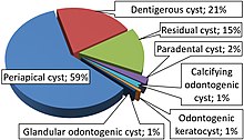
A dentigerous cyst, also known as a follicular cyst, is an epithelial-lined developmental cyst formed by accumulation of fluid between the reduced enamel epithelium and the crown of an unerupted tooth. It is formed when there is an alteration in the reduced enamel epithelium and encloses the crown of an unerupted tooth at the cemento-enamel junction. Fluid is accumulated between reduced enamel epithelium and the crown of an unerupted tooth.

Dens invaginatus (DI), also known as tooth within a tooth, is a rare dental malformation and a developmental anomaly where there is an infolding of enamel into dentin. The prevalence of this condition is 0.3 - 10%, affecting males more frequently than females. The condition presents in two forms, coronal involving tooth crown and radicular involving tooth root, with the former being more common.

Dens evaginatus is a rare odontogenic developmental anomaly that is found in teeth where the outer surface appears to form an extra bump or cusp.

<span class="mw-page-title-main">Odontogenic keratocyst</span> Medical condition

An odontogenic keratocyst is a rare and benign but locally aggressive developmental cyst. It most often affects the posterior mandible and most commonly presents in the third decade of life. Odontogenic keratocysts make up around 19% of jaw cysts.

Lateral periodontal cysts (LPCs) are defined as non-keratinised and non-inflammatory developmental cysts located adjacent or lateral to the root of a vital tooth.” LPCs are a rare form of jaw cysts, with the same histopathological characteristics as gingival cysts of adults (GCA). Hence LPCs are regarded as the intraosseous form of the extraosseous GCA. They are commonly found along the lateral periodontium or within the bone between the roots of vital teeth, around mandibular canines and premolars. Standish and Shafer reported the first well-documented case of LPCs in 1958, followed by Holder and Kunkel in the same year although it was called a periodontal cyst. Since then, there has been more than 270 well-documented cases of LPCs in literature.

<span class="mw-page-title-main">Calcifying odontogenic cyst</span> Medical condition

Calcifying odontogenic cyst (COC) is a rare developmental lesion that comes from odontogenic epithelium. It is also known as a calcifying cystic odontogenic tumor, which is a proliferation of odontogenic epithelium and scattered nest of ghost cells and calcifications that may form the lining of a cyst, or present as a solid mass.

<span class="mw-page-title-main">Dental abscess</span> Medical condition

A dental abscess is a localized collection of pus associated with a tooth. The most common type of dental abscess is a periapical abscess, and the second most common is a periodontal abscess. In a periapical abscess, usually the origin is a bacterial infection that has accumulated in the soft, often dead, pulp of the tooth. This can be caused by tooth decay, broken teeth or extensive periodontal disease. A failed root canal treatment may also create a similar abscess.

Apical periodontitis is typically the body's defense response to the threat of microbial invasion from the root canal. Primary among the members of the host defense mechanism is the polymorphonuclear leukocyte, otherwise known as the neutrophil. The task of the neutrophil is to locate and destroy microbes that intrude into the body – anywhere in the body – and they represent the hallmark of acute inflammation.

Dental pertains to the teeth, including dentistry. Topics related to the dentistry, the human mouth and teeth include:

<span class="mw-page-title-main">Tooth resorption</span> Medical condition

Resorption of the root of the tooth, or root resorption, is the progressive loss of dentin and cementum by the action of odontoclasts. Root resorption is a normal physiological process that occurs in the exfoliation of the primary dentition. However, pathological root resorption occurs in the permanent or secondary dentition and sometimes in the primary dentition.

Pulp necrosis is a clinical diagnostic category indicating the death of cells and tissues in the pulp chamber of a tooth with or without bacterial invasion. It is often the result of many cases of dental trauma, caries and irreversible pulpitis.

Odontogenic cyst are a group of jaw cysts that are formed from tissues involved in odontogenesis. Odontogenic cysts are closed sacs, and have a distinct membrane derived from rests of odontogenic epithelium. It may contain air, fluids, or semi-solid material. Intra-bony cysts are most common in the jaws, because the mandible and maxilla are the only bones with epithelial components. That odontogenic epithelium is critical in normal tooth development. However, epithelial rests may be the origin for the cyst lining later. Not all oral cysts are odontogenic cysts. For example, mucous cyst of the oral mucosa and nasolabial duct cyst are not of odontogenic origin.

A cyst is a pathological epithelial lined cavity that fills with fluid or soft material and usually grows from internal pressure generated by fluid being drawn into the cavity from osmosis. The bones of the jaws, the mandible and maxilla, are the bones with the highest prevalence of cysts in the human body. This is due to the abundant amount of epithelial remnants that can be left in the bones of the jaws. The enamel of teeth is formed from ectoderm, and so remnants of epithelium can be left in the bone during odontogenesis. The bones of the jaws develop from embryologic processes which fuse, and ectodermal tissue may be trapped along the lines of this fusion. This "resting" epithelium is usually dormant or undergoes atrophy, but, when stimulated, may form a cyst. The reasons why resting epithelium may proliferate and undergo cystic transformation are generally unknown, but inflammation is thought to be a major factor. The high prevalence of tooth impactions and dental infections that occur in the bones of the jaws is also significant to explain why cysts are more common at these sites.

<span class="mw-page-title-main">Periapical periodontitis</span> Medical condition

Periapical periodontitis or apical periodontitis (AP) is an acute or chronic inflammatory lesion around the apex of a tooth root, most commonly caused by bacterial invasion of the pulp of the tooth. It is a likely outcome of untreated dental caries, and in such cases it can be considered a sequela in the natural history of tooth decay, irreversible pulpitis and pulpal necrosis. Other causes can include occlusal trauma due to 'high spots' after restoration work, extrusion from the tooth of root filling material, or bacterial invasion and infection from the gums. Periapical periodontitis may develop into a periapical abscess, where a collection of pus forms at the end of the root, the consequence of spread of infection from the tooth pulp, or into a periapical cyst, where an epithelial lined, fluid-filled structure forms.

In the dental specialty of endodontics, periradicular surgery is surgery to the external root surface. Examples of periradicular surgery include apicoectomy, root resection, repair of root perforation or resorption defects, removal of broken fragments of the tooth or a filling material, and exploratory surgery to look for root fractures.

Periapical granuloma, also sometimes referred to as a radicular granuloma or apical granuloma, is an inflammation at the tip of a dead (nonvital) tooth. It is a lesion or mass that typically starts out as an epithelial lined cyst, and undergoes an inward curvature that results in inflammation of granulation tissue at the root tips of a dead tooth. This is usually due to dental caries or a bacterial infection of the dental pulp. Periapical granuloma is an infrequent disorder that has an occurrence rate between 9.3 to 87.1 percent. Periapical granuloma is not a true granuloma due to the fact that it does not contain granulomatous inflammation; however, periapical granuloma is a common term used.

References

  1. Menditti, Dardo; Laino, Luigi; Di Domenico, Marina; Troiano, Giuseppe; Guglielmotti, Mario; Sava, Sara; Mezzogiorno, Antonio; Baldi, Alfonso (2018). "Cysts and Pseudocysts of the Oral Cavity: Revision of the Literature and a New Proposed Classification". In Vivo. 32 (5): 999–1007. doi:10.21873/invivo.11340. PMC   6199599 . PMID   30150421.
  2. 1 2 3 4 5 6 7 Scholl, Robert J.; Kellett, Helen M.; Neumann, David P.; Lurie, Alan G. (1999-09-01). "Cysts and Cystic Lesions of the Mandible: Clinical and Radiologic-Histopathologic Review". RadioGraphics. 19 (5): 1107–1124. doi:10.1148/radiographics.19.5.g99se021107. ISSN   0271-5333. PMID   10489168.
  3. 1 2 Dunfee, Brian L.; Sakai, Osamu; Pistey, Robert; Gohel, Anita (2006-11-01). "Radiologic and Pathologic Characteristics of Benign and Malignant Lesions of the Mandible". RadioGraphics. 26 (6): 1751–1768. doi:10.1148/rg.266055189. ISSN   0271-5333. PMID   17102048.
  4. 1 2 3 R., Rajendran (2010). Shafer's textbook of oral pathology. [S.l.]: Reed Elsevier. ISBN   9788131215708. OCLC   682882649.
  5. Torabinejad, M. (February 1983). "The role of immunological reactions in apical cyst formation". Int J Oral Surg. 12 (1): 14–22. doi:10.1016/s0300-9785(83)80075-1. PMID   6406374.
  6. Huang, George T.-J. (2010-10-05). "Apical Cyst Theory: a Missing Link". Dental Hypotheses. 1 (2): 76–84. doi:10.5436/j.dehy.2010.1.00013. ISSN   2155-8213. PMC   4205966 . PMID   25346864.
  7. Webteam, University of Pittsburgh University Marketing Communications. "Bone - Structural Characteristics - School of Dental Medicine - University of Pittsburgh". dental.pitt.edu.
  8. "the definition of unilocular". Dictionary.com.
  9. "the definition of cortication". Dictionary.com.
  10. 1 2 3 "Difference between True and Pseudo Cyst" . Retrieved 2017-12-09.
  11. 1 2 Annie S. Morrison; Kelly Magliocca. "Mandible & maxilla - Odontogenic cysts - Periapical (radicular) cyst". Pathology Outlines. Topic Completed: 1 March 2014. Revised: 13 December 2019
  12. 1 2 "Cystostomy". www.medeco.de.
  13. Kirtaniya, BC; Sachdev, V; Singla, A; Sharma, AK (2010-07-01). "Marsupialization: A conservative approach for treating dentigerous cyst in children in the mixed dentition". Journal of Indian Society of Pedodontics and Preventive Dentistry. 28 (3): 203–8. doi: 10.4103/0970-4388.73795 . PMID   21157055.
  14. Leandro Bezerra Borges; Francisco Vagnaldo Fechine; Mário Rogério Lima Mota; Fabrício Bitu Sousa; Ana Paula Negreiros Nunes Alves (2012). "Odontogenic lesions of the jaw: a clinical-pathological study of 461 cases". Revista Gaúcha de Odontologia. 60 (1).