Ectopic salivary gland tissue

Last updated

Ectopic salivary gland tissue which is located in sites other than the normal location is variously described as aberrant, accessory, ectopic, heterotopic or salivary gland choristoma. [1] [2]

An ectopia is a displacement or malposition of an organ or other body part, which is then referred to as ectopic. Most ectopias are congenital, but some may happen later in life.

Heterotopia (medicine)

In medicine, heterotopia is the presence of a particular tissue type at a non-physiological site, but usually co-existing with original tissue in its correct anatomical location. In other words, it implies ectopic tissue, in addition to retention of the original tissue type. In neuropathology, for example, gray matter heterotopia is the presence of gray matter within the cerebral white matter or ventricles. Heterotopia within the brain is often divided into three groups: subependymal heterotopia, focal cortical heterotopia and band heterotopia. Another example is a Meckel's diverticulum, which may contain heterotopic gastric or pancreatic tissue.

Choristomas, a form of heterotopia, are closely related benign tumors found in abnormal locations.

Contents

Accessory salivary glands

An accessory salivary gland is ectopic salivary gland tissue with a salivary gland duct system. [3] The most common location of accessory salivary gland tissue is an extra major salivary gland in front of the parotid gland. It is typically about 3 cm or less in size, and drains into the parotid duct via a single tributary. [3] Accessory parotid tissue is found in 21-56% of adults. [3] Any disease process which affects the salivary glands, including cancer, may also occur within an accessory salivary gland tissue.

Parotid gland

The parotid gland is a major salivary gland in many animals. In humans, the two parotid glands are present on either side of the mouth and in front of both ears. They are the largest of the salivary glands. Each parotid is wrapped around the mandibular ramus, and secretes serous saliva through the parotid duct into the mouth, to facilitate mastication and swallowing and to begin the digestion of starches. There are also two other types of salivary glands; they are submandibular and sublingual glands. Sometimes accessory parotid glands are found close to the main parotid glands.

Heterotopic salivary gland tissue

Salivary gland heterotopia is where salivary gland acini cells are present in an abnormal location without any duct system. [3] The most common location is the cervical lymph nodes. Other reported sites of heterotopic salivary gland tissue are the middle ear, parathyroid glands, thyroid gland, pituitary gland, cerebellar pontine angle, soft tissue medial to sternocleidomastoid, stomach, rectum and vulva. [3] Salivary gland neoplasm occurrence within heterotopic salivary gland tissue is rare. [3]

Lymph node organ of the lymphatic system

A lymph node or lymph gland is an ovoid or kidney-shaped organ of the lymphatic system, and of the adaptive immune system, that is widely present throughout the body. They are linked by the lymphatic vessels as a part of the circulatory system. Lymph nodes are major sites of B and T lymphocytes, and other white blood cells. Lymph nodes are important for the proper functioning of the immune system, acting as filters for foreign particles and cancer cells. Lymph nodes do not have a detoxification function, which is primarily dealt with by the liver and kidneys.

The middle ear is the portion of the ear internal to the eardrum, and external to the oval window of the inner ear. The mammalian middle ear contains three ossicles, which transfer the vibrations of the eardrum into waves in the fluid and membranes of the inner ear. The hollow space of the middle ear is also known as the tympanic cavity and is surrounded by the tympani bone. The auditory tube joins the tympanic cavity with the nasal cavity (nasopharynx), allowing pressure to equalize between the middle ear and throat.

Parathyroid gland endocrine gland

Parathyroid glands are small endocrine glands in the neck of humans and other tetrapods that produce parathyroid hormone. Humans usually have four parathyroid glands, variably located on the back of the thyroid gland. Parathyroid hormone and calcitonin have key roles in regulating the amount of calcium in the blood and within the bones.

See also

Related Research Articles

Salivary gland exocrine gland that produces saliva also it reduces bleeding in mouth

The salivary glands in mammals are exocrine glands that produce saliva through a system of ducts. Humans have three paired major salivary glands as well as hundreds of minor salivary glands. Salivary glands can be classified as serous, mucous or seromucous (mixed).

Submandibular gland

The paired submandibular glands are major salivary glands located beneath the floor of the mouth. They each weigh about 15 grams and contribute some 60–67% of unstimulated saliva secretion; on stimulation their contribution decreases in proportion as the parotid secretion rises to 50%.

Sublingual gland

The paired sublingual glands are major salivary glands in the mouth. They are the smallest, most diffuse, and the only unencapsulated major salivary glands. They provide only 3-5% of the total salivary volume. There are also two other types of salivary glands; they are submandibular and Parotid glands.

Parotitis parotid disease characterized by the inflammation of one or both parotid glands

Parotitis is an inflammation of one or both parotid glands, the major salivary glands located on either side of the face, in humans. The parotid gland is the salivary gland most commonly affected by inflammation.

Warthins tumor type of benign tumor of the salivary glands

Warthin's tumor, also known as papillary cystadenoma lymphomatosum, is a benign cystic tumor of the salivary glands containing abundant lymphocytes and germinal centers. It is named for pathologist Aldred Scott Warthin, who described two cases in 1929.

Pleomorphic adenoma gastrointestinal benign neoplasm that is a located in the salivary glands

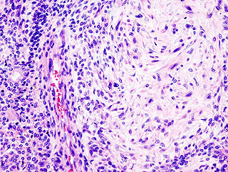
Pleomorphic adenoma is a common benign salivary gland neoplasm characterised by neoplastic proliferation of parenchymatous glandular cells along with myoepithelial components, having a malignant potentiality. It is the most common type of salivary gland tumor and the most common tumor of the parotid gland. It derives its name from the architectural Pleomorphism seen by light microscopy. It is also known as "Mixed tumor, salivary gland type", which refers to its dual origin from epithelial and myoepithelial elements as opposed to its pleomorphic appearance.

Oral mucocele

Oral mucocele is a clinical term for two related phenomena: mucus extravasation phenomenon and mucus retention cyst. Other names include mucous extravasation cyst, mucous cyst of the oral mucosa, and mucous retention and extravasation phenomena.

Oral medicine is a specialty focused on the mouth and nearby structures. It lies at the interface between medicine and dentistry.

Sialadenitis inflammation of a salivary gland

Sialadenitis (sialoadenitis) is inflammation of salivary glands, usually the major ones, the most common being the parotid gland, followed by submandibular and sublingual glands. It should not be confused with sialadenosis (sialosis) which is a non-inflammatory enlargement of the major salivary glands.

Sialolithiasis condition where a calcified mass or sialolith forms within a salivary gland, usually in the duct of the submandibular gland

Sialolithiasis, is a condition where a calcified mass or sialolith forms within a salivary gland, usually in the duct of the submandibular gland. Less commonly the parotid gland or rarely the sublingual gland or a minor salivary gland may develop salivary stones.

Salivary gland tumour human disease

Salivary gland tumours or neoplasms are tumours that form in the tissues of salivary glands. The salivary glands are classified as major or minor. The major salivary glands consist of the parotid, submandibular, and sublingual glands. The minor salivary glands consist of 800-1000 small mucus-secreting glands located throughout the lining of the oral cavity.

Salivary gland pathology Human disease

Salivary gland diseases (SGD) are multiple and varied in cause.

A parotidectomy is the surgical excision (removal) of the parotid gland, the major and largest of the salivary glands. The procedure is most typically performed due to neoplasms (tumors), which are growths of rapidly and abnormally dividing cells. Neoplasms can be benign (non-cancerous) or malignant (cancerous). The majority of parotid gland tumors are benign, however 20% of parotid tumors are found to be malignant. A parotidectomy is performed mostly by the oral and maxillofacial surgeons and otolaryngologist.

Chronic sclerosing sialadenitis is a chronic (long-lasting) inflammatory condition affecting the salivary gland. Relatively rare in occurrence, this condition is benign, but presents as hard, indurated and enlarged masses that are clinically indistinguishable from salivary gland neoplasms or tumors. It is now regarded as a manifestation of IgG4-related disease.

Human digestive system combination of anatomical organs that are responsible for digestive function

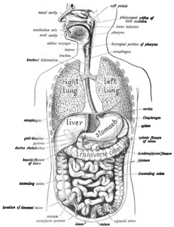
The human digestive system consists of the gastrointestinal tract plus the accessory organs of digestion. Digestion involves the breakdown of food into smaller and smaller components, until they can be absorbed and assimilated into the body. The process of digestion has many stages. The first stage is the cephalic phase of digestion which begins with gastric secretions in response to the sight and smell of food. The next stage starts in the mouth.

A salivary gland fistula is a fistula involving a salivary gland or duct.

Salivary duct stricture is narrowing of the duct of a major salivary gland.

Basal cell adenoma is a rare, low-grade benign salivary gland neoplasm.

The parotid fascia in human anatomy is a fascia that builds a closed membrane together with the masseteric fascia. This common membrane sheaths the parotid gland, its excretory duct and the passing out branches of the facial nerve as well. The parotid fascia proceeds of the superficial layer of the deep cervical fascia that splits to cover the gland. At the lateral side of the gland this fascia is called the parotid fascia. The fascia itself is made of two layers: A superficial layer that passes cranial into the temporal fascia and lateral into the masseteric fascia, and a deeper layer that covers the Stylohyoid muscle, the styloglossus and the Musculus stylopharyngeus. The superficial layer is attached to the zygomatic arch above and to the mandible below.

References

  1. Barnes L (2008). Surgical pathology of the head and neck (3rd ed.). New York: Informa Healthcare. pp. 477–478. ISBN   9780849390234.
  2. Ghom AG; Ghom SA (1 July 2014). Textbook of Oral Medicine. JP Medical Ltd. p. 605. ISBN   978-93-5152-303-1.
  3. 1 2 3 4 5 6 Witt RL (1 January 2011). Salivary Gland Diseases: Surgical and Medical Management. Thieme. pp. 50–51. ISBN   978-1-60406-537-4.