Buccal exostosis

Last updated
Buccal exostosis
Other namesAlveolar exostosis [1]
Buccal exostosis.jpg
Buccal exostosis along upper left alveolar ridge
Specialty Oral medicine

A buccal exostosis is an exostosis (bone prominence) on the buccal surface (cheek side) of the alveolar ridge of the maxilla or mandible. More commonly seen in the maxilla than the mandible, buccal exostoses are considered to be site specific. [2] Existing as asymptomatic bony nodules, [3] buccal exostoses don’t usually present until adult life, [4] and some consider buccal exostoses to be a variation of normal anatomy rather than disease. Bone is thought to become hyperplastic, consisting of mature cortical and trabecular bone with a smooth outer surface. [3] They are less common when compared with mandibular tori. [3]

Contents

Signs and symptoms

Buccal exostoses are bony hamartomas, which are non- malignant, exophytic nodular outgrowths of dense cortical bone that are relatively avascular.’ [5]

Symptoms:

Buccal exostoses generally tend to be asymptomatic and are usually painless. However, they may increase patient concern about poor aesthetics, inability to perform oral hygiene procedures due to difficulty in cleaning around the area with a toothbrush, and compromised periodontal health by causing food lodgement, which could lead to patients reporting increased bleeding when tooth brushing.

Signs:

The overlying mucosa appears to be stretched but intact and normal in colour.

Causes

Why buccal exostoses form is unclear, [8] but it may involve bruxism (tooth clenching and grinding), [8] and genetic factors. [9] Typically they first appear in early adulthood. [10]

Diagnosis

The presence of buccal exostosis can be diagnosed by both clinical examination and radiological interpretation of the oral cavity.

Clinically, buccal exostoses appear as single, broad-based masses, usually situated bilaterally in the premolar and molar region on the facial surface of the maxillary alveolar bone. [11] The mass is generally smooth although in some cases a sharp, bony prominence may be present resulting in tenderness beneath the mucosa. [12] Initial presentation usually occurs in early adolescence and the lesions may slowly enlarge up to 3–4 cm in diameter with time, however there is no malignant potential. [6] Despite buccal exostoses being generally painless and self-limiting, they may cause patient concern regarding poor aesthetics, food lodgement and compromised oral hygiene. Their tendency to grow in size may also contribute to periodontal disease as a result of food build up in the area of the lesion. [11]

Radiographically, buccal exostoses can be identified as round, well-defined structures which superimpose the roots of the teeth, normally in the premolar and molar region. Dental panoramic tomography and cone beam tomography can be used to confirm diagnosis. [13]

An additional biopsy for diagnosis of buccal exostosis is not usually recommended, however it is important to rule out the possibility of early osteosarcomas and chondrosarcomas. [6] In addition, it is recommended that patients who present with multiple growths showing similar characteristics but not in the classic exostoses locations should be evaluated for Gardner syndrome. [7]

Management

Currently, buccal exostoses do not commonly require treatment. If there is neither pain nor sensitivity, the buccal exostosis simply needs to be monitored with routine dental check-ups. Patients are given oral hygiene advice and are advised to cleanse above and below the growth with a mouthwash once a day to remove any food debris. [14] Due to it being difficult to clean around the exostosis, periodontal disease can often occur as a result, and so this should be treated by the dentist or dental hygienist/therapist. There are, however, some occasions were treatment is required, for example;

If the growth needs to be removed then a simple surgical procedure can be carried out under local anaesthetic with no recurrence in the long-term follow up. [15] This is done by reflecting the mucoperiosteal flap in order to expose the extended bone for complete excision. This bone will then be sent for histopathological examination to differentiate from benign to malignant neoplastic processes. Routine post-operative instructions are given to the patient and occasionally antibiotics may be prescribed. [16] The patient will then be recalled after 1 week in order to remove the sutures and often after a few months for post-operative follow up.

Epidemiology

They are more common in males than females, occurring in a ratio of about 5:1. [9] They are strongly associated with the presence of torus mandibularis and torus palatinus. [9]

Related Research Articles

<span class="mw-page-title-main">Human tooth</span> Calcified whitish structure in humans mouths used to break down food

Human teeth function to mechanically break down items of food by cutting and crushing them in preparation for swallowing and digesting. As such, they are considered part of the human digestive system. Humans have four types of teeth: incisors, canines, premolars, and molars, which each have a specific function. The incisors cut the food, the canines tear the food and the molars and premolars crush the food. The roots of teeth are embedded in the maxilla or the mandible and are covered by gums. Teeth are made of multiple tissues of varying density and hardness.

<span class="mw-page-title-main">Gums</span> Mouth anatomy

The gums or gingiva consist of the mucosal tissue that lies over the mandible and maxilla inside the mouth. Gum health and disease can have an effect on general health.

<span class="mw-page-title-main">Alveolar osteitis</span> Medical condition

Alveolar osteitis, also known as dry socket, is inflammation of the alveolar bone. Classically, this occurs as a postoperative complication of tooth extraction.

<span class="mw-page-title-main">Human tooth development</span> Process by which teeth form

Tooth development or odontogenesis is the complex process by which teeth form from embryonic cells, grow, and erupt into the mouth. For human teeth to have a healthy oral environment, all parts of the tooth must develop during appropriate stages of fetal development. Primary (baby) teeth start to form between the sixth and eighth week of prenatal development, and permanent teeth begin to form in the twentieth week. If teeth do not start to develop at or near these times, they will not develop at all, resulting in hypodontia or anodontia.

<span class="mw-page-title-main">Maxillary sinus</span> Largest of the paranasal sinuses, and drains into the middle meatus of the nose

The pyramid-shaped maxillary sinus is the largest of the paranasal sinuses, located in the maxilla. It drains into the middle meatus of the nose through the semilunar hiatus. It is located to the side of the nasal cavity, and below the orbit.

<span class="mw-page-title-main">Dental follicle</span>

The dental follicle, also known as dental sac, is made up of mesenchymal cells and fibres surrounding the enamel organ and dental papilla of a developing tooth. It is a vascular fibrous sac containing the developing tooth and its odontogenic organ. The dental follicle (DF) differentiates into the periodontal ligament. In addition, it may be the precursor of other cells of the periodontium, including osteoblasts, cementoblasts and fibroblasts. They develop into the alveolar bone, the cementum with Sharpey's fibers and the periodontal ligament fibers respectively. Similar to dental papilla, the dental follicle provides nutrition to the enamel organ and dental papilla and also have an extremely rich blood supply.

<span class="mw-page-title-main">Dental extraction</span> Operation to remove a tooth

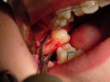
A dental extraction is the removal of teeth from the dental alveolus (socket) in the alveolar bone. Extractions are performed for a wide variety of reasons, but most commonly to remove teeth which have become unrestorable through tooth decay, periodontal disease, or dental trauma, especially when they are associated with toothache. Sometimes impacted wisdom teeth cause recurrent infections of the gum (pericoronitis), and may be removed when other conservative treatments have failed. In orthodontics, if the teeth are crowded, healthy teeth may be extracted to create space so the rest of the teeth can be straightened.

<span class="mw-page-title-main">Alveolar process</span> Bulge on jaws holding teeth

The alveolar process or alveolar bone is the thickened ridge of bone that contains the tooth sockets on the jaw bones. The structures are covered by gums as part of the oral cavity.

<span class="mw-page-title-main">Veterinary dentistry</span> Branch of veterinary medicine

Veterinary dentistry is the field of dentistry applied to the care of animals. It is the art and science of prevention, diagnosis, and treatment of conditions, diseases, and disorders of the oral cavity, the maxillofacial region, and its associated structures as it relates to animals.

<span class="mw-page-title-main">Torus palatinus</span> Medical condition

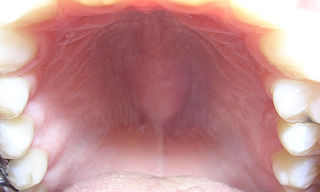
A torus palatinus, or palatal torus, is a bony protrusion on the palate. Palatal tori are usually present on the midline of the hard palate. Most palatal tori are less than 2 cm in diameter, but their size can change throughout life.

<span class="mw-page-title-main">Dentigerous cyst</span> Medical condition

A dentigerous cyst, also known as a follicular cyst, is an epithelial-lined developmental cyst formed by accumulation of fluid between the reduced enamel epithelium and the crown of an unerupted tooth. It is formed when there is an alteration in the reduced enamel epithelium and encloses the crown of an unerupted tooth at the cemento-enamel junction. Fluid is accumulated between reduced enamel epithelium and the crown of an unerupted tooth.

<span class="mw-page-title-main">Calcifying odontogenic cyst</span> Medical condition

Calcifying odontogenic cyst (COC) is a rare developmental lesion that comes from odontogenic epithelium. It is also known as a calcifying cystic odontogenic tumor, which is a proliferation of odontogenic epithelium and scattered nest of ghost cells and calcifications that may form the lining of a cyst, or present as a solid mass.

<span class="mw-page-title-main">Cementoma</span> Medical condition

Cementoma is an odontogenic tumor of cementum. It is usually observed as a benign spherical mass of hard tissue fused to the root of a tooth. It is found most commonly in the mandible in the region of the lower molar teeth, occurring between the ages of 8 and 30 in both sexes with equal frequency. It causes distortion of surrounding areas but is usually a painless growth, at least initially. Considerable thickening of the cementum can often be observed. A periapical form is also recognized. Cementoma is not exclusive to the mandible as it can infrequently occur in the maxilla and other parts of the body such as the long bones.

Dental pertains to the teeth, including dentistry. Topics related to the dentistry, the human mouth and teeth include:

Oral and maxillofacial pathology refers to the diseases of the mouth, jaws and related structures such as salivary glands, temporomandibular joints, facial muscles and perioral skin. The mouth is an important organ with many different functions. It is also prone to a variety of medical and dental disorders.

<span class="mw-page-title-main">Furcation defect</span>

In dentistry, a furcation defect is bone loss, usually a result of periodontal disease, affecting the base of the root trunk of a tooth where two or more roots meet. The extent and configuration of the defect are factors in both diagnosis and treatment planning.

Aggressive periodontitis describes a type of periodontal disease and includes two of the seven classifications of periodontitis as defined by the 1999 classification system:

  1. Localized aggressive periodontitis (LAP)
  2. Generalized aggressive periodontitis (GAP)

A cyst is a pathological epithelial lined cavity that fills with fluid or soft material and usually grows from internal pressure generated by fluid being drawn into the cavity from osmosis. The bones of the jaws, the mandible and maxilla, are the bones with the highest prevalence of cysts in the human body. This is due to the abundant amount of epithelial remnants that can be left in the bones of the jaws. The enamel of teeth is formed from ectoderm, and so remnants of epithelium can be left in the bone during odontogenesis. The bones of the jaws develop from embryologic processes which fuse, and ectodermal tissue may be trapped along the lines of this fusion. This "resting" epithelium is usually dormant or undergoes atrophy, but, when stimulated, may form a cyst. The reasons why resting epithelium may proliferate and undergo cystic transformation are generally unknown, but inflammation is thought to be a major factor. The high prevalence of tooth impactions and dental infections that occur in the bones of the jaws is also significant to explain why cysts are more common at these sites.

Tooth ankylosis refers to a fusion between a tooth and underlying bony support tissues. In some species, this is a normal process that occurs during the formation or maintenance of the dentition. By contrast, in humans tooth ankylosis is pathological, whereby a fusion between alveolar bone and the cementum of a tooth occurs.

Alveoloplasty is a surgical pre-prosthetic procedure performed to facilitate removal of teeth, and smoothen or reshape the jawbone for prosthetic and cosmetic purposes. In this procedure, the bony edges of the alveolar ridge and its surrounding structures is made smooth, redesigned or recontoured so that a well-fitting, comfortable, and esthetic prosthesis may be fabricated or implants may be surgically inserted. This pre-prosthetic surgery which may include bone grafting prepares the mouth to receive a prosthesis or implants by improving the condition and quality of the supporting structures so they can provide support, better retention and stability to the prosthesis.

References

  1. Sakai O (24 May 2011). Head and Neck Imaging Cases. McGraw Hill Professional. p. 910. ISBN   978-0-07-154372-9.
  2. Mehra, D'Innocenzo, Pushkar, Richard (2015). Manual of Minor Oral Surgery for the General Dentist (2nd ed.). John Wiley & Sons, 2015. pp. 91–93. ISBN   978-1-118-93848-5.{{cite book}}: CS1 maint: multiple names: authors list (link)
  3. 1 2 3 Regezi, Joseph A.; Sciubba, James; Jordan, Richard C. K. (2012). Oral Pathology - E-Book: Clinical Pathologic Correlations. Elsevier Health Sciences. p. 311. ISBN   978-1-4557-0269-5.
  4. Gnepp, Douglas R. (2009-04-07). Diagnostic Surgical Pathology of the Head and Neck E-Book: Expert Consult (2nd ed.). Elsevier Health Sciences, 2009. p. 1224. ISBN   978-1-4377-1951-2.
  5. Pynn, BR; Kurys-Kos, NS; Walker, DA; Mayhall, JT (December 1995). "Tori mandibularis: a case report and review of the literature". Journal of the Canadian Dental Association. 61 (12): 1057–8, 1063–6. PMID   8536198.
  6. 1 2 3 4 Medsinge, Sonali V; Kohad, Ramesh; Budhiraja, Harmeeta; Singh, Atamjeet; Gurha, Shradha; Sharma, Akash (2015). "Buccal Exostosis: A Rare Entity". Journal of International Oral Health. 7 (5): 62–64. PMC   4441241 . PMID   26028907.
  7. 1 2 3 Jainkittivong, Aree; Langlais, Robert P. (July 2000). "Buccal and palatal exostoses: Prevalence and concurrence with tori". Oral Surgery, Oral Medicine, Oral Pathology, Oral Radiology, and Endodontology. 90 (1): 48–53. doi:10.1067/moe.2000.105905. PMID   10884635. S2CID   17172819.
  8. 1 2 Mehra P; D'Innocenzo R (18 May 2015). Manual of Minor Oral Surgery for the General Dentist. John Wiley & Sons. p. 300. ISBN   978-1-118-93843-0.
  9. 1 2 3 Woo SB (2012). Oral Pathology: A Comprehensive Atlas and Text. Elsevier Health Sciences. p. 383. ISBN   978-1-4377-2226-0.
  10. Scully C (2013). Oral and Maxillofacial Medicine, The Basis of Diagnosis and Treatment,3: Oral and Maxillofacial Medicine. Elsevier Health Sciences. p. 122. ISBN   978-0-7020-4948-4.
  11. 1 2 Horning, Gregory M.; Cohen, Mark E.; Neils, Todd A. (June 2000). "Buccal Alveolar Exostoses: Prevalence, Characteristics, and Evidence for Buttressing Bone Formation". Journal of Periodontology. 71 (6): 1032–1042. doi:10.1902/jop.2000.71.6.1032. PMID   10914809.
  12. Bouquot JE (1988). Bond's Book of Oral Disease (4th ed.). Philadelphia: Churchill-Livingstone. p. [ page needed ].
  13. Curé, Joel K.; Vattoth, Surjith; Shah, Ritu (November 2012). "Radiopaque Jaw Lesions: An Approach to the Differential Diagnosis". RadioGraphics. 32 (7): 1909–1925. doi:10.1148/rg.327125003. PMID   23150848. S2CID   207553947.
  14. 1 2 Boge, RDH, BS, MPA, E. (2019). Buccal Exostosis: Causes, Treatment and Care | Colgate®. [online] Colgate.com. Available at: https://www.colgate.com/en-us/oral-health/basics/mouth-and-teeth-anatomy/buccal-exostosis--causes--treatment-and-care [Accessed 14 Feb. 2019].
  15. Chandna, Shalu; Sachdeva, Surinder; Kochar, Deepak; Kapil, Harsh (2015). "Surgical management of the bilateral maxillary buccal exostosis". Journal of Indian Society of Periodontology. 19 (3): 352–5. doi: 10.4103/0972-124X.152412 . PMC   4520128 . PMID   26229284.
  16. Kukuła, K; Plakwicz, P (22 July 2016). "Oral pathology: Exostosis deforming face features". British Dental Journal. 221 (2): 50–51. doi:10.1038/sj.bdj.2016.514. PMID   27444586.