Anterior cruciate ligament reconstruction

Last updated
Anterior cruciate ligament reconstruction
Anterior cruciate ligament repair 3 legend.jpg
Arthroscopic anterior cruciate ligament (ACL) reconstruction (right knee). The tendon of the semitendinosus muscle was prelevated, folded and used as an autograft (1). It appears through the remnant of the injured original ACL (3). The autograft then courses upwardly and backwardly in front of the posterior cruciate ligament (2).
Other namesACL reconstruction
ICD-9-CM 81.45
MedlinePlus 007208

Anterior cruciate ligament reconstruction (ACL reconstruction) is a surgical tissue graft replacement of the anterior cruciate ligament, located in the knee, to restore its function after an injury. [1] The torn ligament can either be removed from the knee (most common), or preserved (where the graft is passed inside the preserved ruptured native ligament) before reconstruction through an arthroscopic procedure. ACL repair is also a surgical option. This involves repairing the ACL by re-attaching it, instead of performing a reconstruction. Theoretical advantages of repair include faster recovery [2] and a lack of donor site morbidity, but randomised controlled trials and long-term data regarding re-rupture rates using contemporary surgical techniques are lacking.

Contents

Background

The Anterior Cruciate Ligament is the ligament that keeps the knee stable. [3] Anterior Cruciate Ligament damage is a very common injury, especially among athletes. Anterior Cruciate Ligament Reconstruction (ACL) surgery is a common intervention. 1 in every 3,000 American ruptures their ACL and between 100,000 and 300,000 reconstruction surgeries will be performed each year in the United States. [4] [5] Around $500 million health care dollar will come from ACL injuries. ACL injuries can be categorized into groups- contact and non-contact based on the nature of the injury [6] Contact injuries occur when a person or object come into contact with the knee causing the ligament to tear. However, non-contact tears typically occur during the following movements: decelerating, cutting, or landing from a jump. ACL injury is 4-6 times higher in females than in males. ACL injuries account for a quarter of all knee injuries in the high school population. [7] An increased Q angle and hormonal differences are a few examples of the gender disparity in ACL tear rates. [8]

Types of grafts

Grafts are inserted through a tunnel that is drilled through the shin bone (tibia) and thigh bone (femur). The graft is then pulled through the tunnel and fixated with screws. The two bright objects in this X-ray are screws in the thigh bone (above) and shin bone (below). LCA-RX2.jpg
Grafts are inserted through a tunnel that is drilled through the shin bone (tibia) and thigh bone (femur). The graft is then pulled through the tunnel and fixated with screws. The two bright objects in this X-ray are screws in the thigh bone (above) and shin bone (below).

Graft options for ACL reconstruction include:

Autograft

An accessory hamstring or part of the patellar ligament are the most common donor tissues used in autografts. While originally less commonly utilized, the quadriceps tendon has become a more popular graft. [9]

Because the tissue used in an autograft is the patient's own, the risk of rejection is minimal. The retear rate in young, active individuals has been shown to be lower when using autograft as compared to allograft.

Hamstring tendon

Left knee following hamstring autograft ACL reconstruction, partial meniscectomy and medial meniscus repair. "Socks" are actually post-op pressure stockings. ACL reconstruction hamstring autograft.jpg
Left knee following hamstring autograft ACL reconstruction, partial meniscectomy and medial meniscus repair. "Socks" are actually post-op pressure stockings.

Hamstring autografts are made with the semitendinosus tendon, either alone or accompanied by the gracilis tendon for a stronger graft. The semitendinosus is an accessory hamstring (the primary hamstrings are left intact), and the gracilis is not a hamstring, but an accessory adductor (the primary adductors are left intact as well). The two tendons are commonly combined and referred to as a four-strand hamstring graft, made by a long piece (about 25 cm) removed from each tendon. The tendon segments are folded and braided together to form a tendon of quadruple thickness for the graft. The braided segment is threaded through the heads of the tibia and femur, and its ends are fixed with screws on the opposite sides of the two bones.[ citation needed ]

Unlike the patellar ligament, the hamstring tendon's fixation to the bone can be affected by motion after surgery. Therefore, a brace is often used to immobilize the knee for one to two weeks. Evidence suggests that the hamstring tendon graft does as well, or nearly as well, as the patellar ligament graft in the long term. [10] A Cochrane review in 2011 found insufficient evidence to suggest whether a hamstring versus patellar ligament graft was superior. [11] It found that individuals receiving hamstring autografts had reduced flexion (bending knee) range of motion and strength. [11] Common problems during recovery include strengthening of the quadriceps, IT-band, and calf muscles.[ citation needed ]

The main surgical wound is over the upper proximal tibia, which prevents the typical pain experienced when kneeling after surgery. The wound is typically smaller than that of a patellar ligament graft, and so causes less post-operative pain. Another option first described by Kodkani et al in 2004, a minimally invasive technique for harvesting from the back of the knee (Posterior Mini-incision), is faster, produces a significantly smaller wound, avoids the complications of graft harvesting from the anterior incision, and decreases the risk of nerve injury. [12]

There is some controversy as to how well a hamstring tendon regenerates after the harvesting. Most studies suggest that the tendon can be regenerated at least partially, though it will still be weaker than the original tendon. [13] [14]

Advantages of hamstring grafts include their high "load to failure" strength, the stiffness of the graft, and the low postoperative morbidity. The natural ACL can withstand a load of up to 2,160 newtons. With a hamstring graft, this number doubles, decreasing the risk of re-injury. The stiffness of a hamstring graft—quadruple that of the natural ACL (Bartlett, Clatworthy and Ngugen, 2001)—also reduces the risk of re-injury.[ citation needed ]

Patellar tendon

Knees following ACL reconstruction surgery. A patellar tendon graft was used. Discoloration of the left leg is from swelling that drained from the knee to the shin. ACLReconstruction.jpg
Knees following ACL reconstruction surgery. A patellar tendon graft was used. Discoloration of the left leg is from swelling that drained from the knee to the shin.

The patellar tendon connects the patella (kneecap) to the tibia (shin). The graft is normally taken from the injured knee, but in some circumstances, such as a second operation, the other knee may be used. The middle third of the tendon is used, with bone fragments removed on each end. The graft is then threaded through holes drilled in the tibia and femur, and screwed into place. It is slightly larger than a hamstring graft.[ citation needed ]

A 2011 Cochrane review, found no significant difference in long term outcome between patellar and hamstring autografts. [11] Those receiving patellar autografts had improved static stability but a loss of extension (straightening knee) range of motion and strength. [11]

Disadvantages compared with a hamstring graft include:

  • Increased wound pain
  • Increased scar formation
  • Risk of fracturing the patella during harvesting of the graft
  • Increased risk of tendinitis.
  • Increased pain levels, even years after surgery, with activities that require kneeling. [15]

Some or all of these disadvantages may be attributable to post-operative patellar tendon shortening. [16]

Contralateral Patellar Tendon ACL Revision

The rehabilitation after the surgery is different for each knee. The beginning rehab for the ACL graft knee is focused on reducing swelling, gaining full range of motion, and stimulating the leg muscles. The goal for the graft donor need is to immediately start high repetition strength training exercises. [17]

Allograft

The patellar ligament, tibialis anterior tendon, or Achilles tendon may be recovered from a cadaver and used in ACL reconstruction. The Achilles tendon, because of its large size, must be shaved to fit within the joint cavity.

Choice of graft

Type

Typically, age and lifestyle help determine the type of graft used for ACL reconstruction. [15] The biggest factors in knee stability are correct graft placement by the surgeon and treatment of other menisco-ligament injuries in the knee, rather than type of graft. However, with the current literature, only KT-1000 arthrometer assessment demonstrated more laxity with allograft reconstruction. [18] Bone-patellar tendon-bone grafts have resulted fewer failures and more stability on KT-1000 arthrometer testing. [19] [20]

Site

No ideal graft site for ACL reconstruction exists. Surgeons have historically regarded patellar tendon grafts as the "gold standard" for knee stability. [15]

Hamstring autografts have failed at a higher rate than bone-tendon-bone autografts, after short- to mid-term followup of primary ACL reconstruction. However, the observed difference in failure rates is small enough that both are still regarded as viable options for primary ACL reconstruction. [21]

Hamstring grafts historically had problems with fixation slippage and stretching out over time. Modern fixation methods avoid graft slippage and produce similarly stable outcomes with easier rehabilitation, less anterior knee pain and less joint stiffness.

Although there is less experience with the use of tibialis anterior grafts, preliminary data has shown no difference in short-term subjective outcomes between tibialis anterior allografts and patellar tendon allografts. [22]

The quadriceps tendon, while historically reserved for revision reconstructions, has enjoyed a renewed focus as a versatile and durable graft for primary reconstructions. Use of the quadriceps tendon usually does not result in the same degree of anterior knee pain postoperatively, and quadriceps tendon harvest produces a reliably thick, robust graft. The quadriceps tendon has approximately 20% greater collagen per cross-sectional area than the patellar tendon, and a greater diameter of usable soft tissue is available.[ medical citation needed ]

Bridge Enhanced ACL Restoration (BEAR Implant)

A new approach to treating ACL tears was developed at Boston Children's Hospital and is currently in clinical trials. [23] The Bridge Enhanced ACL Restoration (BEAR) implant is a bio-engineered bridging implant that is injected with a small amount of patient's own blood to stimulate healing and reconnection of the ACL. Results from the first-in-human study published in March 2019 in the Orthopedic Journal of Sports Medicine showed the 10 patients who received the BEAR implant had similar clinical, functional and patient-reported outcomes as the 10 patients undergoing autograft ACL reconstruction. [24] Additional clinical studies are underway. [25] In a study by the American Journal of Sports Medicine, they looked at a young and active population two years post surgery using the BEAR technique. The results showed that the BEAR technique was non-inferior to the autograft ACLR, and that it can also show an improvement in hamstring muscle strength at a two year follow up. [26]

Ligament Advanced Reinforcement System (LARS)

The anterior cruciate ligament (ACL) of the knee is commonly injured. There is insufficient re-vascularization of the ligament after complete rupture, which limits its ability to heal and necessitates reconstruction surgery. Within the last 20 years, new types of synthetic ligaments have been developed. The Ligament Advanced Reinforcement System (LARS), is one of these new synthetic ligaments that has recently gained popularity. There is evidence that supports LARS as a viable option for reconstruction surgery in regards to complication rates and high patient satisfaction scores, when compared to traditional surgical techniques. However, systematic reviews of the LARS in regarding graft stability and long term functional outcomes, have highlighted several important gaps in existing literature that requires future investigation. The necessity of rehabilitation following LARS is well recognized, but there is limited evidence available that guide rehabilitation protocols. [27]

Stem cell treatment

Autologous stem-cell transplantation using mesenchymal stem cells (MSCs) has been used to improve recovery time from ACL surgery, especially for athletes. MSCs are multipotent stem cells, meaning they can differentiate into multiple cell types. In the case of mesenchymal stem cells, these cell types include osteoblasts (bone cells), adipocytes (fat cells), and chondrocytes (cartilage cells). Ligament tissue mainly consists of fibroblasts and extracellular matrix. Ligament cells differ in size, respond to different cues in the cell environment, and express different cell surface markers, limiting the number of clinical treatments for accelerated repair of ACL tissue to MSCs and primary fibroblasts obtained from other ACL tissue. Therefore, most modern stem cell injections use MSCs to promote faster repair of the ACL and allow people such as athletes to return to their previous form faster.[ citation needed ]

In order for MSCs to differentiate into an ACL, they must be placed in a proper scaffold on which to grow, and must be in a bioreactor that maintains a normal physiological environment for the cells to reproduce and proliferate effectively. [28] The scaffold must have the mechanical properties of a healthy ACL to sustain the ligament while it is in its primary form and maintain normal knee movement. Scaffolds that are used for ACL growth include collagen, silk, gelatin, polylactic acid, and glycosaminoglycans. [29] Mechanical properties of the scaffolds are further enhanced through braiding and twisting of the scaffold materials.

The bioreactor must have similar properties to a knee joint so that when the ACL is inserted into the body, it is not rejected as foreign, which could cause infection. Therefore, it has to have compatible pH levels, oxygen concentration levels, metabolite levels and temperature, in addition to being sterile. [30]

Recovery

Initial physical therapy consists of range of motion (ROM) exercises, often with the guidance of a physical therapist. Range of motion exercises are used to regain the flexibility of the ligament, prevent or break down scar tissue from forming and reduce loss of muscle tone. Range of motion exercise examples include: quadriceps contractions and straight leg raises. In some cases, a continuous passive motion (CPM) device is used immediately after surgery to help with flexibility. The preferred method of preventing muscle loss is isometric exercises that put zero strain on the knee. Knee extension within two weeks is important with many rehab guidelines.

Perturbation training can help improve gait asymmetries of the knee joint. [31] [32]

Approximately six weeks is required for the bone to attach to the graft. However, the patient can typically walk on their own and perform simple physical tasks prior to this with caution, relying on the surgical fixation of the graft until true healing (graft attachment to bone) has taken place. At this stage, the first round of physical therapy can begin. This usually consists of careful exercises to regain flexibility and small amounts of strength. One of the more important benchmarks in recovery is the twelve weeks post-surgery period. After this, the patient can typically begin a more aggressive regimen of exercises involving stress on the knee, and increasing resistance. Jogging may be incorporated as well.

After four months, more intense activities such as running are possible without risk. After five months, light ball work may commence as the ligament is nearly regenerated. After six months, the reconstructed ACL is generally at full strength (ligament tissue has fully regrown), and the patient may return to activities involving cutting and twisting if a brace is worn. Recovery varies highly from case to case, and sometimes resumption of stressful activities may take a year or longer.

Risks

If the proper rehabilitation procedure is not followed out post surgery, the ACL becomes less mobile and the bones begin to rub against each other. The abnormal bone movement can also damage the tissue, this damage can lead to osteoarthritis. If the proper rehabilitation regiment is not followed, chances of reinjury increase. Correlational analysis studies show that greater attendance at rehab sessions correspond with fewer reported symptoms in the surgical knee. However, this does also depend on the quality of the physical therapist or athletic trainer. Fear is a known factor in recovery and return to sport as well, with studies showing that greater self-reported levels of fear in an athlete while rehabbing had lower scores on hop tests and quadriceps strength symmetry, increasing the risk for reinjury. [33] </ref> [34] </ref> [35]

Rehabilitation

The recovery process for the ACL is usually broken down into different phases of rehabilitation. Each phase has its own objectives, however is intertwined with other phases since the goals are as progressive as the recovery itself. The rehabilitation process is at the pace of the patient. It is also important to take the patients mental health into account. The rehab and recovery is very demanding. With this being said, it can often lead to depressive disorders, mood changes, and low self-esteem. [36] Timelines are sometimes given to help give an idea of where one can be during rehabilitation. Timelines are not used to discourage or encourage those who aren't ready to advance their recovery process. Such acts may cause serious injury or re-injury of the ACL.

Pre-rehabilitation

Pre-rehabilitation before ACL reconstruction surgery has been shown to help with recovery post operation. Increased knee extensor strength and range of motion for those who participated in a pre-rehabilitation program in the first 3 to 6 weeks, but no significant change at 3 to 6 months.

[37]

Phase 1

This phase begins immediately post surgery while the patient is still on crutches and in a removable knee brace, which they're projected to be using for seven to ten days. During this phase the patient will begin seeing a physical therapist that will discuss the main goals of rehabilitation. Some of these goals include: reducing pain and inflammation, increasing range of motion, strengthening surrounding muscles, and beginning weight bearing exercises. Generally, in Phase 1 strengthening consists of isometric exercises. [38] Extension deficit is a frequent issue after surgery and is often related to arthrogenic muscle inhibition. [39] Specific exercises and cryotherapy are proven to be effective in addressing arthrogenic muscle inhibition. [40] If the patient used a patellar tendon graft for their reconstructed ACL, therapist will also work on mobilizing the patellar tendon to keep it from shortening.[ medical citation needed ]

Some equipment that can be used and exercises that can be performed are:

- provides cold compression
- stand against wall, push extended knee against rolled towel
- progress to straight leg raised to 30deg.
- To increase knee flexion
- Sit in chair, squeeze rolled towel between knees for 5 seconds. Relax & repeat.

This particular swimming technique encompasses all the muscles in the knee and will increase not only mobility but also the strength of the surrounding muscles, which include the quadriceps, hamstrings, gastrocnemius, tibialis anterior (shin muscle), abductor hallucis, abductor digiti minimi, and flexor digitorum brevis (foot muscles).

Phase 2

Many of the goals from phase I will be continued to the following phases until they have been reached. Some of these goals are reducing pain, swelling, and increasing the knee's range of motion is still crucial during this phase. Physical therapist may begin to incorporate core exercises as well as light weight exercises to strengthen the surrounding muscles and hips. Some examples of these exercises include the usage of resistance/stretch bands, stationary biking, and elliptical. During this phase the patient may begin performing more strenuous exercises such as half-squatting and partial lunges.[ medical citation needed ]

Some exercises that can be performed are:

- Progress to full squats → single-leg half squat
- Progress to full lunges
- Double-leg → single
- Floor → Swiss ball
- Assisted → un-assisted → eyes closed (assisted → unassisted)
- Assisted → un-assisted → eyes closed (assisted → unassisted) → unstable surface
- Walking
- Bike
- Roman Chair

Phase 3

Patients will continue to work on decreasing pain/swelling and building up their strength. Lateral movement, jogging in a straight line, single-leg squats and exercises will start to be incorporated as patient begins to regain confidence in the knee. Building strength in the hips with lateral stepping as well as step-ups and step-downs will still be a strong focus in this phase.[ medical citation needed ]

Some exercises that can be performed are:

- Jump from block & stick landing
- Double-leg landing → single-leg
- Jumping over blocks, sideways & forward
- Hopping up & down steps/stairs

Phase 4

By this time the range of motion should be greater than 110 degrees flexed and the patient's bodily mechanics like walking and light jogging should be back to normal (before operation). Single leg exercises will be continued as well as balancing activities to strengthen the core and lower body. Stamina and endurance should be improved for exercises such as: biking, jogging, and step-ups/downs. If by this time the patient does not have 110 degrees of flexion in the knee, they are advised to see their therapist or surgeon. There's a chance that the knee could need another operation to increase the elasticity of the ligament. [ medical citation needed ]

The goal of this phase is a return to activity, however it requires an ability to perform some functional performance tests such as:

These tests are used to test the ability of the knee to withstand cutting and planting maneuvers.
  • Single Leg Step-Down Tests
    • These tests can be used to identify any hip and core musculature weaknesses before cleared for return-to-play.
The patient jumps straight in the air from a standing start and lands on two feet as stable as possible.
The patient jumps as far as possible with the uninjured leg and lands on the injured leg. A patient's ability to stick the landing is indicative of good knee function.
This is used to evaluate muscle strength.
The individual should have at least 90% quadricep strength of the uninjured leg.
They should also have equal hamstring strength to their uninjured leg as well.

Phase 5

This is the last phase of the recovery rehabilitation. Phase V includes returning to sports after being cleared by therapist or surgeon. In order for this to happen the patient must have full range of motion, continue maintaining strength and endurance, and be able to increase proprioception with agility drills. Patient is still to be aware that going down hill or down stairs while the knee is aggravated may cause further injury like a meniscus tear.[ medical citation needed ]

Cost of procedure

The cost of an ACL reconstruction surgery will vary due to a few different reasons such as where a patient lives, which graft is used, if the meniscus is also torn, and the coverage of the patient's insurance. A study has shown in 2016 that metropolitan areas, of at least one million residents, located on the western coast of the United States of America and areas like Minnesota, Indiana, and Michigan were more expensive than the East and South East coast of the United States. [41] Another study, conducted by Baylor University, found that ACL reconstruction procedures using the bone-patella tendon-bone technique took 2.5 hours longer than using a hamstring graft. The operation room costs and hospital charges for that amount of extra time came to about $1,580 more expensive. [42] This also applies to having a torn meniscus during the procedure. Fixing the torn cartilage will increase the procedure time, increasing cost. Insurance plays the biggest role in cost for an ACL reconstruction since that it will be covering majority of the costs. The coverage of a patient's plan, deductibles, and insurance company will determine how much the patient will pay in copays. [43]

Despite the complexity of the procedure and numerous doctor's visits involved, 80–90% of patients who have had the surgery said they had favorable results. [44]

Related Research Articles

<span class="mw-page-title-main">Knee</span> Leg joint in primates

In humans and other primates, the knee joins the thigh with the leg and consists of two joints: one between the femur and tibia, and one between the femur and patella. It is the largest joint in the human body. The knee is a modified hinge joint, which permits flexion and extension as well as slight internal and external rotation. The knee is vulnerable to injury and to the development of osteoarthritis.

<span class="mw-page-title-main">Posterior cruciate ligament</span> One of four major ligaments of the knee

The posterior cruciate ligament (PCL) is a ligament in each knee of humans and various other animals. It works as a counterpart to the anterior cruciate ligament (ACL). It connects the posterior intercondylar area of the tibia to the medial condyle of the femur. This configuration allows the PCL to resist forces pushing the tibia posteriorly relative to the femur.

<span class="mw-page-title-main">Anterior cruciate ligament</span> Type of cruciate ligament in the human knee

The anterior cruciate ligament (ACL) is one of a pair of cruciate ligaments in the human knee. The two ligaments are also called "cruciform" ligaments, as they are arranged in a crossed formation. In the quadruped stifle joint, based on its anatomical position, it is also referred to as the cranial cruciate ligament. The term cruciate translates to cross. This name is fitting because the ACL crosses the posterior cruciate ligament to form an "X". It is composed of strong, fibrous material and assists in controlling excessive motion. This is done by limiting mobility of the joint. The anterior cruciate ligament is one of the four main ligaments of the knee, providing 85% of the restraining force to anterior tibial displacement at 30 and 90° of knee flexion. The ACL is the most injured ligament of the four located in the knee.

Allotransplant is the transplantation of cells, tissues, or organs to a recipient from a genetically non-identical donor of the same species. The transplant is called an allograft, allogeneic transplant, or homograft. Most human tissue and organ transplants are allografts.

<span class="mw-page-title-main">Osgood–Schlatter disease</span> Inflammation of the patellar ligament

Osgood–Schlatter disease (OSD) is inflammation of the patellar ligament at the tibial tuberosity (apophysitis) usually affecting adolescents during growth spurts. It is characterized by a painful bump just below the knee that is worse with activity and better with rest. Episodes of pain typically last a few weeks to months. One or both knees may be affected and flares may recur.

<span class="mw-page-title-main">Semitendinosus muscle</span> One of the hamstring muscles; posterior part of the thigh

The semitendinosus is a long superficial muscle in the back of the thigh. It is so named because it has a very long tendon of insertion. It lies posteromedially in the thigh, superficial to the semimembranosus.

<span class="mw-page-title-main">Patellar tendon rupture</span> Medical condition

Patellar tendon rupture is a tear of the tendon that connects the knee cap (patella) to the tibia. Often there is sudden onset of pain and walking is difficult. In a complete rupture, the ability to extend that knee is decreased. A pop may be felt when it occurs.

<span class="mw-page-title-main">Anterior cruciate ligament injury</span> Ligament injury near the knee

An anterior cruciate ligament injury occurs when the anterior cruciate ligament (ACL) is either stretched, partially torn, or completely torn. The most common injury is a complete tear. Symptoms include pain, an audible cracking sound during injury, instability of the knee, and joint swelling. Swelling generally appears within a couple of hours. In approximately 50% of cases, other structures of the knee such as surrounding ligaments, cartilage, or meniscus are damaged.

<span class="mw-page-title-main">Patellar tendon</span> Tendon in the human knee

The patellar tendon is the distal portion of the common tendon of the quadriceps femoris, which is continued from the patella to the tibial tuberosity. It is also sometimes called the patellar ligament as it forms a bone to bone connection when the patella is fully ossified.

<span class="mw-page-title-main">Unhappy triad</span> Medical condition of the knee

The unhappy triad, also known as a blown knee among other names, is an injury to the anterior cruciate ligament, medial collateral ligament, and meniscus. Analysis during the 1990s indicated that this 'classic' O'Donoghue triad is actually an unusual clinical entity among athletes with knee injuries. Some authors mistakenly believe that in this type of injury, "combined anterior cruciate and medial collateral ligament disruptions that were incurred during athletic endeavors" always present with concomitant medial meniscus injury. However, the 1990 analysis showed that lateral meniscus tears are more common than medial meniscus tears in conjunction with sprains of the ACL.

<span class="mw-page-title-main">Meniscus tear</span> Rupturing of the fibrocartilage strips in the knee called menisci

A tear of a meniscus is a rupturing of one or more of the fibrocartilage strips in the knee called menisci. When doctors and patients refer to "torn cartilage" in the knee, they actually may be referring to an injury to a meniscus at the top of one of the tibiae. Menisci can be torn during innocuous activities such as walking or squatting. They can also be torn by traumatic force encountered in sports or other forms of physical exertion. The traumatic action is most often a twisting movement at the knee while the leg is bent. In older adults, the meniscus can be damaged following prolonged 'wear and tear'. Especially acute injuries can lead to displaced tears which can cause mechanical symptoms such as clicking, catching, or locking during motion of the joint. The joint will be in pain when in use, but when there is no load, the pain goes away.

<span class="mw-page-title-main">Patellar dislocation</span> Medical condition

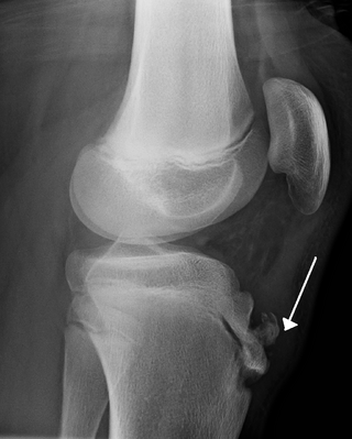
A patellar dislocation is a knee injury in which the patella (kneecap) slips out of its normal position. Often the knee is partly bent, painful and swollen. The patella is also often felt and seen out of place. Complications may include a patella fracture or arthritis.

<span class="mw-page-title-main">Medial patellofemoral ligament</span> Ligament of the knee

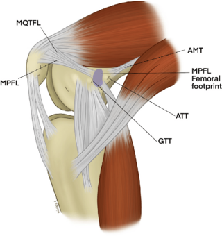
The medial patellofemoral ligament (MPFL) is one of several ligaments on the medial aspect of the knee. It originates in the superomedial aspect of the patella and inserts in the space between the adductor tubercle and the medial femoral epicondyle. The ligament itself extends from the femur to the superomedial patella, and its shape is similar to a trapezoid. It keeps the patella in place, but its main function is to prevent lateral displacement of the patella.

<span class="mw-page-title-main">Knee pain</span> Medical condition

Knee pain is pain in or around the knee.

Eccentric training is a type of strength training that involves using the target muscles to control weight as it moves in a downward motion. This type of training can help build muscle, improve athletic performance, and reduce the risk of injury. An eccentric contraction is the motion of an active muscle while it is lengthening under load. Eccentric training is repetitively doing eccentric muscle contractions. For example, in a biceps curl the action of lowering the dumbbell back down from the lift is the eccentric phase of that exercise – as long as the dumbbell is lowered slowly rather than letting it drop.

Posterolateral corner injuries of the knee are injuries to a complex area formed by the interaction of multiple structures. Injuries to the posterolateral corner can be debilitating to the person and require recognition and treatment to avoid long term consequences. Injuries to the PLC often occur in combination with other ligamentous injuries to the knee; most commonly the anterior cruciate ligament (ACL) and posterior cruciate ligament (PCL). As with any injury, an understanding of the anatomy and functional interactions of the posterolateral corner is important to diagnosing and treating the injury.

<span class="mw-page-title-main">Medial knee injuries</span> Medical condition

Medial knee injuries are the most common type of knee injury. The medial ligament complex of the knee consists of:

<span class="mw-page-title-main">Posterior cruciate ligament injury</span> Medical condition

The function of the posterior cruciate ligament (PCL) is to prevent the femur from sliding off the anterior edge of the tibia and to prevent the tibia from displacing posterior to the femur. Common causes of PCL injuries are direct blows to the flexed knee, such as the knee hitting the dashboard in a car accident or falling hard on the knee, both instances displacing the tibia posterior to the femur.

<span class="mw-page-title-main">Labral reconstruction</span> Medical procedure

Labral reconstruction is a type of hip arthroscopy in which the patient's native labrum is partially or completely removed and reconstructed using either autograft or allograft tissue. Originally described in 2009 using the ligamentum teres capitis, arthroscopic labral reconstruction using a variety of graft tissue has demonstrated promising short and mid-term clinical outcomes. Most importantly, labral reconstruction has demonstrated utility when the patient's native labral tissue is far too damaged for debridement or repair.

Artificial ligaments are devices used to replace damaged ligaments. Today, the most common use of artificial ligaments is in anterior cruciate ligament reconstruction. Although autotransplantation remains the most common method of ligament reconstruction, numerous materials and structures were developed to optimize the artificial ligament since its creation in the World War I era. Many modern artificial ligaments are made of synthetic polymers, such as polyethylene terephthalate. Various coatings have been added to improve the biocompatibility of the synthetic polymers. Early artificial ligaments developed in the 1980s were ineffective due to material deterioration. Currently, the Ligament Advanced Reinforcement System (LARS) artificial ligament has been utilized extensively in clinical applications. Tissue engineering is a growing area of research which aims to regenerate and restore ligament function.

References

  1. "Anterior Cruciate Ligament (ACL) Injuries". www.webmd.com. Retrieved 25 April 2016.
  2. Praz C, Kandhari VK, Saithna A, Sonnery-Cottet B (March 2019). "ACL rupture in the immediate build-up to the Olympic Games: return to elite alpine ski competition 5 months after injury and ACL repair". BMJ Case Reports. 12 (3): e227735. doi:10.1136/bcr-2018-227735. PMC   6424301 . PMID   30878956.
  3. "Anterior Cruciate Ligament (ACL) Injuries – Topic Overview". WebMD . Retrieved 22 April 2015.
  4. Baer GS, Harner CD (October 2007). "Clinical outcomes of allograft versus autograft in anterior cruciate ligament reconstruction". Clinics in Sports Medicine. 26 (4): 661–81. doi:10.1016/j.csm.2007.06.010. PMID   17920959.
  5. Cohen SB, Yucha DT, Ciccotti MC, Goldstein DT, Ciccotti MA, Ciccotti MG (September 2009). "Factors affecting patient selection of graft type in anterior cruciate ligament reconstruction". Arthroscopy. 25 (9): 1006–10. doi:10.1016/j.arthro.2009.02.010. PMID   19732639.
  6. "Stats on ACL injuries".
  7. Wong SE, Feeley BT, Pandya NK (September 2019). "Complications After Pediatric ACL Reconstruction: A Meta-analysis". Journal of Pediatric Orthopedics. 39 (8): e566–e571. doi:10.1097/BPO.0000000000001075. PMID   31393290. S2CID   199503697.
  8. "Anatomy and Gender Disparity of ACL injuries". Archived from the original on 2 April 2015.
  9. Ouabo EC, Gillain L, Saithna A, Blanchard J, Siegrist O, Sonnery-Cottet B (August 2019). "Combined Anatomic Anterior Cruciate and Anterolateral Ligament Reconstruction With Quadriceps Tendon Autograft and Gracilis Allograft Through a Single Femoral Tunnel". Arthroscopy Techniques. 8 (8): e827–e834. doi:10.1016/j.eats.2019.03.021. PMC   6823836 . PMID   31700778.
  10. Pinczewski LA, Lyman J, Salmon LJ, Russell VJ, Roe J, Linklater J (April 2007). "A 10-year comparison of anterior cruciate ligament reconstructions with hamstring tendon and patellar tendon autograft: a controlled, prospective trial". The American Journal of Sports Medicine. 35 (4): 564–574. doi:10.1177/0363546506296042. PMID   17261567. S2CID   73233440.
  11. 1 2 3 4 Mohtadi NG, Chan DS, Dainty KN, Whelan DB (September 2011). Cochrane Bone, Joint and Muscle Trauma Group (ed.). "Patellar tendon versus hamstring tendon autograft for anterior cruciate ligament rupture in adults". The Cochrane Database of Systematic Reviews. 2011 (9): CD005960. doi:10.1002/14651858.CD005960.pub2. PMC   6465162 . PMID   21901700.
  12. Kodkani PS, Govekar DP, Patankar HS (October 2004). "A new technique of graft harvest for anterior cruciate ligament reconstruction with quadruple semitendinosus tendon autograft". Arthroscopy. 20 (8): e101-4. doi:10.1016/j.arthro.2004.07.016. PMID   15483531.
  13. Okahashi K, Sugimoto K, Iwai M, Oshima M, Samma M, Fujisawa Y, Takakura Y (June 2006). "Regeneration of the hamstring tendons after harvesting for arthroscopic anterior cruciate ligament reconstruction: a histological study in 11 patients". Knee Surgery, Sports Traumatology, Arthroscopy. 14 (6): 542–5. doi:10.1007/s00167-006-0068-z. PMID   16525795. S2CID   7147812.
  14. Gill SS, Turner MA, Battaglia TC, Leis HT, Balian G, Miller MD (July 2004). "Semitendinosus regrowth: biochemical, ultrastructural, and physiological characterization of the regenerate tendon". The American Journal of Sports Medicine. 32 (5). The American Orthopedic Society for Sports Medicine: 1173–81. doi:10.1177/0363546503262159. PMID   15262639. S2CID   28512769.
  15. 1 2 3 Kraeutler MJ, Bravman JT, McCarty EC (October 2013). "Bone-patellar tendon-bone autograft versus allograft in outcomes of anterior cruciate ligament reconstruction: a meta-analysis of 5182 patients". The American Journal of Sports Medicine. 41 (10): 2439–48. doi:10.1177/0363546513484127. PMID   23585484. S2CID   7445341.
  16. Marrale J, Morrissey MC, Haddad FS (June 2007). "A literature review of autograft and allograft anterior cruciate ligament reconstruction". Knee Surgery, Sports Traumatology, Arthroscopy. 15 (6): 690–704. doi:10.1007/s00167-006-0236-1. PMID   17429611. S2CID   12469855.
  17. Shelbourne KD, Vanadurongwan B, Gray T (October 2007). "Primary anterior cruciate ligament reconstruction using contralateral patellar tendon autograft". Clinics in Sports Medicine. 26 (4): 549–65. doi:10.1016/j.csm.2007.06.008. PMID   17920952.
  18. Tibor LM, Long JL, Schilling PL, Lilly RJ, Carpenter JE, Miller BS (January 2010). "Clinical outcomes after anterior cruciate ligament reconstruction: a meta-analysis of autograft versus allograft tissue". Sports Health. 2 (1): 56–72. doi:10.1177/1941738109347984. PMC   3438864 . PMID   23015924.
  19. Spindler KP, Huston LJ, Wright RW, Kaeding CC, Marx RG, Amendola A, Parker RD, Andrish JT, Reinke EK, Harrell FE, Dunn WR (February 2011). "The prognosis and predictors of sports function and activity at minimum 6 years after anterior cruciate ligament reconstruction: a population cohort study". The American Journal of Sports Medicine. 39 (2): 348–59. doi:10.1177/0363546510383481. PMC   3692351 . PMID   21084660.
  20. Li X, Orvets N (February 2016). "Arthroscopic ACL Reconstruction with Bone Patellar Bone Graft using Anteromedial Technique". Journal of Medical Insight. 2016 (45). doi:10.24296/jomi/45.
  21. Samuelsen BT, Webster KE, Johnson NR, Hewett TE, Krych AJ (October 2017). "Hamstring Autograft versus Patellar Tendon Autograft for ACL Reconstruction: Is There a Difference in Graft Failure Rate? A Meta-analysis of 47,613 Patients". Clinical Orthopaedics and Related Research. 475 (10): 2459–2468. doi:10.1007/s11999-017-5278-9. PMC   5599382 . PMID   28205075.
  22. O'Brien DF, Kraeutler MJ, Koyonos L, Flato RR, Ciccotti MG, Cohen SB (March 2014). "Allograft anterior cruciate ligament reconstruction in patients younger than 30 years: a matched-pair comparison of bone-patellar tendon-bone and tibialis anterior". American Journal of Orthopedics. 43 (3): 132–6. PMID   24660179.
  23. "ACL Program - Bridge-Enhanced ACL Repair (BEAR) Clinical Trial". www.childrenshospital.org. Archived from the original on 20 May 2017. Retrieved 25 April 2016.
  24. Murray MM, Kalish LA, Fleming BC, Flutie B, Freiberger C, Henderson RN, et al. (March 2019). "Bridge-Enhanced Anterior Cruciate Ligament Repair: Two-Year Results of a First-in-Human Study". Orthopaedic Journal of Sports Medicine. 7 (3): 2325967118824356. doi:10.1177/2325967118824356. PMC   6431773 . PMID   30923725.
  25. "A Comparison of ACL Repair With BEAR Device vs. Autograft Patellar Tendon ACL Reconstruction - Full Text View - ClinicalTrials.gov". clinicaltrials.gov. Retrieved 2019-10-22.
  26. Murray MM, Fleming BC, Badger GJ, Freiberger C, Henderson R, Barnett S, et al. (May 2020). "Bridge-Enhanced Anterior Cruciate Ligament Repair Is Not Inferior to Autograft Anterior Cruciate Ligament Reconstruction at 2 Years: Results of a Prospective Randomized Clinical Trial". The American Journal of Sports Medicine. 48 (6): 1305–1315. doi:10.1177/0363546520913532. PMC   7227128 . PMID   32298131.
  27. Machotka Z, Scarborough I, Duncan W, Kumar S, Perraton L (December 2010). "Anterior cruciate ligament repair with LARS (ligament advanced reinforcement system): a systematic review". Sports Medicine, Arthroscopy, Rehabilitation, Therapy & Technology. 2 (1): 29. doi: 10.1186/1758-2555-2-29 . PMC   3016369 . PMID   21138589. CC-BY icon.svg Text was copied from this source, which is available under a Creative Commons Attribution 2.0 (CC BY 2.0) license.
  28. King JA, Miller WM (August 2007). "Bioreactor development for stem cell expansion and controlled differentiation". Current Opinion in Chemical Biology. 11 (4): 394–8. doi:10.1016/j.cbpa.2007.05.034. PMC   2038982 . PMID   17656148.
  29. Farrell E, O'Brien FJ, Doyle P, Fischer J, Yannas I, Harley BA, et al. (March 2006). "A collagen-glycosaminoglycan scaffold supports adult rat mesenchymal stem cell differentiation along osteogenic and chondrogenic routes". Tissue Engineering. 12 (3): 459–68. doi:10.1089/ten.2006.12.459. PMID   16579679.
  30. Yates EW, Rupani A, Foley GT, Khan WS, Cartmell S, Anand SJ (2012). "Ligament tissue engineering and its potential role in anterior cruciate ligament reconstruction". Stem Cells International. 2012: 438125. doi: 10.1155/2012/438125 . PMC   3255293 . PMID   22253633.
  31. Arundale AJ, Cummer K, Capin JJ, Zarzycki R, Snyder-Mackler L (October 2017). "Report of the Clinical and Functional Primary Outcomes in Men of the ACL-SPORTS Trial: Similar Outcomes in Men Receiving Secondary Prevention With and Without Perturbation Training 1 and 2 Years After ACL Reconstruction". Clinical Orthopaedics and Related Research. 475 (10): 2523–2534. doi:10.1007/s11999-017-5280-2. PMC   5599384 . PMID   28224443.
  32. Capin JJ, Zarzycki R, Arundale A, Cummer K, Snyder-Mackler L (October 2017). "Report of the Primary Outcomes for Gait Mechanics in Men of the ACL-SPORTS Trial: Secondary Prevention With and Without Perturbation Training Does Not Restore Gait Symmetry in Men 1 or 2 Years After ACL Reconstruction". Clinical Orthopaedics and Related Research. 475 (10): 2513–2522. doi:10.1007/s11999-017-5279-8. PMC   5599383 . PMID   28224442.
  33. BW Brewer, AE Cornelius , JL Van Raalte , JC Brickner , JH Sklar , JR Corsetti , MH Pohlman , TD Ditmar & K Emery, (2004). Rehabilitation adherence and anterior cruciate ligament reconstruction outcome, Psychology, Health & Medicine, 9:2, 163-175, DOI: 10.1080/13548500410001670690
  34. Paterno MV, Flynn K, Thomas S, Schmitt LC. Self-Reported Fear Predicts Functional Performance and Second ACL Injury After ACL Reconstruction and Return to Sport: A Pilot Study. Sports Health. 2018;10(3):228-233. doi:10.1177/1941738117745806
  35. "Anterior Cruciate Ligament (ACL) Injuries - Topic Overview". www.webmd.com. Retrieved 25 April 2016.
  36. "ACL Surgery Can Affect Emotional Health". Powerplay. 7 August 2023. Retrieved September 12, 2023.
  37. Cunha J, Solomon DJ (January 2022). "ACL Prehabilitation Improves Postoperative Strength and Motion and Return to Sport in Athletes". Arthroscopy, Sports Medicine, and Rehabilitation. 4 (1): e65–e69. doi:10.1016/j.asmr.2021.11.001. PMC   8811524 . PMID   35141537.
  38. Andrade R, Pereira R, van Cingel R, Staal JB, Espregueira-Mendes J (May 2020). "How should clinicians rehabilitate patients after ACL reconstruction? A systematic review of clinical practice guidelines (CPGs) with a focus on quality appraisal (AGREE II)". British Journal of Sports Medicine. 54 (9): 512–519. doi: 10.1136/bjsports-2018-100310 . PMID   31175108.
  39. Sonnery-Cottet B, Saithna A, Quelard B, Daggett M, Borade A, Ouanezar H, et al. (March 2019). "Arthrogenic muscle inhibition after ACL reconstruction: a scoping review of the efficacy of interventions". British Journal of Sports Medicine. 53 (5): 289–298. doi:10.1136/bjsports-2017-098401. PMC   6579490 . PMID   30194224.
  40. Delaloye JR, Murar J, Sánchez MG, Saithna A, Ouanezar H, Thaunat M, et al. (June 2018). "How to Rapidly Abolish Knee Extension Deficit After Injury or Surgery: A Practice-Changing Video Pearl From the Scientific Anterior Cruciate Ligament Network International (SANTI) Study Group". Arthroscopy Techniques. 7 (6): e601–e605. doi:10.1016/j.eats.2018.02.006. PMC   6019855 . PMID   30013901.
  41. Vanvuren C (2016-11-29). "How much does ACL surgery cost?". Amino Blog: Telling Health Care Stories with Data. Retrieved 2017-05-15.
  42. Bonsell S (October 2000). "Financial analysis of anterior cruciate ligament reconstruction at Baylor University Medical Center". Proceedings. 13 (4): 327–30. doi:10.1080/08998280.2000.11927697. PMC   1312224 . PMID   16389334.
  43. Lewis S (2016-10-17). "How Much Does ACL Surgery Cost?". Healthgrades Operating Company, Inc. Retrieved 2017-05-15.
  44. "Anterior Cruciate Ligament (ACL) Surgery". WebMD. Retrieved 25 April 2016.