Discectomy

Last updated
Discectomy
Gray301.png
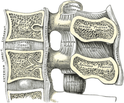
Median sagittal section of two lumbar vertebrae and their ligaments. ("Intervertebral fibrocartilage", an old name for the intervertebral disc, labeled at center left.)
ICD-9-CM 80.51
MeSH D017586
MedlinePlus 007250
Illustration depicting a surgical discectomy Blausen 0319 Discectomy.png
Illustration depicting a surgical discectomy

A discectomy (also called open discectomy, if done through a 1/2 inch or larger skin opening) is the surgical removal of abnormal disc material that presses on a nerve root or the spinal cord. The procedure involves removing a portion of an intervertebral disc, which causes pain, weakness or numbness by stressing the spinal cord or radiating nerves. The traditional open discectomy, or Love's technique, was published by Ross and Love in 1971. Advances have produced visualization improvements to traditional discectomy procedures (e.g. microdiscectomy, an open discectomy using an external microscope typically done through a 1-inch or larger skin opening), or endoscopic discectomy (the scope passes internally and typically done through a 2 mm skin opening or larger, up to 12 mm). In conjunction with the traditional discectomy or microdiscectomy, a laminotomy is often involved to permit access to the intervertebral disc. Laminotomy means a significant amount of typically normal bone (the lamina) is removed from the vertebra, allowing the surgeon to better see and access the area of disc herniation.

Contents

Endoscopic discectomy

Small or ultra-small endoscopic discectomy (called Nano Endoscopic Discectomy) does not have internal cutting or bone removal and, due to the small size, is not called "open". These procedures do not cause post-laminectomy syndrome (failed back syndrome). [1] [2]

Microdiscectomy

Microdiscectomy is a spine operation with a smaller incision than traditional discectomy, in which a portion of a herniated nucleus pulposus is removed by way of a surgical instrument, while using an external operating microscope for lighting and magnification. They may be "open", i.e., with a larger incision, or minimally invasive, i.e., with a 1.5 to 2.0 cm surgical incision. [3] [4]

Indications

Microdiscectomy may be a surgical option for patients with a single-level disc herniation and evidence of nerve root compression with residual unremitting radicular symptoms after failed conservative treatment. Cauda equina syndrome and progressive or new motor deficits are among the urgent surgical indications for microdiscectomy. [3]

Contraindications

Contraindications include additional pathologies, including infection, tumor, or segmental instability or vertebral fracture where fusion or instrumentation may be required. However, segmental instability and spondylolisthesis may be considered relative contraindications by some physicians. [3]

Discectomy for athletes

Degeneration caused by years of repetitive mechanical stress can cause the disc of an athlete to be herniated. Lumbar disc herniation (LDH) is a critical injury for elite athletes that could cause extreme pain and significantly hinder performance. To relieve the pain, athletes usually go through microdiscectomy. However, the results of treatments in elite athletes differ due to the demand for optimal treatment, short recovery period, and high performance after the operation.[ citation needed ]

Most athletes return to their pre-surgery level after a discectomy. A systematic review of 450 athletes shows that 75–100% of athletes return to play after surgery. [5] The average recovery period ranged from 2.8 to 8.7 months. Athletes recovered an average of 64.4% to 103.6% of their preoperative performance and had reported career longevity of 2.6 to 4.8 years post-return. [5] There are unsuccessful cases of discectomy for certain athletes like Tiger Woods, a world-famous PGA Tour golfer. Woods underwent three microdiscectomy procedures from 2014 to 2015 which failed to alleviate his pain. [6] The removal of disc material due to discectomy meant that Woods eventually had to go through spinal fusion to recover.

In the US

In the U.S., it has been estimated that the Medicare system spends over $300 million annually on lumbar discectomies. [7]

See also

Related Research Articles

<span class="mw-page-title-main">Lumbar disc disease</span> Medical condition

Lumbar disc disease is the drying out of the spongy interior matrix of an intervertebral disc in the spine. Many physicians and patients use the term lumbar disc disease to encompass several different causes of back pain or sciatica. In this article, the term is used to describe a lumbar herniated disc. It is thought that lumbar disc disease causes about one-third of all back pain.

<span class="mw-page-title-main">Intervertebral disc</span> Cartilage of spine

An intervertebral disc lies between adjacent vertebrae in the vertebral column. Each disc forms a fibrocartilaginous joint, to allow slight movement of the vertebrae, to act as a ligament to hold the vertebrae together, and to function as a shock absorber for the spine.

<span class="mw-page-title-main">Sciatica</span> Lower back pain that extends down leg

Sciatica is pain going down the leg from the lower back. This pain may go down the back, outside, or front of the leg. Onset is often sudden following activities like heavy lifting, though gradual onset may also occur. The pain is often described as shooting. Typically, symptoms are only on one side of the body. Certain causes, however, may result in pain on both sides. Lower back pain is sometimes present. Weakness or numbness may occur in various parts of the affected leg and foot.

<span class="mw-page-title-main">Schmorl's nodes</span> Medical condition

Schmorl's nodes are protrusions of the nucleus pulposus of the intervertebral disc through the vertebral body endplate and into the adjacent vertebra.

<span class="mw-page-title-main">Laminectomy</span> Surgical removal of a lamina

A laminectomy is a surgical procedure that removes a portion of a vertebra called the lamina, which is the roof of the spinal canal. It is a major spine operation with residual scar tissue and may result in postlaminectomy syndrome. Depending on the problem, more conservative treatments may be viable.

<span class="mw-page-title-main">Back injury</span> Damage or wear to bones, muscles or other tissues of the back

Back injuries result from damage, wear, or trauma to the bones, muscles, or other tissues of the back. Common back injuries include sprains and strains, herniated discs, and fractured vertebrae. The lumbar spine is often the site of back pain. The area is susceptible because of its flexibility and the amount of body weight it regularly bears. It is estimated that low-back pain may affect as much as 80 to 90 percent of the general population in the United States.

<span class="mw-page-title-main">Spondylosis</span> Degeneration of the vertebral column

Spondylosis is the degeneration of the vertebral column from any cause. In the more narrow sense it refers to spinal osteoarthritis, the age-related degeneration of the spinal column, which is the most common cause of spondylosis. The degenerative process in osteoarthritis chiefly affects the vertebral bodies, the neural foramina and the facet joints. If severe, it may cause pressure on the spinal cord or nerve roots with subsequent sensory or motor disturbances, such as pain, paresthesia, imbalance, and muscle weakness in the limbs.

<span class="mw-page-title-main">Degenerative disc disease</span> Medical condition

Degenerative disc disease (DDD) is a medical condition typically brought on by the normal aging process in which there are anatomic changes and possibly a loss of function of one or more intervertebral discs of the spine. DDD can take place with or without symptoms, but is typically identified once symptoms arise. The root cause is thought to be loss of soluble proteins within the fluid contained in the disc with resultant reduction of the oncotic pressure, which in turn causes loss of fluid volume. Normal downward forces cause the affected disc to lose height, and the distance between vertebrae is reduced. The anulus fibrosus, the tough outer layers of a disc, also weakens. This loss of height causes laxity of the longitudinal ligaments, which may allow anterior, posterior, or lateral shifting of the vertebral bodies, causing facet joint malalignment and arthritis; scoliosis; cervical hyperlordosis; thoracic hyperkyphosis; lumbar hyperlordosis; narrowing of the space available for the spinal tract within the vertebra ; or narrowing of the space through which a spinal nerve exits with resultant inflammation and impingement of a spinal nerve, causing a radiculopathy.

<span class="mw-page-title-main">Spinal fusion</span> Immobilization or ankylosis of two or more vertebrae by fusion of the vertebral bodies

Spinal fusion, also called spondylodesis or spondylosyndesis, is a neurosurgical or orthopedic surgical technique that joins two or more vertebrae. This procedure can be performed at any level in the spine and prevents any movement between the fused vertebrae. There are many types of spinal fusion and each technique involves using bone grafting—either from the patient (autograft), donor (allograft), or artificial bone substitutes—to help the bones heal together. Additional hardware is often used to hold the bones in place while the graft fuses the two vertebrae together. The placement of hardware can be guided by fluoroscopy, navigation systems, or robotics.

Failed back syndrome or post-laminectomy syndrome is a condition characterized by chronic pain following back surgeries. Many factors can contribute to the onset or development of FBS, including residual or recurrent spinal disc herniation, persistent post-operative pressure on a spinal nerve, altered joint mobility, joint hypermobility with instability, scar tissue (fibrosis), depression, anxiety, sleeplessness, spinal muscular deconditioning and even Cutibacterium acnes infection. An individual may be predisposed to the development of FBS due to systemic disorders such as diabetes, autoimmune disease and peripheral blood vessels (vascular) disease.

<span class="mw-page-title-main">Spinal disc herniation</span> Injury to the connective tissue between spinal vertebrae

A spinal disc herniation is an injury to the cushioning and connective tissue between vertebrae, usually caused by excessive strain or trauma to the spine. It may result in back pain, pain or sensation in different parts of the body, and physical disability. The most conclusive diagnostic tool for disc herniation is MRI, and treatment may range from painkillers to surgery. Protection from disc herniation is best provided by core strength and an awareness of body mechanics including posture.

<span class="mw-page-title-main">Radiculopathy</span> Medical condition

Radiculopathy, also commonly referred to as pinched nerve, refers to a set of conditions in which one or more nerves are affected and do not work properly. Radiculopathy can result in pain, weakness, altered sensation (paresthesia) or difficulty controlling specific muscles. Pinched nerves arise when surrounding bone or tissue, such as cartilage, muscles or tendons, put pressure on the nerve and disrupt its function.

<span class="mw-page-title-main">Anterior cervical discectomy and fusion</span> Surgical procedure

Anterior cervical discectomy and fusion (ACDF) is a surgical procedure to treat nerve root or spinal cord compression by decompressing the spinal cord and nerve roots of the cervical spine with a discectomy, followed by inter-vertebral fusion to stabilize the corresponding vertebrae. This procedure is used when other non-surgical treatments have failed.

<span class="mw-page-title-main">Laminotomy</span> Surgical procedure

A laminotomy is an orthopaedic neurosurgical procedure that removes part of the lamina of a vertebral arch in order to relieve pressure in the vertebral canal. A laminotomy is less invasive than conventional vertebral column surgery techniques, such as laminectomy because it leaves more ligaments and muscles attached to the spinous process intact and it requires removing less bone from the vertebra. As a result, laminotomies typically have a faster recovery time and result in fewer postoperative complications. Nevertheless, possible risks can occur during or after the procedure like infection, hematomas, and dural tears. Laminotomies are commonly performed as treatment for lumbar spinal stenosis and herniated disks. MRI and CT scans are often used pre- and post surgery to determine if the procedure was successful.

<span class="mw-page-title-main">Spinal decompression</span> Relief of pressure on the spinal cord or compressed nerve roots

Spinal decompression is the relief of pressure on the spinal cord or on one or more compressed nerve roots passing through or exiting the spinal column. Decompression of the spinal neural elements is a key component in treating spinal radiculopathy, myelopathy and claudication.

<span class="mw-page-title-main">Lumbar provocative discography</span>

Lumbar provocative discography is an invasive diagnostic procedure for evaluation for intervertebral disc pathology. It is usually reserved for persons with persistent, severe low back pain (LBP) who have abnormal spaces between vertebrae on magnetic resonance imaging (MRI), where other diagnostic tests have failed to reveal clear confirmation of a suspected disc as the source of pain, and surgical intervention is being considered.

The TESSYS method is a minimally-invasive, endoscopic spinal procedure for the treatment of a herniated disc. It was a further development of the YESS method by the Dutch Dr Thomas Hoogland in the Alpha Klinik in Munich in 1989 and was first called THESSYS. The procedure involves performing a small foramenotomy and removal of soft tissue compressing the nerve root.

Minimally invasive spine surgery, also known as MISS, has no specific meaning or definition. It implies a lack of severe surgical invasion. The older style of open-spine surgery for a relatively small disc problem used to require a 5-6 inch incision and a month in the hospital. MISS techniques utilize more modern technology, advanced imaging techniques and special medical equipment to reduce tissue trauma, bleeding, radiation exposure, infection risk, and decreased hospital stays by minimizing the size of the incision. Modern endoscopic procedures can be done through a 2 to 5 mm skin opening. By contrast, procedures done with a microscope require skin openings of approximately one inch, or more.

<span class="mw-page-title-main">Parviz Kambin</span> American-Iranian medical doctor and orthopaedic surgeon (1931–2020)

Parviz Kambin was an American-Iranian medical doctor and orthopaedic surgeon. He was a Professor of Orthopaedic Surgery and has established an Endowed Chair of Spinal Surgery Research at Drexel University College of Medicine. He published more than 55 articles in peer-reviewed journals, edited two textbooks and contributed chapters in spinal surgery textbooks. He lectured worldwide in the field of minimally invasive spinal surgery. His research and development in this specialty began in 1970.

The Philadelphia Surgery Center is a medical facility in Narberth, Pennsylvania, that specializes in small-scale endoscopic spine surgery for the treatment of spinal stenosis and herniated or fragmented spinal discs.

References

  1. Book Chapter - Decision Making in Spinal Care - Chapter 61; Copyright 2013 by Thieme
  2. "ISASS17 - Regular Poster Presentation Abstracts - Endoscopic Surgery - 455 - Nano Endoscopic Approach for Central Lumbar Disc Herniations".
  3. 1 2 3 Dowling, Thomas J.; Dowling, Thomas J. (2021), "Microdiscectomy", StatPearls, Treasure Island (FL): StatPearls Publishing, PMID   32310444 , retrieved 2021-05-07
  4. "Nucleus Pulposus - an overview | ScienceDirect Topics". www.sciencedirect.com. Retrieved 2021-05-07.
  5. 1 2 Nair, Rueben; Kahlenberg, Cynthia A.; Hsu, Wellington K. (2015-06-01). "Outcomes of Lumbar Discectomy in Elite Athletes: The Need for High-level Evidence". Clinical Orthopaedics and Related Research. 473 (6): 1971–1977. doi:10.1007/s11999-014-3762-z. ISSN   1528-1132. PMC   4419017 . PMID   25002213.
  6. O'Sullivan, Peter (2015-07-01). "Common misconceptions about back pain in sport: Tiger Woods' case brings five fundamental questions into sharp focus". British Journal of Sports Medicine. 49 (14): 905–907. doi:10.1136/bjsports-2014-094542. ISSN   0306-3674. PMID   25807161. S2CID   43137403.
  7. Schoenfeld, A. J.; Weiner, B. K. (2010). "Treatment of lumbar disc herniation: Evidence-based practice". International Journal of General Medicine . 3: 209–214. doi: 10.2147/ijgm.s12270 . PMC   2915533 . PMID   20689695.