In anatomy, the atlas (C1) is the most superior (first) cervical vertebra of the spine and is located in the neck.

The medulla oblongata or simply medulla is a long stem-like structure which makes up the lower part of the brainstem. It is anterior and partially inferior to the cerebellum. It is a cone-shaped neuronal mass responsible for autonomic (involuntary) functions, ranging from vomiting to sneezing. The medulla contains the cardiac, respiratory, vomiting and vasomotor centers, and therefore deals with the autonomic functions of breathing, heart rate and blood pressure as well as the sleep–wake cycle.

The scapula, also known as the shoulder blade, is the bone that connects the humerus with the clavicle. Like their connected bones, the scapulae are paired, with each scapula on either side of the body being roughly a mirror image of the other. The name derives from the Classical Latin word for trowel or small shovel, which it was thought to resemble.

The facial nerve, also known as the seventh cranial nerve, cranial nerve VII, or simply CN VII, is a cranial nerve that emerges from the pons of the brainstem, controls the muscles of facial expression, and functions in the conveyance of taste sensations from the anterior two-thirds of the tongue. The nerve typically travels from the pons through the facial canal in the temporal bone and exits the skull at the stylomastoid foramen. It arises from the brainstem from an area posterior to the cranial nerve VI and anterior to cranial nerve VIII.

The glossopharyngeal nerve, also known as the ninth cranial nerve, cranial nerve IX, or simply CN IX, is a cranial nerve that exits the brainstem from the sides of the upper medulla, just anterior to the vagus nerve. Being a mixed nerve (sensorimotor), it carries afferent sensory and efferent motor information. The motor division of the glossopharyngeal nerve is derived from the basal plate of the embryonic medulla oblongata, whereas the sensory division originates from the cranial neural crest.

The tibia, also known as the shinbone or shankbone, is the larger, stronger, and anterior (frontal) of the two bones in the leg below the knee in vertebrates ; it connects the knee with the ankle. The tibia is found on the medial side of the leg next to the fibula and closer to the median plane. The tibia is connected to the fibula by the interosseous membrane of leg, forming a type of fibrous joint called a syndesmosis with very little movement. The tibia is named for the flute tibia. It is the second largest bone in the human body, after the femur. The leg bones are the strongest long bones as they support the rest of the body.

The fornix is a C-shaped bundle of nerve fibers in the brain that acts as the major output tract of the hippocampus. The fornix also carries some afferent fibers to the hippocampus from structures in the diencephalon and basal forebrain. The fornix is part of the limbic system. While its exact function and importance in the physiology of the brain are still not entirely clear, it has been demonstrated in humans that surgical transection—the cutting of the fornix along its body—can cause memory loss. There is some debate over what type of memory is affected by this damage, but it has been found to most closely correlate with recall memory rather than recognition memory. This means that damage to the fornix can cause difficulty in recalling long-term information such as details of past events, but it has little effect on the ability to recognize objects or familiar situations.

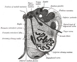
In anatomy, the olivary bodies or simply olives are a pair of prominent oval structures in the medulla oblongata, the lower portion of the brainstem. They contain the olivary nuclei.

The lentiform nucleus are the putamen (laterally) and the globus pallidus (medially), collectively. Due to their proximity, these two structures were formerly considered one, however, the two are separated by a thin layer of white matter - the external medullary lamina - and are functionally and connectionally distinct.

The spinocerebellar tract is a nerve tract originating in the spinal cord and terminating in the same side (ipsilateral) of the cerebellum.

In the medulla oblongata, the arcuate nucleus is a group of neurons located on the anterior surface of the medullary pyramids. These nuclei are the extension of the pontine nuclei. They receive fibers from the corticospinal tract and send their axons through the anterior external arcuate fibers and medullary striae to the cerebellum via the inferior cerebellar peduncle.

The ischium forms the lower and back region of the hip bone.

The squamous part of temporal bone, or temporal squama, forms the front and upper part of the temporal bone, and is scale-like, thin, and translucent.

The wing(ala)of ilium is the large expanded portion of the ilium, the bone which bounds the greater pelvis laterally. It presents for examination two surfaces—an external and an internal—a crest, and two borders—an anterior and a posterior.

The zygomatic processes are three processes (protrusions) from other bones of the skull which each articulate with the zygomatic bone. The three processes are:

The olfactory tract is a bilateral bundle of afferent nerve fibers from the mitral and tufted cells of the olfactory bulb that connects to several target regions in the brain, including the piriform cortex, amygdala, and entorhinal cortex. It is a narrow white band, triangular on coronal section, the apex being directed upward.
In neuroanatomy, the medullary pyramids are paired white matter structures of the brainstem's medulla oblongata that contain motor fibers of the corticospinal and corticobulbar tracts – known together as the pyramidal tracts. The lower limit of the pyramids is marked when the fibers cross (decussate).

The anterior median fissure contains a fold of pia mater, and extends along the entire length of the medulla oblongata: It ends at the lower border of the pons in a small triangular expansion, termed the foramen cecum.

Each vertebra is an irregular bone with a complex structure composed of bone and some hyaline cartilage, that make up the vertebral column or spine, of vertebrates. The proportions of the vertebrae differ according to their spinal segment and the particular species.