The medulla oblongata or simply medulla is a long stem-like structure which makes up the lower part of the brainstem. It is anterior and partially inferior to the cerebellum. It is a cone-shaped neuronal mass responsible for autonomic (involuntary) functions, ranging from vomiting to sneezing. The medulla contains the cardiac, respiratory, vomiting and vasomotor centers, and therefore deals with the autonomic functions of breathing, heart rate and blood pressure as well as the sleep–wake cycle. "Medulla" is from Latin, ‘pith or marrow’. And "oblongata" is from Latin, ‘lengthened or longish or elongated'.
Pronator quadratus is a square-shaped muscle on the distal forearm that acts to pronate the hand.
The brainstem is the stalk-like part of the brain that connects the forebrain with the spinal cord. In the human brain, the brainstem is composed of the midbrain, the pons, and the medulla oblongata. The midbrain is continuous with the thalamus of the diencephalon through the tentorial notch.

In neuroanatomy, the trigeminal nerve (lit. triplet nerve), also known as the fifth cranial nerve, cranial nerve V, or simply CN V, is a cranial nerve responsible for sensation in the face and motor functions such as biting and chewing; it is the most complex of the cranial nerves. Its name (trigeminal, from Latin tri- 'three' and -geminus 'twin') derives from each of the two nerves (one on each side of the pons) having three major branches: the ophthalmic nerve (V1), the maxillary nerve (V2), and the mandibular nerve (V3). The ophthalmic and maxillary nerves are purely sensory, whereas the mandibular nerve supplies motor as well as sensory (or "cutaneous") functions. Adding to the complexity of this nerve is that autonomic nerve fibers as well as special sensory fibers (taste) are contained within it.

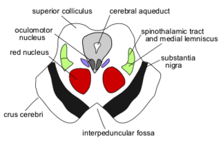
The spinothalamic tract is a nerve tract in the anterolateral system in the spinal cord. This tract is an ascending sensory pathway to the thalamus. From the ventral posterolateral nucleus in the thalamus, sensory information is relayed upward to the somatosensory cortex of the postcentral gyrus.

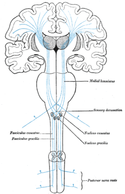
The dorsal column–medial lemniscus pathway (DCML) (also known as the posterior column-medial lemniscus pathway is a sensory pathway of the central nervous system that conveys sensations of fine touch, vibration, two-point discrimination, and proprioception from the skin and joints. It transmits this information to the somatosensory cortex of the postcentral gyrus in the parietal lobe of the brain. The pathway receives information from sensory receptors throughout the body, and carries this in the gracile fasciculus and the cuneate fasciculus, tracts that make up the dorsal columns. The dorsal columns are carried in the white matter of the posterior funiculus of the spinal cord. At the level of the medulla oblongata, the fibers of the tracts decussate and are continued in the medial lemniscus, on to the thalamus and relayed from there through the internal capsule and transmitted to the somatosensory cortex. The name dorsal-column medial lemniscus comes from the two structures that carry the sensory information: the dorsal columns of the spinal cord, and the medial lemniscus in the brainstem.

The lateral lemniscus is a tract of axons in the brainstem that carries information about sound from the cochlear nucleus to various brainstem nuclei and ultimately the contralateral inferior colliculus of the midbrain. Three distinct, primarily inhibitory, cellular groups are located interspersed within these fibers, and are thus named the nuclei of the lateral lemniscus.

The inferior colliculus (IC) is the principal midbrain nucleus of the auditory pathway and receives input from several peripheral brainstem nuclei in the auditory pathway, as well as inputs from the auditory cortex. The inferior colliculus has three subdivisions: the central nucleus, a dorsal cortex by which it is surrounded, and an external cortex which is located laterally. Its bimodal neurons are implicated in auditory-somatosensory interaction, receiving projections from somatosensory nuclei. This multisensory integration may underlie a filtering of self-effected sounds from vocalization, chewing, or respiration activities.

The corticobulbartract is a two-neuron white matter motor pathway connecting the motor cortex in the cerebral cortex to the medullary pyramids, which are part of the brainstem's medulla oblongata region, and are primarily involved in carrying the motor function of the non-oculomotor cranial nerves. The corticobulbar tract is one of the pyramidal tracts, the other being the corticospinal tract.

The pontine nuclei are all neurons of the ventral pons collectively. Corticopontine fibres project from the primary motor cortex to the ipsilateral pontine nucleus; pontocerebellar fibers then relay the information to the contralateral cerebellum via the middle cerebellar peduncle.

The spinocerebellar tract is a nerve tract originating in the spinal cord and terminating in the same side (ipsilateral) of the cerebellum.

The cochlear nucleus (CN) or cochlear nuclear complex comprises two cranial nerve nuclei in the human brainstem, the ventral cochlear nucleus (VCN) and the dorsal cochlear nucleus (DCN). The ventral cochlear nucleus is unlayered whereas the dorsal cochlear nucleus is layered. Auditory nerve fibers, fibers that travel through the auditory nerve carry information from the inner ear, the cochlea, on the same side of the head, to the nerve root in the ventral cochlear nucleus. At the nerve root the fibers branch to innervate the ventral cochlear nucleus and the deep layer of the dorsal cochlear nucleus. All acoustic information thus enters the brain through the cochlear nuclei, where the processing of acoustic information begins. The outputs from the cochlear nuclei are received in higher regions of the auditory brainstem.

In neuroanatomy, the internal arcuate fibers or internal arcuate tract are the axons of second-order sensory neurons that compose the gracile and cuneate nuclei of the medulla oblongata. These second-order neurons begin in the gracile and cuneate nuclei in the medulla. They receive input from first-order sensory neurons, which provide sensation to many areas of the body and have cell bodies in the dorsal root ganglia of the dorsal root of the spinal nerves. Upon decussation from one side of the medulla to the other, also known as the sensory decussation, they are then called the medial lemniscus.
The dorsal column nuclei are a pair of nuclei in the dorsal columns of the dorsal column–medial lemniscus pathway (DCML) in the brainstem. The name refers collectively to the cuneate nucleus and gracile nucleus, which are situated at the lower end of the medulla oblongata. Both nuclei contain second-order neurons of the DCML, which convey fine touch and proprioceptive information from the body to the brain via the thalamus.

The sensory decussation or decussation of the lemnisci is a decussation of axons from the gracile nucleus and cuneate nucleus, known together as the dorsal column nuclei. The dorsal column nuclei are responsible for fine touch, vibration, proprioception and two-point discrimination.

The ventral posteromedial nucleus (VPM) is a nucleus of the thalamus and serves an analogous somatosensory relay role for the ascending trigeminothalamic tracts as its lateral neighbour the ventral posterolateral nucleus serves for dorsal column–medial lemniscus pathway 2nd-order neurons.
The ventral trigeminal tract, ventral trigeminothalamic tract, anterior trigeminal tract, or anterior trigeminothalamic tract, is a tract composed of second-order neuronal axons. These afferent fibers carry sensory information about discriminative and crude touch, conscious proprioception, pain, and temperature from the head, face, and oral cavity. The ventral trigeminal tract connects the two major components of the brainstem trigeminal complex – the principal, or main sensory nucleus and the spinal trigeminal nucleus, to the ventral posteromedial nucleus of the thalamus.
The trigeminal lemniscus or the trigeminothalamic tracts is a somatosensory tract containing second-order neuron fibers of the trigeminal system. It consists of the ventral and dorsal trigeminal tracts. Its second-order sensory axons convey tactile, pain, and temperature impulses from the skin of the face, the mucous membranes of the nasal and oral cavities, and the eye, as well as proprioceptive information from the facial and masticatory muscles.
The central tegmental tract is a structure in the midbrain and pons.

The spinal cord is a long, thin, tubular structure made up of nervous tissue that extends from the medulla oblongata in the brainstem to the lumbar region of the vertebral column (backbone) of vertebrate animals. The center of the spinal cord is hollow and contains a structure called the central canal, which contains cerebrospinal fluid. The spinal cord is also covered by meninges and enclosed by the neural arches. Together, the brain and spinal cord make up the central nervous system.