Mechanoreceptor

Last updated

A mechanoreceptor, also called mechanoceptor, is a sensory receptor that responds to mechanical pressure or distortion. Mechanoreceptors are innervated by sensory neurons that convert mechanical pressure into electrical signals that, in animals, are sent to the central nervous system.

Contents

Vertebrate mechanoreceptors

Cutaneous mechanoreceptors

Cutaneous mechanoreceptors respond to mechanical stimuli that result from physical interaction, including pressure and vibration. They are located in the skin, like other cutaneous receptors. They are all innervated by Aβ fibers, except the mechanorecepting free nerve endings, which are innervated by Aδ fibers. Cutaneous mechanoreceptors can be categorized by what kind of sensation they perceive, by the rate of adaptation, and by morphology. Furthermore, each has a different receptive field.

Tactile receptors. Blausen 0809 Skin TactileReceptors.png
Tactile receptors.

By sensation

  • The Slowly Adapting type 1 (SA1) mechanoreceptor, with the Merkel corpuscle end-organ (also known as Merkel discs) detect sustained pressure and underlies the perception of form and roughness on the skin. [1] They have small receptive fields and produce sustained responses to static stimulation.
  • The Slowly Adapting type 2 (SA2) mechanoreceptors, with the Ruffini corpuscle end-organ (also known as the bulbous corpuscles), detect tension deep in the skin and fascia and respond to skin stretch, but have not been closely linked to either proprioceptive or mechanoreceptive roles in perception. [2] They also produce sustained responses to static stimulation, but have large receptive fields.
  • The Rapidly Adapting (RA) or Meissner corpuscle end-organ mechanoreceptor (also known as the tactile corpuscles) underlies the perception of light touch such as flutter [3] and slip on the skin. [4] It adapts rapidly to changes in texture (vibrations around 50 Hz). They have small receptive fields and produce transient responses to the onset and offset of stimulation.
  • The Pacinian corpuscle or Vater-Pacinian corpuscles or Lamellar corpuscles [5] in the skin and fascia detect rapid vibrations of about 200–300 Hz. [3] [6] They also produce transient responses, but have large receptive fields.
  • Free nerve endings detect touch, pressure, stretching, as well as the tickle and itch sensations. Itch sensations are caused by stimulation of free nerve ending from chemicals. [7]
  • Hair follicle receptors called hair root plexuses sense when a hair changes position. Indeed, the most sensitive mechanoreceptors in humans are the hair cells in the cochlea of the inner ear (no relation to the follicular receptors – they are named for the hair-like mechanosensory stereocilia they possess); these receptors transduce sound for the brain. [7]

By rate of adaptation

Cutaneous mechanoreceptors can also be separated into categories based on their rates of adaptation. When a mechanoreceptor receives a stimulus, it begins to fire impulses or action potentials at an elevated frequency (the stronger the stimulus, the higher the frequency). The cell, however, will soon "adapt" to a constant or static stimulus, and the pulses will subside to a normal rate. Receptors that adapt quickly (i.e., quickly return to a normal pulse rate) are referred to as "phasic". Those receptors that are slow to return to their normal firing rate are called tonic. Phasic mechanoreceptors are useful in sensing such things as texture or vibrations, whereas tonic receptors are useful for temperature and proprioception among others.

By receptive field

Cutaneous mechanoreceptors with small, accurate receptive fields are found in areas needing accurate taction (e.g. the fingertips). In the fingertips and lips, innervation density of slowly adapting type I and rapidly adapting type I mechanoreceptors are greatly increased. These two types of mechanoreceptors have small discrete receptive fields and are thought to underlie most low-threshold use of the fingers in assessing texture, surface slip, and flutter. Mechanoreceptors found in areas of the body with less tactile acuity tend to have larger receptive fields.

Lamellar corpuscles

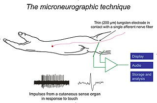
Lamellar corpuscles, or Pacinian corpuscles or Vater-Pacini corpuscle, are deformation or pressure receptors located in the skin and also in various internal organs. [8] Each is connected to a sensory neuron. Because of its relatively large size, a single lamellar corpuscle can be isolated and its properties studied. Mechanical pressure of varying strength and frequency can be applied to the corpuscle by stylus, and the resulting electrical activity detected by electrodes attached to the preparation.

Deforming the corpuscle creates a generator potential in the sensory neuron arising within it. This is a graded response: the greater the deformation, the greater the generator potential. If the generator potential reaches threshold, a volley of action potentials (nerve impulses) are triggered at the first node of Ranvier of the sensory neuron.

Once threshold is reached, the magnitude of the stimulus is encoded in the frequency of impulses generated in the neuron. So the more massive or rapid the deformation of a single corpuscle, the higher the frequency of nerve impulses generated in its neuron.

The optimal sensitivity of a lamellar corpuscle is 250 Hz, the frequency range generated upon finger tips by textures made of features smaller than 200  micrometres. [9]

Ligamentous mechanoreceptors

There are four types of mechanoreceptors embedded in ligaments. As all these types of mechanoreceptors are myelinated, they can rapidly transmit sensory information regarding joint positions to the central nervous system. [10]

Type II and Type III mechanoreceptors in particular are believed to be linked to one's sense of proprioception.

Other mechanoreceptors

Other mechanoreceptors than cutaneous ones include the hair cells, which are sensory receptors in the vestibular system of the inner ear, where they contribute to the auditory system and equilibrioception. Baroreceptors are a type of mechanoreceptor sensory neuron that is excited by stretch of the blood vessel. There are also juxtacapillary (J) receptors, which respond to events such as pulmonary edema, pulmonary emboli, pneumonia, and barotrauma.

Muscle spindles and the stretch reflex

The knee jerk is the popularly known stretch reflex (involuntary kick of the lower leg) induced by tapping the knee with a rubber-headed hammer. The hammer strikes a tendon that inserts an extensor muscle in the front of the thigh into the lower leg. Tapping the tendon stretches the thigh muscle, which activates stretch receptors within the muscle called muscle spindles. Each muscle spindle consists of sensory nerve endings wrapped around special muscle fibers called intrafusal muscle fibers. Stretching an intrafusal fiber initiates a volley of impulses in the sensory neuron (a I-a neuron) attached to it. The impulses travel along the sensory axon to the spinal cord where they form several kinds of synapses:

  1. Some of the branches of the I-a axons synapse directly with alpha motor neurons. These carry impulses back to the same muscle causing it to contract. The leg straightens.
  2. Some of the branches of the I-a axons synapse with inhibitory interneurons in the spinal cord. These, in turn, synapse with motor neurons leading back to the antagonistic muscle, a flexor in the back of the thigh. By inhibiting the flexor, these interneurons aid contraction of the extensor.
  3. Still other branches of the I-a axons synapse with interneurons leading to brain centers, e.g., the cerebellum, that coordinate body movements. [11]

Mechanism of sensation

In somatosensory transduction, the afferent neurons transmit messages through synapses in the dorsal column nuclei, where second-order neurons send the signal to the thalamus and synapse with third-order neurons in the ventrobasal complex. The third-order neurons then send the signal to the somatosensory cortex.

More recent work has expanded the role of the cutaneous mechanoreceptors for feedback in fine motor control. [12] Single action potentials from Meissner's corpuscle, Pacinian corpuscle and Ruffini ending afferents are directly linked to muscle activation, whereas Merkel cell-neurite complex activation does not trigger muscle activity. [13]

Invertebrate mechanoreceptors

Insect and arthropod mechanoreceptors include: [14]

Plant mechanoreceptors

Mechanoreceptors are also present in plant cells where they play an important role in normal growth, development and the sensing of their environment. [20] Mechanoreceptors aid the Venus flytrap (Dionaea muscipula Ellis) in capturing large [21] prey. [22]

Molecular biology

Mechanoreceptor proteins are ion channels whose ion flow is induced by touch. Early research showed that touch transduction in the nematode Caenorhabditis elegans was found to require a two transmembrane, amiloride-sensitive ion channel protein related to epithelial sodium channels (ENaCs). [23] This protein, called MEC-4, forms a heteromeric Na+-selective channel together with MEC-10. Related genes in mammals are expressed in sensory neurons and were shown to be gated by low pH. The first of such receptor was ASIC1a, named so because it is an acid sensing ion channel (ASIC). [24]

See also

Related Research Articles

<span class="mw-page-title-main">Motor neuron</span> Nerve cell sending impulse to muscle

A motor neuron is a neuron whose cell body is located in the motor cortex, brainstem or the spinal cord, and whose axon (fiber) projects to the spinal cord or outside of the spinal cord to directly or indirectly control effector organs, mainly muscles and glands. There are two types of motor neuron – upper motor neurons and lower motor neurons. Axons from upper motor neurons synapse onto interneurons in the spinal cord and occasionally directly onto lower motor neurons. The axons from the lower motor neurons are efferent nerve fibers that carry signals from the spinal cord to the effectors. Types of lower motor neurons are alpha motor neurons, beta motor neurons, and gamma motor neurons.

<span class="mw-page-title-main">Trigeminal nerve</span> Cranial nerve responsible for the faces senses and motor functions

In neuroanatomy, the trigeminal nerve (lit. triplet nerve), also known as the fifth cranial nerve, cranial nerve V, or simply CN V, is a cranial nerve responsible for sensation in the face and motor functions such as biting and chewing; it is the most complex of the cranial nerves. Its name (trigeminal, from Latin tri- 'three', and -geminus 'twin') derives from each of the two nerves (one on each side of the pons) having three major branches: the ophthalmic nerve (V1), the maxillary nerve (V2), and the mandibular nerve (V3). The ophthalmic and maxillary nerves are purely sensory, whereas the mandibular nerve supplies motor as well as sensory (or "cutaneous") functions. Adding to the complexity of this nerve is that autonomic nerve fibers as well as special sensory fibers (taste) are contained within it.

<span class="mw-page-title-main">Sensory nervous system</span> Part of the nervous system

The sensory nervous system is a part of the nervous system responsible for processing sensory information. A sensory system consists of sensory neurons, neural pathways, and parts of the brain involved in sensory perception and interoception. Commonly recognized sensory systems are those for vision, hearing, touch, taste, smell, balance and visceral sensation. Sense organs are transducers that convert data from the outer physical world to the realm of the mind where people interpret the information, creating their perception of the world around them.

<span class="mw-page-title-main">Stimulus (physiology)</span> Detectable change in the internal or external surroundings

Template:Microbiology

<span class="mw-page-title-main">Free nerve ending</span> Type of nerve fiber carrying sensory signals

A free nerve ending (FNE) or bare nerve ending, is an unspecialized, afferent nerve fiber sending its signal to a sensory neuron. Afferent in this case means bringing information from the body's periphery toward the brain. They function as cutaneous nociceptors and are essentially used by vertebrates to detect noxious stimuli that often result in pain.

Stimulus modality, also called sensory modality, is one aspect of a stimulus or what is perceived after a stimulus. For example, the temperature modality is registered after heat or cold stimulate a receptor. Some sensory modalities include: light, sound, temperature, taste, pressure, and smell. The type and location of the sensory receptor activated by the stimulus plays the primary role in coding the sensation. All sensory modalities work together to heighten stimuli sensation when necessary.

A cutaneous receptor is the type of sensory receptor found in the skin. They are a part of the somatosensory system. Cutaneous receptors include mechanoreceptors, nociceptors (pain), and thermoreceptors (temperature).

<span class="mw-page-title-main">Tactile corpuscle</span> Type of mechanoreceptor that detects light touch

Tactile corpuscles or Meissner's corpuscles are a type of mechanoreceptor discovered by anatomist Georg Meissner (1829–1905) and Rudolf Wagner. This corpuscle is a type of nerve ending in the skin that is responsible for sensitivity to pressure. In particular, they have their highest sensitivity when sensing vibrations between 10 and 50 hertz. They are rapidly adaptive receptors. They are most concentrated in thick hairless skin, especially at the finger pads.

<span class="mw-page-title-main">Sensory neuron</span> Nerve cell that converts environmental stimuli into corresponding internal stimuli

Sensory neurons, also known as afferent neurons, are neurons in the nervous system, that convert a specific type of stimulus, via their receptors, into action potentials or graded receptor potentials. This process is called sensory transduction. The cell bodies of the sensory neurons are located in the dorsal ganglia of the spinal cord.

<span class="mw-page-title-main">Caridoid escape reaction</span> Innate escape mechanism by crustaceans

The caridoid escape reaction, also known as lobstering or tail-flipping, refers to an innate escape mechanism in marine and freshwater crustaceans such as lobsters, krill, shrimp and crayfish.

<span class="mw-page-title-main">Pacinian corpuscle</span> Type of mechanoreceptor cell in hairless mammals

The Pacinian corpuscle, lamellar corpuscle or Vater-Pacini corpuscle is one of the four major types of mechanoreceptors for mechanical sensation) found in mammalian skin. This type of mechanoreceptor is found in both hairy, and hairless skin, viscera, joints, and attached to the periosteum of bone, primarily responsible for sensitivity to vibration. A few are also sensitive to quasi-static or low frequency pressure stimuli. Most of them respond only to sudden disturbances and are especially sensitive to vibration of a few hundreds hertz. The vibrational role may be used for detecting surface texture, such as rough and smooth. Most of the Pacinian corpuscles act as rapidly adapting mechanoreceptors. Groups of corpuscles respond to pressure changes, such as on grasping or releasing an object.

Merkel nerve endings are mechanoreceptors, a type of sensory receptor, that are found in the basal epidermis and hair follicles. They are nerve endings and provide information on mechanical pressure, position, and deep static touch features, such as shapes and edges.

<span class="mw-page-title-main">Tactile corpuscles of Grandry</span>

The tactile corpuscles of Grandry or Grandry corpuscles are mechanoreceptors found in the beak skin and oral mucosa of aquatic birds. They were first described by Grandry in 1869 in the bill skin of ducks and geese. Their general structure includes the flattened endings of an afferent nerve fiber sandwiched between two or more somewhat flattened sensory cells called Grandry cells, all surrounded by a layer of satellite cells and a partial capsule of collagen protein. Electrophysiological studies have shown that Grandry corpuscles function as rapidly adapting velocity detectors. In birds, Grandry and Merkel corpuscles share many morphological similarities, which has led to some confusion in the literature over their classification.

<span class="mw-page-title-main">Microneurography</span>

Microneurography is a neurophysiological method employed to visualize and record the traffic of nerve impulses that are conducted in peripheral nerves of waking human subjects. It can also be used in animal recordings. The method has been successfully employed to reveal functional properties of a number of neural systems, e.g. sensory systems related to touch, pain, and muscle sense as well as sympathetic activity controlling the constriction state of blood vessels. To study nerve impulses of an identified nerve, a fine tungsten needle microelectrode is inserted into the nerve and connected to a high input impedance differential amplifier. The exact position of the electrode tip within the nerve is then adjusted in minute steps until the electrode discriminates nerve impulses of interest. A unique feature and a significant strength of the microneurography method is that subjects are fully awake and able to cooperate in tests requiring mental attention, while impulses in a representative nerve fibre or set of nerve fibres are recorded, e.g. when cutaneous sense organs are stimulated or subjects perform voluntary precision movements.

Cutaneous innervation refers to an area of the skin which is supplied by a specific cutaneous nerve.

Type II sensory fiber is a type of sensory fiber, the second of the two main groups of touch receptors. The responses of different type Aβ fibers to these stimuli can be subdivided based on their adaptation properties, traditionally into rapidly adapting (RA) or slowly adapting (SA) neurons. Type II sensory fibers are slowly-adapting (SA), meaning that even when there is no change in touch, they keep respond to stimuli and fire action potentials. In the body, Type II sensory fibers belong to pseudounipolar neurons. The most notable example are neurons with Merkel cell-neurite complexes on their dendrites and Ruffini endings. Under pathological conditions they may become hyper-excitable leading to stimuli that would usually elicit sensations of tactile touch causing pain. These changes are in part induced by PGE2 which is produced by COX1, and type II fibers with free nerve endings are likely to be the subdivision of fibers that carry out this function.

Eimer's organs are sensory organs in which the epidermis is modified to form bulbous papillae. First isolated by Theodor Eimer from the European mole in 1871, these organs are present in many moles, and are particularly common in the star-nosed mole, which bears 25,000 of them on its unique tentacled snout. The organs are formed from a stack of epidermal cells, which is innervated by nerve processes from myelinated fibers in the dermis, which form terminal swellings just below the outer keratinized layer of epidermis. They contain a Merkel cell-neurite complex in the epidermis and a lamellated corpuscle in the dermal connective tissue.

Pallesthesia, or vibratory sensation, is the ability to perceive vibration. This sensation, often conducted through skin and bone, is usually generated by mechanoreceptors such as Pacinian corpuscles, Merkel disk receptors, and tactile corpuscles. All of these receptors stimulate an action potential in afferent nerves found in various layers of the skin and body. The afferent neuron travels to the spinal column and then to the brain where the information is processed. Damage to the peripheral nervous system or central nervous system can result in a decline or loss of pallesthesia.

<span class="mw-page-title-main">Axon reflex</span>

The axon reflex is the response stimulated by peripheral nerves of the body that travels away from the nerve cell body and branches to stimulate target organs. Reflexes are single reactions that respond to a stimulus making up the building blocks of the overall signaling in the body's nervous system. Neurons are the excitable cells that process and transmit these reflex signals through their axons, dendrites, and cell bodies. Axons directly facilitate intercellular communication projecting from the neuronal cell body to other neurons, local muscle tissue, glands and arterioles. In the axon reflex, signaling starts in the middle of the axon at the stimulation site and transmits signals directly to the effector organ skipping both an integration center and a chemical synapse present in the spinal cord reflex. The impulse is limited to a single bifurcated axon, or a neuron whose axon branches into two divisions and does not cause a general response to surrounding tissue.

<span class="mw-page-title-main">Somatosensory system</span> Nerve system for sensing touch, temperature, body position, and pain

Touch is perceiving the environment using skin. Specialized receptors in the skin send signals to the brain indicating light and soft pressure, hot and cold, body position and pain. It is a subset of the sensory nervous system, which also includes the visual, auditory, olfactory, gustatory and vestibular senses.

References

  1. Johnson KO, Hsiao SS (1992). "Neural mechanisms of tactual form and texture perception". Annual Review of Neuroscience. 15: 227–50. doi:10.1146/annurev.ne.15.030192.001303. PMID   1575442.
  2. Torebjörk HE, Ochoa JL (December 1980). "Specific sensations evoked by activity in single identified sensory units in man". Acta Physiologica Scandinavica. 110 (4): 445–7. doi:10.1111/j.1748-1716.1980.tb06695.x. PMID   7234450.
  3. 1 2 Talbot WH, Darian-Smith I, Kornhuber HH, Mountcastle VB (March 1968). "The sense of flutter-vibration: comparison of the human capacity with response patterns of mechanoreceptive afferents from the monkey hand". Journal of Neurophysiology. 31 (2): 301–34. doi:10.1152/jn.1968.31.2.301. PMID   4972033.
  4. Johansson RS, Westling G (1987). "Signals in tactile afferents from the fingers eliciting adaptive motor responses during precision grip". Experimental Brain Research. 66 (1): 141–54. doi:10.1007/bf00236210. PMID   3582528. S2CID   22450227.
  5. Biswas A, Manivannan M, Srinivasan MA (2015). "Multiscale layered biomechanical model of the pacinian corpuscle". IEEE Transactions on Haptics. 8 (1): 31–42. doi:10.1109/TOH.2014.2369416. PMID   25398182. S2CID   24658742.
  6. Biswas A, Manivannan M, Srinivasan MA (2015). "Vibrotactile sensitivity threshold: nonlinear stochastic mechanotransduction model of the Pacinian Corpuscle". IEEE Transactions on Haptics. 8 (1): 102–13. doi:10.1109/TOH.2014.2369422. PMID   25398183. S2CID   15326972.
  7. 1 2 Tortora GJ (2019). Principles of anatomy and physiology. John Wiley & Sons Australia, Limited. ISBN   978-0-7303-5500-7. OCLC   1059417106.
  8. Biswas A (2015). Characterization and Modeling of Vibrotactile Sensitivity Threshold of Human Finger Pad and the Pacinian Corpuscle (PhD). Indian Institute of Technology Madras, Tamil Nadu, India. doi:10.13140/RG.2.2.18103.11687.
  9. Scheibert J, Leurent S, Prevost A, Debrégeas G (March 2009). "The role of fingerprints in the coding of tactile information probed with a biomimetic sensor". Science. 323 (5920): 1503–6. arXiv: 0911.4885 . Bibcode:2009Sci...323.1503S. doi:10.1126/science.1166467. PMID   19179493. S2CID   14459552.
  10. Michelson JD, Hutchins C (March 1995). "Mechanoreceptors in human ankle ligaments". The Journal of Bone and Joint Surgery. British Volume. 77 (2): 219–24. doi: 10.1302/0301-620X.77B2.7706334 . PMID   7706334.
  11. Kimball JW (2011). "Mechanoreceptors". Kimball's Biology Pages. Archived from the original on 27 February 2011.
  12. Johansson RS, Flanagan JR (May 2009). "Coding and use of tactile signals from the fingertips in object manipulation tasks". Nature Reviews. Neuroscience. 10 (5): 345–59. doi:10.1038/nrn2621. PMID   19352402. S2CID   17298704.
  13. McNulty PA, Macefield VG (December 2001). "Modulation of ongoing EMG by different classes of low-threshold mechanoreceptors in the human hand". The Journal of Physiology. 537 (Pt 3): 1021–32. doi:10.1111/j.1469-7793.2001.01021.x. PMC   2278990 . PMID   11744774.
  14. Tuthill JC, Wilson RI (October 2016). "Mechanosensation and Adaptive Motor Control in Insects". Current Biology. 26 (20): R1022–R1038. doi:10.1016/j.cub.2016.06.070. PMC   5120761 . PMID   27780045.
  15. Bässler, U. (1977-06-01). "Sensory control of leg movement in the stick insect Carausius morosus". Biological Cybernetics. 25 (2): 61–72. doi:10.1007/BF00337264. ISSN   1432-0770. PMID   836915. S2CID   2634261.
  16. Mamiya, Akira; Gurung, Pralaksha; Tuthill, John C. (2018-11-07). "Neural Coding of Leg Proprioception in Drosophila". Neuron. 100 (3): 636–650.e6. doi:10.1016/j.neuron.2018.09.009. ISSN   0896-6273. PMC   6481666 . PMID   30293823. S2CID   52927792.
  17. Tuthill, John C.; Wilson, Rachel I. (2016-02-25). "Parallel Transformation of Tactile Signals in Central Circuits of Drosophila". Cell. 164 (5): 1046–1059. doi:10.1016/j.cell.2016.01.014. ISSN   0092-8674. PMC   4879191 . PMID   26919434.
  18. Corfas, G; Dudai, Y (1990-02-01). "Adaptation and fatigue of a mechanosensory neuron in wild-type Drosophila and in memory mutants". The Journal of Neuroscience. 10 (2): 491–499. doi:10.1523/JNEUROSCI.10-02-00491.1990. ISSN   0270-6474. PMC   6570162 . PMID   2154560.
  19. Li, Jiefu; Zhang, Wei; Guo, Zhenhao; Wu, Sophia; Jan, Lily Yeh; Jan, Yuh-Nung (2016-11-02). "A Defensive Kicking Behavior in Response to Mechanical Stimuli Mediated by Drosophila Wing Margin Bristles". Journal of Neuroscience. 36 (44): 11275–11282. doi:10.1523/JNEUROSCI.1416-16.2016. ISSN   0270-6474. PMC   5148243 . PMID   27807168. S2CID   2187830.
  20. Monshausen GB, Haswell ES (November 2013). "A force of nature: molecular mechanisms of mechanoperception in plants". Journal of Experimental Botany. 64 (15): 4663–80. doi:10.1093/jxb/ert204. PMC   3817949 . PMID   23913953.
  21. Chamovitz D (2012). What a plant knows : a field guide to the senses (1st ed.). New York: Scientific American/Farrar, Straus and Giroux. ISBN   9780374533885. OCLC   755641050.
  22. Volkov AG, Forde-Tuckett V, Volkova MI, Markin VS (2014-02-10). "Morphing structures of the Dionaea muscipula Ellis during the trap opening and closing". Plant Signaling & Behavior. 9 (2): e27793. Bibcode:2014PlSiB...9E7793V. doi:10.4161/psb.27793. PMC   4091236 . PMID   24618927.
  23. Driscoll, Monica; Chalfie, Martin (February 1991). "The mec-4 gene is a member of a family of Caenorhabditis elegans genes that can mutate to induce neuronal degeneration". Nature. 349 (6310): 588–593. Bibcode:1991Natur.349..588D. doi:10.1038/349588a0. ISSN   0028-0836. PMID   1672038. S2CID   4334128.
  24. Omerbašić, Damir; Schuhmacher, Laura-Nadine; Bernal Sierra, Yinth-Andrea; Smith, Ewan St. John; Lewin, Gary R. (2015-07-01). "ASICs and mammalian mechanoreceptor function". Neuropharmacology. Acid-Sensing Ion Channels in the Nervous System. 94: 80–86. doi: 10.1016/j.neuropharm.2014.12.007 . ISSN   0028-3908. PMID   25528740. S2CID   6721868.