Optic nerve

Last updated
Optic nerve
Gray773.png
The left optic nerve and the optic tracts.
Details
Innervates Vision
Identifiers
Latin nervus opticus
MeSH D009900
NeuroNames 289
TA98 A14.2.01.006
A15.2.04.024
TA2 6183
FMA 50863
Anatomical terms of neuroanatomy

In neuroanatomy, the optic nerve, also known as the second cranial nerve, cranial nerve II, or simply CN II, is a paired cranial nerve that transmits visual information from the retina to the brain. In humans, the optic nerve is derived from optic stalks during the seventh week of development and is composed of retinal ganglion cell axons and glial cells; it extends from the optic disc to the optic chiasma and continues as the optic tract to the lateral geniculate nucleus, pretectal nuclei, and superior colliculus. [1] [2]

Contents

Structure

The optic nerve has been classified as the second of twelve paired cranial nerves, but it is technically a myelinated tract of the central nervous system, rather than a classical nerve of the peripheral nervous system because it is derived from an out-pouching of the diencephalon (optic stalks) during embryonic development. As a consequence, the fibers of the optic nerve are covered with myelin produced by oligodendrocytes, rather than Schwann cells of the peripheral nervous system, and are encased within the meninges. [3] Peripheral neuropathies like Guillain–Barré syndrome do not affect the optic nerve. However, most typically, the optic nerve is grouped with the other eleven cranial nerves and is considered to be part of the peripheral nervous system.

The optic nerve is ensheathed in all three meningeal layers (dura, arachnoid, and pia mater) rather than the epineurium, perineurium, and endoneurium found in peripheral nerves. Fiber tracts of the mammalian central nervous system have only limited regenerative capabilities compared to the peripheral nervous system. [4] Therefore, in most mammals, optic nerve damage results in irreversible blindness. The fibers from the retina run along the optic nerve to nine primary visual nuclei in the brain, from which a major relay inputs into the primary visual cortex.

A fundus photograph showing the back of the retina. The white circle is the beginning of the optic nerve. Fundus photograph of normal left eye.jpg
A fundus photograph showing the back of the retina. The white circle is the beginning of the optic nerve.

The optic nerve is composed of retinal ganglion cell axons and glia. Each human optic nerve contains between 770,000 and 1.7 million nerve fibers, [5] which are axons of the retinal ganglion cells of one retina. In the fovea, which has high acuity, these ganglion cells connect to as few as 5 photoreceptor cells; in other areas of the retina, they connect to thousands of photoreceptors.

The optic nerve leaves the orbit (eye socket) via the optic canal, running postero-medially towards the optic chiasm, where there is a partial decussation (crossing) of fibers from the temporal visual fields (the nasal hemi-retina) of both eyes. The proportion of decussating fibers varies between species, and is correlated with the degree of binocular vision enjoyed by a species. [6] Most of the axons of the optic nerve terminate in the lateral geniculate nucleus from where information is relayed to the visual cortex, while other axons terminate in the pretectal area [7] and are involved in reflexive eye movements. Other axons terminate in the suprachiasmatic nucleus and are involved in regulating the sleep-wake cycle. Its diameter increases from about 1.6 mm within the eye to 3.5 mm in the orbit to 4.5 mm within the cranial space. The optic nerve component lengths are 1 mm in the globe, 24 mm in the orbit, 9 mm in the optic canal, and 16 mm in the cranial space before joining the optic chiasm. There, partial decussation occurs, and about 53% of the fibers cross to form the optic tracts. Most of these fibers terminate in the lateral geniculate body. [1]

Based on this anatomy, the optic nerve may be divided into four parts as indicated in the image at the top of this section (this view is from above as if you were looking into the orbit after the top of the skull had been removed): 1. the optic head (which is where it begins in the eyeball (globe) with fibers from the retina); 2. orbital part (which is the part within the orbit); 3. intracanicular part (which is the part within a bony canal known as the optic canal); and, 4. cranial part (the part within the cranial cavity, which ends at the optic chiasm). [2]

From the lateral geniculate body, fibers of the optic radiation pass to the visual cortex in the occipital lobe of the brain. In more specific terms, fibers carrying information from the contralateral superior visual field traverse Meyer's loop to terminate in the lingual gyrus below the calcarine fissure in the occipital lobe, and fibers carrying information from the contralateral inferior visual field terminate more superiorly, to the cuneus. [8]

Function

The optic nerve transmits all visual information including brightness perception, color perception and contrast (visual acuity). It also conducts the visual impulses that are responsible for two important neurological reflexes: the light reflex and the accommodation reflex. The light reflex refers to the constriction of both pupils that occurs when light is shone into either eye. The accommodation reflex refers to the swelling of the lens of the eye that occurs when one looks at a near object (for example: when reading, the lens adjusts to near vision). [1]

The eye's blind spot is a result of the absence of photoreceptors in the area of the retina where the optic nerve leaves the eye. [1]

Clinical significance

Disease

Damage to the optic nerve typically causes permanent and potentially severe loss of vision, as well as an abnormal pupillary reflex, which is important for the diagnosis of nerve damage.

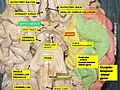
The type of visual field loss will depend on which portions of the optic nerve were damaged. In general, the location of the damage in relation to the optic chiasm (see diagram above) will affect the areas of vision loss. Damage to the optic nerve that is anterior, or in front of the optic chiasm (toward the face) causes loss of vision in the eye on the same side as the damage. Damage at the optic chiasm itself typically causes loss of vision laterally in both visual fields or bitemporal hemianopsia (see image to the right). Such damage may occur with large pituitary tumors, such as pituitary adenoma. Finally, damage to the optic tract, which is posterior to, or behind the chiasm, causes loss of the entire visual field from the side opposite the damage, e.g. if the left optic tract were cut, there would be a loss of vision from the entire right visual field.

Injury to the optic nerve can be the result of congenital or inheritable problems like Leber's hereditary optic neuropathy, glaucoma, trauma, toxicity, inflammation, ischemia, infection (very rarely), or compression from tumors or aneurysms. By far, the three most common injuries to the optic nerve are from glaucoma; optic neuritis, especially in those younger than 50 years of age; and anterior ischemic optic neuropathy, usually in those older than 50.

Glaucoma is a group of diseases involving loss of retinal ganglion cells causing optic neuropathy in a pattern of peripheral vision loss, initially sparing central vision. Glaucoma is frequently associated with increased intraocular pressure that damages the optic nerve as it exits the eyeball. The trabecular meshwork assists the drainage of aqueous humor fluid. The presence of excess aqueous humor, increases IOP, yielding the diagnosis and symptoms of glaucoma. [9]

Optic neuritis is inflammation of the optic nerve. It is associated with a number of diseases, the most notable one being multiple sclerosis. The patient will likely experience varying vision loss and eye pain. The condition tends to be episodic.

Anterior ischemic optic neuropathy is commonly known as a "stroke of the optic nerve" and affects the optic nerve head (where the nerve exits the eyeball). There is usually a sudden loss of blood supply and nutrients to the optic nerve head. Vision loss is typically sudden and most commonly occurs upon waking up in the morning. This condition is most common in diabetic patients 40–70 years old.

Other optic nerve problems are less common. Optic nerve hypoplasia is the underdevelopment of the optic nerve resulting in little to no vision in the affected eye. Tumors, especially those of the pituitary gland, can put pressure on the optic nerve causing various forms of visual loss. Similarly, cerebral aneurysms, a swelling of blood vessel(s), can also affect the nerve. Trauma can cause serious injury to the nerve. Direct optic nerve injury can occur from a penetrating injury to the orbit, but the nerve can also be injured by indirect trauma in which severe head impact or movement stretches or even tears the nerve. [1]

Ophthalmologists and optometrists can detect and diagnose some optic nerve diseases but neuro-ophthalmologists are often best suited to diagnose and treat diseases of the optic nerve. The International Foundation for Optic Nerve Diseases (IFOND) sponsors research and provides information on a variety of optic nerve disorders.

Additional images

See also

Related Research Articles

<span class="mw-page-title-main">Cranial nerves</span> Nerves that emerge directly from the brain and the brainstem

Cranial nerves are the nerves that emerge directly from the brain, of which there are conventionally considered twelve pairs. Cranial nerves relay information between the brain and parts of the body, primarily to and from regions of the head and neck, including the special senses of vision, taste, smell, and hearing.

<span class="mw-page-title-main">Optic chiasm</span> Part of the brain where the optic nerves cross

In neuroanatomy, the optic chiasm, or optic chiasma, is the part of the brain where the optic nerves cross. It is located at the bottom of the brain immediately inferior to the hypothalamus. The optic chiasm is found in all vertebrates, although in cyclostomes, it is located within the brain.

<span class="mw-page-title-main">Abducens nerve</span> Cranial nerve VI, for eye movements

The abducens nerve or abducent nerve, also known as the sixth cranial nerve, cranial nerve VI, or simply CN VI, is a cranial nerve in humans and various other animals that controls the movement of the lateral rectus muscle, one of the extraocular muscles responsible for outward gaze. It is a somatic efferent nerve.

Articles related to anatomy include:

<span class="mw-page-title-main">Oculomotor nerve</span> Cranial nerve III, for eye movements

The oculomotor nerve, also known as the third cranial nerve, cranial nerve III, or simply CN III, is a cranial nerve that enters the orbit through the superior orbital fissure and innervates extraocular muscles that enable most movements of the eye and that raise the eyelid. The nerve also contains fibers that innervate the intrinsic eye muscles that enable pupillary constriction and accommodation. The oculomotor nerve is derived from the basal plate of the embryonic midbrain. Cranial nerves IV and VI also participate in control of eye movement.

<span class="mw-page-title-main">Visual system</span> Body parts responsible for vision

The visual system comprises the sensory organ and parts of the central nervous system which gives organisms the sense of vision as well as enabling the formation of several non-image photo response functions. It detects and interprets information from the optical spectrum perceptible to that species to "build a representation" of the surrounding environment. The visual system carries out a number of complex tasks, including the reception of light and the formation of monocular neural representations, colour vision, the neural mechanisms underlying stereopsis and assessment of distances to and between objects, the identification of a particular object of interest, motion perception, the analysis and integration of visual information, pattern recognition, accurate motor coordination under visual guidance, and more. The neuropsychological side of visual information processing is known as visual perception, an abnormality of which is called visual impairment, and a complete absence of which is called blindness. Non-image forming visual functions, independent of visual perception, include the pupillary light reflex and circadian photoentrainment.

<span class="mw-page-title-main">Lateral geniculate nucleus</span> Component of the visual system in the brains thalamus

In neuroanatomy, the lateral geniculate nucleus is a structure in the thalamus and a key component of the mammalian visual pathway. It is a small, ovoid, ventral projection of the thalamus where the thalamus connects with the optic nerve. There are two LGNs, one on the left and another on the right side of the thalamus. In humans, both LGNs have six layers of neurons alternating with optic fibers.

<span class="mw-page-title-main">Pupillary light reflex</span> Eye reflex which alters the pupils size in response to light intensity

The pupillary light reflex (PLR) or photopupillary reflex is a reflex that controls the diameter of the pupil, in response to the intensity (luminance) of light that falls on the retinal ganglion cells of the retina in the back of the eye, thereby assisting in adaptation of vision to various levels of lightness/darkness. A greater intensity of light causes the pupil to constrict, whereas a lower intensity of light causes the pupil to dilate. Thus, the pupillary light reflex regulates the intensity of light entering the eye. Light shone into one eye will cause both pupils to constrict.

<span class="mw-page-title-main">Retinal ganglion cell</span> Type of cell within the eye

A retinal ganglion cell (RGC) is a type of neuron located near the inner surface of the retina of the eye. It receives visual information from photoreceptors via two intermediate neuron types: bipolar cells and retina amacrine cells. Retina amacrine cells, particularly narrow field cells, are important for creating functional subunits within the ganglion cell layer and making it so that ganglion cells can observe a small dot moving a small distance. Retinal ganglion cells collectively transmit image-forming and non-image forming visual information from the retina in the form of action potential to several regions in the thalamus, hypothalamus, and mesencephalon, or midbrain.

The visual field is "that portion of space in which objects are visible at the same moment during steady fixation of the gaze in one direction"; in ophthalmology and neurology the emphasis is on the structure inside the visual field and it is then considered “the field of functional capacity obtained and recorded by means of perimetry”.

<span class="mw-page-title-main">Optic tract</span> Neural pathway within the human visual system

In neuroanatomy, the optic tract is a part of the visual system in the brain. It is a continuation of the optic nerve that relays information from the optic chiasm to the ipsilateral lateral geniculate nucleus (LGN), pretectal nuclei, and superior colliculus.

<span class="mw-page-title-main">Accommodation reflex</span> Reflex action of the human eye

The accommodation reflex is a reflex action of the eye, in response to focusing on a near object, then looking at a distant object, comprising coordinated changes in vergence, lens shape (accommodation) and pupil size. It is dependent on cranial nerve II, superior centers (interneuron) and cranial nerve III. The change in the shape of the lens is controlled by ciliary muscles inside the eye. Changes in contraction of the ciliary muscles alter the focal distance of the eye, causing nearer or farther images to come into focus on the retina; this process is known as accommodation. The reflex, controlled by the parasympathetic nervous system, involves three responses: pupil constriction, lens accommodation, and convergence.

<span class="mw-page-title-main">Ciliary ganglion</span> Bundle of nerves, parasympathetic ganglion

The ciliary ganglion is a bundle of nerves, parasympathetic ganglion located just behind the eye in the posterior orbit. It is 1–2 mm in diameter and in humans contains approximately 2,500 neurons. The ganglion contains postganglionic parasympathetic neurons. These neurons supply the pupillary sphincter muscle, which constricts the pupil, and the ciliary muscle which contracts to make the lens more convex. Both of these muscles are involuntary since they are controlled by the parasympathetic division of the autonomic nervous system.

Dominant optic atrophy (DOA), or autosomal dominant optic atrophy (ADOA), (Kjer's type) is an autosomally inherited disease that affects the optic nerves, causing reduced visual acuity and blindness beginning in childhood. However, the disease can seem to re-present a second time with further vision loss due to the early onset of presbyopia symptoms (i.e., difficulty in viewing objects up close). DOA is characterized as affecting neurons called retinal ganglion cells (RGCs). This condition is due to mitochondrial dysfunction mediating the death of optic nerve fibers. The RGCs axons form the optic nerve. Therefore, the disease can be considered of the central nervous system. Dominant optic atrophy was first described clinically by Batten in 1896 and named Kjer’s optic neuropathy in 1959 after Danish ophthalmologist Poul Kjer, who studied 19 families with the disease. Although dominant optic atrophy is the most common autosomally inherited optic neuropathy (i.e., disease of the optic nerves), it is often misdiagnosed.

Optic neuropathy is damage to the optic nerve from any cause. The optic nerve is a bundle of millions of fibers in the retina that sends visual signals to the brain. [1].

<span class="mw-page-title-main">Chiasmal syndrome</span> Set of signs and symptoms that are associated with lesions of the optic chiasm

Chiasmal syndrome is the set of signs and symptoms that are associated with lesions of the optic chiasm, manifesting as various impairments of the affected's visual field according to the location of the lesion along the optic nerve. Pituitary adenomas are the most common cause; however, chiasmal syndrome may be caused by cancer, or associated with other medical conditions such as multiple sclerosis and neurofibromatosis.

<span class="mw-page-title-main">Cranial nerve examination</span> Type of neurological examination

The cranial nerve exam is a type of neurological examination. It is used to identify problems with the cranial nerves by physical examination. It has nine components. Each test is designed to assess the status of one or more of the twelve cranial nerves (I-XII). These components correspond to testing the sense of smell (I), visual fields and acuity (II), eye movements and pupils, sensory function of face (V), strength of facial (VII) and shoulder girdle muscles (XI), hearing and balance, taste, pharyngeal movement and reflex, tongue movements (XII).

<span class="mw-page-title-main">Anatomical terms of neuroanatomy</span> Terminology used to describe the central and peripheral nervous systems

This article describes anatomical terminology that is used to describe the central and peripheral nervous systems - including the brain, brainstem, spinal cord, and nerves.

<span class="mw-page-title-main">Outline of the human nervous system</span> Overview of and topical guide to the human nervous system

The following diagram is provided as an overview of and topical guide to the human nervous system:

<span class="mw-page-title-main">Visual pathway lesions</span> Overview about the lesions of visual pathways

The visual pathway consists of structures that carry visual information from the retina to the brain. Lesions in that pathway cause a variety of visual field defects. In the visual system of human eye, the visual information processed by retinal photoreceptor cells travel in the following way:
Retina→Optic nerve→Optic chiasma →Optic tract→Lateral geniculate body→Optic radiation→Primary visual cortex

References

  1. 1 2 3 4 5 Vilensky, Joel; Robertson, Wendy; Suarez-Quian, Carlos (2015). The Clinical Anatomy of the Cranial Nerves: The Nerves of "On Olympus Towering Top". Ames, Iowa: Wiley-Blackwell. ISBN   978-1118492017.
  2. 1 2 Selhorst, John; Chen, Yanjun (February 2009). "The Optic Nerve". Seminars in Neurology. 29 (1): 029–035. doi:10.1055/s-0028-1124020. ISSN   0271-8235. PMID   19214930.
  3. Smith, Austen M.; Czyz, Craig N. (2021). "Neuroanatomy, Cranial Nerve 2 (Optic)". StatPearls. Treasure Island (FL): StatPearls Publishing. PMID   29939684 . Retrieved 14 June 2021.
  4. Benowitz, Larry; Yin, Yuqin (August 2010). "Optic Nerve Regeneration". Archives of Ophthalmology. 128 (8): 1059–1064. doi:10.1001/archophthalmol.2010.152. ISSN   0003-9950. PMC   3072887 . PMID   20697009.
  5. Jonas, Jost B.; et al. (May 1992). "Human optic nerve fiber count and optic disc size". Investigative Ophthalmology & Visual Science. 33 (6): 2012–8. PMID   1582806.
  6. Textbook of Veterinary Anatomy, 4th Edition. Dyce, Sack and Wensing
  7. Belknap, Dianne B.; McCrea, Robert A. (1988-02-01). "Anatomical connections of the prepositus and abducens nuclei in the squirrel monkey". The Journal of Comparative Neurology. 268 (1): 13–28. doi:10.1002/cne.902680103. ISSN   0021-9967. PMID   3346381. S2CID   21565504.
  8. "Vision". casemed.case.edu. Archived from the original on 2020-01-26. Retrieved 2020-01-23.
  9. "The Eye's Drainage System, the Trabecular Meshwork & Glaucoma | BrightFocus Foundation". www.brightfocus.org. Retrieved 2022-01-10.