
The tongue is a muscular organ in the mouth of most vertebrates that manipulates food for mastication, and is used in the act of swallowing. It is of importance in the digestive system and is the primary organ of taste in the gustatory system. The tongue's upper surface (dorsum) is covered by taste buds housed in numerous lingual papillae. It is sensitive and kept moist by saliva, and is richly supplied with nerves and blood vessels. The tongue also serves as a natural means of cleaning the teeth. A major function of the tongue is the enabling of speech in humans and vocalization in other animals.

The glossopharyngeal nerve, known as the ninth cranial nerve, is a mixed nerve that carries afferent sensory and efferent motor information. It exits the brainstem out from the sides of the upper medulla, just rostral to the vagus nerve. The motor division of the glossopharyngeal nerve is derived from the basal plate of the embryonic medulla oblongata, while the sensory division originates from the cranial neural crest.

The hypoglossal nerve is the twelfth cranial nerve, and innervates all the extrinsic and intrinsic muscles of the tongue, except for the palatoglossus which is innervated by the vagus nerve. It is a nerve with a solely motor function. The nerve arises from the hypoglossal nucleus in the brain stem as a number of small rootlets, passes through the hypoglossal canal and down through the neck, and eventually passes up again over the tongue muscles it supplies into the tongue. There are two hypoglossal nerves in the body: one on the left, and one on the right.

The mandibular nerve (V3) is the largest of the three divisions of the trigeminal nerve, the fifth cranial nerve (CN V).

The paired submandibular glands are major salivary glands located beneath the floor of the mouth. They each weigh about 15 grams and contribute some 60–67% of unstimulated saliva secretion; on stimulation their contribution decreases in proportion as the parotid secretion rises to 50%.

The paired sublingual glands are major salivary glands in the mouth. They are the smallest, most diffuse, and the only unencapsulated major salivary glands. They provide only 3-5% of the total salivary volume. There are also two other types of salivary glands; they are submandibular and Parotid glands.
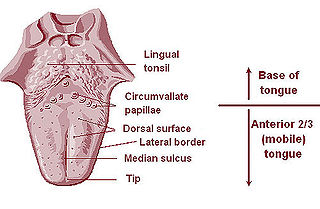
The lingual tonsils are two small mounds of lymphatic tissue located at the back of the base of the tongue, one on either side. They are composed of lymphatic tissue that functions to assist the immune system in the production of antibodies in response to invading pathogenic bacteria or viruses.

The palatoglossus, glossopalatinus, or palatoglossal muscle is a small fleshy fasciculus, narrower in the middle than at either end, forming, with the mucous membrane covering its surface, the glossopalatine arch.
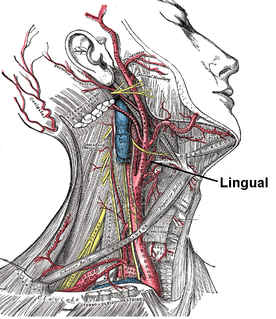
The lingual artery arises from the external carotid between the superior thyroid artery and facial artery. It can be located easily in the tongue.
The superior laryngeal nerve is a branch of the vagus nerve. It arises from the middle of the inferior ganglion of vagus nerve and in its course receives a branch from the superior cervical ganglion of the sympathetic nervous system.
One branch of the pterygopalatine ganglion, longer and larger than the others, is named the nasopalatine nerve.
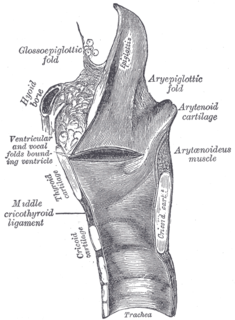
The anterior or lingual surface of the epiglottis is curved forward, and covered on its upper, free part by mucous membrane which is reflected on to the sides and root of the tongue, forming a median and two lateral glossoepiglottic folds; the lateral folds are partly attached to the wall of the pharynx.

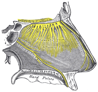
The greater palatine nerve is a branch of the pterygopalatine ganglion that carries both general sensory fibres from the maxillary nerve and parasympathetic fibers from the nerve of the pterygoid canal. It descends through the greater palatine canal, emerges upon the hard palate through the greater palatine foramen, and passes forward in a groove in the hard palate, nearly as far as the incisor teeth.

The intermediate nerve, nervus intermedius, nerve of Wrisberg or Glossopalatine nerve, is the part of the facial nerve located between the motor component of the facial nerve and the vestibulocochlear nerve. It contains the sensory and parasympathetic fibers of the facial nerve. Upon reaching the facial canal, it joins with the motor root of the facial nerve at the geniculate ganglion.
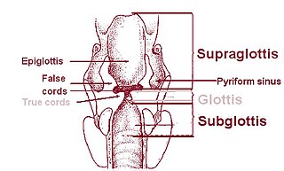
On either side of the laryngeal orifice in humans is a recess, termed the piriform sinus, which is bounded medially by the aryepiglottic fold, laterally by the thyroid cartilage and thyrohyoid membrane. The fossae are involved in speech.

The deep branch of the perineal nerve are distributed to the muscles of the perineum. These include the superficial transverse perineal muscle, bulbospongiosus, ischiocavernosus, and Sphincter urethræ.
Inferior alveolar nerve block is a nerve block technique which induces anesthesia (numbness) in the areas of the mouth and face innervated by one of the inferior alveolar nerves which are paired on the left and right side. These areas are the skin and mucous membranes of the lower lip, the skin of the chin, the lower teeth and the labial gingiva of the anterior teeth, all unilaterally to the midline of the side on which the block is administered. However, depending on technique, the long buccal nerve may not be anesthetized by an IANB and therefore an area of buccal gingiva adjacent to the lower posterior teeth will retain normal sensation unless that nerve is anesthetized separately, via a (long) buccal nerve block. The inferior alveolar nerve is a branch of the mandibular nerve, the third division of the trigeminal nerve. This procedure attempts to anaesthetise the inferior alveolar nerve prior to it entering the mandibular foramen on the medial surface of the mandibular ramus.

In human anatomy, the mouth is the first portion of the alimentary canal that receives food and produces saliva. The oral mucosa is the mucous membrane epithelium lining the inside of the mouth.

Lingual papillae are the small, nipple-like structures on the upper surface of the tongue that give it its characteristic rough texture. The four types of papillae on the human tongue have different structures and are accordingly classfied as circumvallate, fungiform, filiform, and foliate. All except the filiform papillae are associated with taste buds.


